



Teplizumab
Based on 1 publication(s) in Google Scholar
Teplizumab (MGA-031) is a Fc receptor non-binding anti-human CD3 monoclonal antibody. Teplizumab reduces the loss of beta-cell function. Teplizumab can be used in the research of type 1 diabetes.
For research use only. We do not sell to patients.
- Purity : 99.73%
- CAS No.: 876387-05-2
- Molecular Weight:150 kDa
-
Storage:
Please store the product under the recommended conditions in the Certificate of Analysis.


To place orders, for customer services and technical support, please contact: MedChemExpress USA
Tel: 609-228-6898 E-mail: [email protected] [email protected]


-
 Biological Activity
-
Chemical Information
- Protocols
- Purity & Documentation
- References
-
 Help & FAQs


-

HY-P99211
OtelixizumabAnti-CD3 Antibody -

HY-P99199
ForalumabCD3 Inhibitor -

HY-P99152
MuromonabCD3 Monoclonal Antibody -

HY-P9913
RituximabCD20 Inhibitor -

HY-P9904
AtezolizumabAnti-PD-L1 Antibody -

HY-P99539
UbamatamabAnti-MUC16/CD3 Antibody -

HY-P99038
OdronextamabCD20/CD3 Inhibitor -

HY-108829
AbataceptImmunomodulator -
HY-B0579
Cyclosporin ACalcineurin Inhibitor -

HY-P99392
TeclistamabBCMA/CD3 Dual Antibodies -

HY-P9908
AdalimumabTNF-α Inhibitor -
HY-B0490
Hygromycin BAntibiotic
Publications Citing Use of MedChemExpress (MCE) Teplizumab
More

Biological Activity
Description
Isotype
Human IgG1 (L234A/L235A) kappa
Recommend Isotype Controls
Species Reactivity
Human
IC50 & Target
CD3E
In Vitro
Teplizumab (10 ng/mL-10 μg/mL, 5 days) induces human CD8+ T-cell proliferation[3].
Teplizumab (6 days) increases the expression of CD25 and intracellular CTLA4 on CD8+ cells (freshly isolated PBMCs)[3].
MedChemExpress (MCE) has not independently confirmed the accuracy of these methods. They are for reference only. Further protocols information, click here.
-
Cell Line:Human PBMCs
-
Concentration:10 ng/mL, 100 ng/mL, 1 μg/mL, 10 μg/mL.
-
Incubation Time:5 days
-
Result:Induced cell proliferation (measured by incorporation of [3H]thymidine in a 5-day assay.
In Vivo
MedChemExpress (MCE) has not independently confirmed the accuracy of these methods. They are for reference only.
-
Animal Model:NOD/SCID IL2γc-/- (NSG) mice (a tolergenic humanized mouse model)[1]
-
Dosage:0.24 mg/kg, 5 μg
-
Administration:Intraperitoneal injection (i.p.)
-
Result:Decreased CD4:CD8 ratio in the peripheral blood from 2.03 to 1.01, p < 0.05.
Decreased circulating hCD4 cells in the peripheral blood.
Decreased in the proportion of T cells in the bone marrow and lung.
Increased in the total number of human CD45+ cells infiltrating the lamina propria of the small intestine.
Clinical Trial
| NCT Number | Sponsor | Condition | Start Date |
Phase

|
|---|---|---|---|---|
| NCT01329991 | Plexxikon| | 2011-05 | PHASE1 |
Gene ID
Accession
Conjugated
Unconjugated
Reconsititution
The product can be reconstituted/diluted with sterile PBS or saline.
Format
-
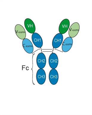
Human IgG1 kappa
Application
ELISA, FACS, Functional assay
Verified Bioactivity
-
 Immobilized Human CD3D&CD3E can bind Teplizumab. The EC50 for this effect is 102.5 ng/mL.
 -
 Flow cytometric analysis of 106 Jurkat cells with Teplizumab (HY-P99222, red). Cells were fixed with 4% paraformaldehyde. Then stained with the primary antibody at 1/200 dilution for an hour at 4℃. Alexa Fluor® 488-conjugated AffiniPure Goat Anti-Human IgG H&L (HY-P83776) was used as the secondary antibody at 1/1,000 dilution for 30 minutes at 4℃. Human IgG1 kappa (HY-P99001, blue) was used as the isotype control.


Chemical Information
-
CAS No. 876387-05-2
-
Appearance Liquid
-
Molecular Weight 150 kDa
-
Color Colorless to light yellow
-
SMILES
[Teplizumab]
-
Synonyms
MGA-031; PRV-031
-
Shipping
Shipping with dry ice.
-
Formulation
Please refer to the lot-specific COA for specific buffer information.
-
Storage
Please store the product under the recommended conditions in the Certificate of Analysis.


Publications (1)
-
Journal Impact Factor
-
Most Recent

Protocols
-
Research Protocol for Metabolic Diseases
AMP-activated protein kinase, AMPK, is a conserved cellular energy sensor that responds to reduced cellular energy status and coordinates metabolism by increasing ATP-generating catabolic pathways while suppressing ATP-consuming anabolic processes. In metabolic disease research, the AMPK pathway is experimentally relevant because it regulates hepatic lipid synthesis, fatty acid oxidation, glucose production, skeletal-muscle glucose disposal, mTORC1-linked biosynthesis, autophagy, mitochondrial homeostasis, and whole-body energy balance. The central pathway logic is that energy stress, metformin, exercise-like stimulation, or direct AMPK activators increase AMPKα Thr172 phosphorylation and downstream substrate phosphorylation, including ACC and RAPTOR. Phosphorylation of ACC suppresses lipogenesis and supports fatty acid oxidation, whereas phosphorylation of RAPTOR suppresses mTORC1 signaling and links cellular energy status to growth and protein synthesis control. The pathway is linked


Purity & Documentation
-
Data Sheet (262 KB)
-
SDS (252 KB)
- English - EN (252 KB)
- Français - FR (252 KB)
- Deutsch - DE (252 KB)
- Norwegian - NO (252 KB)
- Español - ES (252 KB)
- Swedish - SV (252 KB)
- Italian - IT (252 KB)
- Korean - KR (252 KB)
- Portuguese - PT (252 KB)
-
Inhibitory Antibodies User Guide (603 KB)
References
[1]. Kevan C Herold, et al. An Anti-CD3 Antibody, Teplizumab, in Relatives at Risk for Type 1 Diabetes. N Engl J Med. 2019 Aug 15;381(7):603-613. [Content Brief]
[2]. Frank Waldron-Lynch, et al. I Analysis of FcR non-binding anti-CD3 mAb in humanized mice identifies novel human gut tropic cells with regulatory function that are found in patients. Sci Transl Med. 2012 Jan 25;4(118):118ra12. [Content Brief]
[3]. Brygida Bisikirska, et al. TCR stimulation with modified anti-CD3 mAb expands CD8+ T cell population and induces CD8+CD25+ Tregs. J Clin Invest. 2005 Oct;115(10):2904-13. [Content Brief]

Calculators
Concentration (start) × Volume (start) = Concentration (final) × Volume (final)


Teplizumab Related Classifications
HY-P99222 Related Classifications
 Powered by Bioz