



Teprotumumab
Based on 1 publication(s) in Google Scholar
Teprotumumab is an IGF-1 receptor (IGF-1R) blocking human monoclonal antibody. Teprotumumab binds to the ligand binding extracellular α-subunit domain of IGF-1R. Teprotumumab inhibits TSH and IGF-1 action in fibrocytes. Teprotumumab attenuates TSH-dependent IL-6 and IL-8 expression and Akt phosphorylation. Teprotumumab can be used for thyroid-associated ophthalmopathy research.
Nos produits utilisent uniquement pour la recherche. Nous ne vendons pas aux patients.
- Pureté : 99.46%
- CAS No.: 1036734-93-6
- Masse moléculaire:145.671 kDa
-
Stockage:
Please store the product under the recommended conditions in the Certificate of Analysis.


To place orders, for customer services and technical support, please contact: MedChemExpress USA
Tel: 609-228-6898 E-mail: [email protected] [email protected]


-
 Activité biologique
-
Chemical Information
- Protocole
- Pureté et documentation
- Références
-
 Help & FAQs


-

HY-P99294
GanitumabAnti-IGF1R Antibody -

HY-P99197
FigitumumabAnti-IGF1R Antibody -

HY-P99274
XentuzumabIGF1/IGF2 Antagonist -

HY-P99218
RobatumumabAnti-human IGF-1R Antibody -

HY-P99001
Human IgG1 kappa, Isotype ControlIgG1κ Isotype Control -

HY-P99189
CixutumumabAnti-IGF-1R mAb -

HY-P990534
Anti-TSHR Antibody (K1-70)Anti-TSHR Antibody -

HY-P99284
DalotuzumabHumanized Anti-IGF1R mAb -

HY-15494
PicropodophyllinApoptosis Inducer -

HY-P9908
AdalimumabTNF-α Inhibitor -
HY-P991202
Anti-TSHR Antibody (M22)Anti-TSHR Antibody -
HY-10191
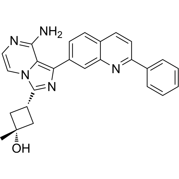
LinsitinibIGF-1R/Insulin Receptor Inhibitor
Publications Citing Use of MedChemExpress (MCE) Teprotumumab
More Customer Validation & Images

-
RT-PCR


Activité biologique
Description
Isotype
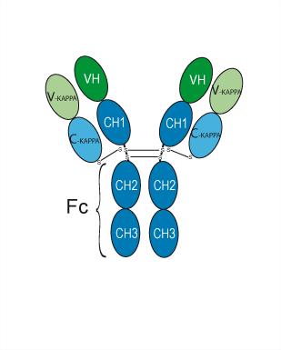
Human IgG1 kappa
Recommend Isotype Controls
Species Reactivity
Human
IC50 & Target
IGF1R/CD221
In Vitro
Essai clinique
| NCT Number | Sponsor | Condition | Start Date |
Phase

|
|---|---|---|---|---|
| NCT01329991 | 2011-05 | PHASE1 |
Gene ID
Accession
Conjugated
Unconjugated
Reconsititution
The product can be reconstituted/diluted with sterile PBS or saline.
Format
-
Human IgG1 kappa
Application
ELISA, FACS, Functional assay
Verified Bioactivity
-
 Immobilized IGF-I R Protein, Human (HEK293, His, HY-P72606) can bind Teprotumumab. The EC50 for this effect is 1.568 ng/mL.
 -
 Loaded Teprotumumab on AHC2 biosensor, can bind IGF-I R Protein, Human (Biotinylated, HEK293, His-Avi, HY-P78142) with an affinity constant of 4.315E-10 M as determined in BLI assay.


Chemical Information
-
CAS No. 1036734-93-6
-
Appearance Liquid
-
Masse moléculaire 145.671 kDa
-
Color Colorless to light yellow
-
SMILES
[Teprotumumab]
-
Livraison
Shipping with dry ice.
-
Formulation
Please refer to the lot-specific COA for specific buffer information.
-
Stockage
Please store the product under the recommended conditions in the Certificate of Analysis.


Publications (1)
-
Journal Impact Factor
-
Most Recent
-
J Clin Invest
Inhibition of aortic CX3CR1+ macrophages mitigates thoracic aortic aneurysm progression in Marfan syndrome in mice. [Abstract]2025 Jan 16;135(2):e178198. PMID: 39817456
Teprotumumab purchased from MedChemExpress. Usage Cited in: J Clin Invest. 2025 Jan 16;135(2):e178198. [Abstract]
Real-time PCR of gene expression in MFS patient-specific iPSC-derived VSMCs with conditioned media treatment in the presence of Teprotumumab (200 μg/mL; 24 h) or IgG. n = 4.


Pureté et documentation
-
Fiche technique (257 KB)
-
SDS (251 KB)
- English - EN (251 KB)
- Français - FR (251 KB)
- Deutsch - DE (251 KB)
- Norwegian - NO (251 KB)
- Español - ES (251 KB)
- Swedish - SV (251 KB)
- Italian - IT (251 KB)
- Korean - KR (251 KB)
- Portuguese - PT (251 KB)
-
Inhibitory Antibodies User Guide (603 KB)
Références

Calculators
Concentration (start) × Volume (start) = Concentration (final) × Volume (final)


Teprotumumab Related Classifications
HY-P99165 Related Classifications
 Powered by Bioz