



Tinurilimab
Based on 2 publication(s) in Google Scholar
Tinurilimab (Bay 1834942) is an anti-CEACAM6 (carcinoembryonic antigen-related cell adhesion molecule 6) humanized IgG2 monoclonal antibody. CEACAM6 is an immune checkpoint regulator suppressing the activity of effector T-cells against tumors. Tinurilimab shows an increased tumor cell killing effect in the tumor-cell/T-cell co-culture system.
For research use only. We do not sell to patients.
- Purity : 98.01%
- CAS No.: 2226224-30-0
- Molecular Weight:144.76 kDa
-
Storage:
Please store the product under the recommended conditions in the Certificate of Analysis.


To place orders, for customer services and technical support, please contact: MedChemExpress USA
Tel: 609-228-6898 E-mail: [email protected] [email protected]


-
 Biological Activity
-
Chemical Information
- Protocols
- Purity & Documentation
- References
-
 Help & FAQs







Publications Citing Use of MedChemExpress (MCE) Tinurilimab
More

Biological Activity
Description
Isotype
Human IgG2 kappa
Recommend Isotype Controls
Species Reactivity
Human
IC50 & Target
CEACAM6/CD66c
In Vitro
Tinurilimab (Bay 1834942) binds to human and cynomolgus monkey CEACAM6 with monovalent dissociation constants of KD of 13 nM and 31 nM, respectively. Tinurilimab is highly selective for human CEACAM6, as it is unable to bind to human CEACAM1, CEACAM3, or CEACAM5[1].
The half-maximal binding EC50 values for Tinurilimab are determined by flow cytometry in a panel of CEACAM6-expressing cancer cell lines. For Tinurilimab, the EC50 values are in the range of 0.15 nM (PA-TU-8902) to 5 nM (HCC2935)[1].
Tinurilimab (0-15 μg/mL) blocks the binding of recombinant CEACAM6-Fc to CEACAM1-coated plates in a dose-dependent manner[1].
Tinurilimab (30 µg/mL) increases type 1 cytokine secretion and the cytotoxic capacity of tumor-specific T cells[1].
Blockade of CEACAM6 by Tinurilimab (0.01-1000 µg/mL) restored T cell activity in a dose-dependent manner, as evidenced by increased IFN-γ secretion by survivin T cells co-incubated with recombinant CEACAM6-loaded beads[1].
MedChemExpress (MCE) has not independently confirmed the accuracy of these methods. They are for reference only. Further protocols information, click here.
Clinical Trial
| NCT Number | Sponsor | Condition | Start Date |
Phase

|
|---|---|---|---|---|
| NCT01329991 | Plexxikon| | 2011-05 | PHASE1 |
Gene ID
Accession
Conjugated
Unconjugated
Reconsititution
The product can be reconstituted/diluted with sterile PBS or saline.
Format
-
Human IgG2 kappa
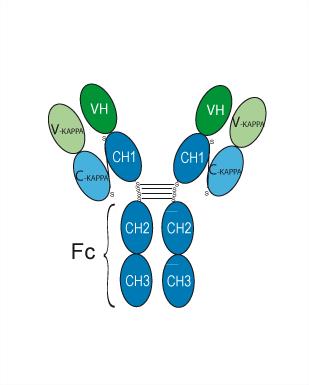
Application
ELISA, FACS, Functional assay
Verified Bioactivity
-
 Flow cytometric analysis of 1.5×10⁶ U937 cells with Satralizumab (HY-P99112, red). Cells were fixed with 4% paraformaldehyde and permeabilised with 90% methanol. Then stained with the primary antibody at 1/200 dilution for an hour at 4℃. Alexa Goat Anti-Human IgG H&L (AF488) (HY-P83776) was used as the secondary antibody at 1/1,000 dilution for 30 minutes at 4℃. Human IgG2 kappa Isotype Control (HY-P99002, blue) was used as the isotype control, cells without incubation with primary antibody were used as the unlabeled control (black).
 -
 Immobilized hu-CEACAM6-ECD-his can bind Tinurilimab. The EC50 for this effect is 6.45 ng/mL.


Chemical Information
-
CAS No. 2226224-30-0
-
Appearance Liquid
-
Molecular Weight 144.76 kDa
-
Color Colorless to light yellow
-
SMILES
[Tinurilimab]
-
Synonyms
BAY 1834942
-
Shipping
Shipping with dry ice.
-
Formulation
Please refer to the lot-specific COA for specific buffer information.
-
Storage
Please store the product under the recommended conditions in the Certificate of Analysis.


Publications (2)
-
Journal Impact Factor
-
Most Recent
-
Cancer Lett
A heavy-chain antibody-drug conjugate targeting glycosylated CEACAM6 inhibits brain metastatic tumor growth. [Abstract]2026 Jun 28:648:218463. PMID: 41912133 -

Protocols
-
Neuron-Astrocyte Co-culture
Neuron-astrocyte co-culture is used to study how astrocytes regulate neuronal survival, synapse formation, dendritic morphology, neuronal activity, and disease-related neurotoxicity. Indirect “sandwich” or insert-based designs physically separate neurons and astrocytes while allowing soluble astrocyte-derived factors to affect neurons, whereas direct co-culture permits cell-contact and network-level readouts.
-
Research Protocol for Cancer Immunology
Cancer immunology studies how the immune system recognizes, suppresses, edits, or fails to eliminate malignant cells through tumor antigen release, antigen presentation, T-cell priming, immune trafficking, tumor-cell killing, and feedback inhibition in the tumor microenvironment. The cancer-immunity cycle links tumor antigenicity, dendritic-cell priming, CD8+ T-cell infiltration, cytotoxic function, and immune-checkpoint regulation to tumor rejection or immune escape. Immune-checkpoint pathways such as PD-1/PD-L1 and CTLA-4 suppress antitumor T-cell activity and can be therapeutically blocked, but many tumors remain resistant because of poor antigen presentation, weak T-cell infiltration, suppressive myeloid cells, regulatory T cells, and tumor-intrinsic immune-exclusion programs. Unresolved questions include which immune-cell states predict response, how tumor-intrinsic pathways exclude immune cells, how myeloid suppression limits checkpoint blockade, and which combination strategies


Purity & Documentation
-
Data Sheet (260 KB)
-
SDS (251 KB)
- English - EN (251 KB)
- Français - FR (251 KB)
- Deutsch - DE (251 KB)
- Norwegian - NO (251 KB)
- Español - ES (251 KB)
- Swedish - SV (251 KB)
- Italian - IT (251 KB)
- Korean - KR (251 KB)
- Portuguese - PT (251 KB)
-
Inhibitory Antibodies User Guide (603 KB)
References

Calculators
Concentration (start) × Volume (start) = Concentration (final) × Volume (final)


Tinurilimab Related Classifications
HY-P99051 Related Classifications
 Powered by Bioz