



Tislelizumab
Based on 3 publication(s) in Google Scholar
Tislelizumab is a monoclonal antibody that specifically binds to programmed cell death receptor 1 (PD-1), blocking its interaction with programmed death ligand 1 (PD-L1) and programmed death ligand 2 (PD-L2). Tislelizumab can reactivate immune cells such as T lymphocytes and enhance anti-tumor activity. Tislelizumab can be used for the research of a variety of tumors including typical Hodgkin's lymphoma, urothelial carcinoma, non-small cell lung cancer and hepatocellular carcinoma.
For research use only. We do not sell to patients.
- Purity : 98.10%
- CAS No.: 1858168-59-8
- Molecular Weight:145 kDa
-
Storage:
Please store the product under the recommended conditions in the Certificate of Analysis.



To place orders, for customer services and technical support, please contact: MedChemExpress USA
Tel: 609-228-6898 E-mail: [email protected] [email protected]


-
 Biological Activity
-
Chemical Information
- Protocols
- Purity & Documentation
- References
-
 Help & FAQs


-

HY-P9902
PembrolizumabPD-1 Inhibitor -

HY-P9903
NivolumabPD-1 Inhibitor -

HY-P9978
ToripalimabPD-1 Inhibitor -

HY-P99048
SintilimabPD-1/PD-L1 Inhibitor -

HY-P9971
CamrelizumabPD-1 Neutralizing Antibody -
HY-P9904
AtezolizumabAnti-PD-L1 Antibody -

HY-P99203
CemiplimabPD-1 Inhibitor -

HY-P9919
DurvalumabPD-L1 Blocking Antibody -

HY-P99118
SerplulimabAnti-PD-1 Antibody -

HY-P99115
EnvafolimabAnti PD-L1 Antibody -

HY-P99345
DostarlimabAnti-PD-1 mAb -

HY-P99108
PenpulimabAnti-PD-1 Monoclonal Antibody
Publications Citing Use of MedChemExpress (MCE) Tislelizumab
More Customer Validation & Images

-
ELISA


Biological Activity
Description
Isotype
Human IgG4 kappa
Recommend Isotype Controls
Species Reactivity
Human
IC50 & Target
PDCD1/PD-1/CD279
In Vivo
MedChemExpress (MCE) has not independently confirmed the accuracy of these methods. They are for reference only.
-
Animal Model:B16F10/GM-CSF tumor cells treated hPD-1 C57BL/6 transgenic mice[2]
-
Dosage:10 mg/kg
-
Administration:Intraperitoneal injection (i.p.); once a week for 4 weeks.
-
Result:Exerted remarkable antitumor efficacy, resulting in approximate 86.7% tumor growth inhibition.
Clinical Trial
| NCT Number | Sponsor | Condition | Start Date |
Phase

|
|---|---|---|---|---|
| NCT01329991 | Plexxikon| | 2011-05 | PHASE1 |
Gene ID
Accession
Conjugated
Unconjugated
Reconsititution
The product can be reconstituted/diluted with sterile PBS or saline.
Format
-
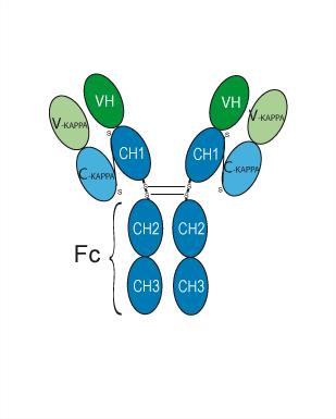
Human IgG4 kappa
Application
ELISA, FACS, Functional assay
Verified Bioactivity


Chemical Information
-
CAS No. 1858168-59-8
-
Appearance Liquid
-
Molecular Weight 145 kDa
-
Color Colorless to light yellow
-
SMILES
[Tislelizumab]
-
Synonyms
BGB-A317
-
Formulation
Please refer to the lot-specific COA for specific buffer information.
-
Storage
Please store the product under the recommended conditions in the Certificate of Analysis.


Publications (3)
-
Journal Impact Factor
-
Most Recent
-
Signal Transduct Target Ther
N-deglycosylation targeting chimera (DGlyTAC): a strategy for immune checkpoint proteins inactivation by specifically removing N-glycan. [Abstract]2025 Apr 28;10(1):139. PMID: 40289109 -
J Pharm Anal
Increasing the tumour targeting of antitumour drugs through anlotinib-mediated modulation of the extracellular matrix and the RhoA/ROCK signalling pathway. [Abstract]2024 Aug;14(8):100984. PMID: 39258171 -
Cancer Cell Int
Blood-based molecular and cellular biomarkers of early response to neoadjuvant PD-1 blockade in patients with non-small cell lung cancer. [Abstract]2024 Jun 29;24(1):225. PMID: 38951894
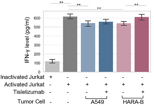
Tislelizumab purchased from MedChemExpress. Usage Cited in: Cancer Cell Int. 2024 Jun 29;24(1):225. [Abstract]
Tislelizumab (50 µg/mL; 24 h). IFN-γ concentration in the supernatant of co-cultured cell lines was assessed by ELISA.

Protocols
-
Apoptosis
Apoptosis, also called programmed cell death, is generally characterized by distinct morphological characteristics.
-
Cell Cytotoxicity Assay
Cytotoxicity assays are usually based on the assessment of cell membrane damage, which can also be indirectly detected by measuring cell viability. Detection methods include MTT assay, CKK-8 assay, LDH assay and ATP assay, etc.
-
Mammalian live/dead viability and cytotoxicity staining
Live/dead viability and cytotoxicity staining assays are based on the simultaneous detection of intracellular esterase activity in metabolically active (viable) cells and membrane integrity loss in non-viable cells. In commonly used dual-staining approaches, membrane-permeant fluorogenic substrates are converted by intracellular esterases into fluorescent products in live cells, while impermeant DNA-binding dyes selectively enter cells with compromised plasma membranes and label nucleic acids in dead or dying cells, enabling discrimination between viable and non-viable populations by fluorescence microscopy or flow cytometry.
-
Apoptosis Solutions
Apoptosis is a regulated, generally non-lytic cell-death pathway that removes unwanted, damaged, infected, or abnormal cells through coordinated morphological changes, caspase activation, DNA fragmentation, and membrane remodeling. The intrinsic apoptosis pathway is controlled mainly by mitochondrial outer membrane permeabilization, BCL-2 family proteins, cytochrome c release, apoptosome formation, caspase-9 activation, and downstream executioner caspase-3/7 activation. The extrinsic apoptosis pathway is initiated by death receptors such as Fas, TNFR, and TRAIL receptors, which recruit adaptor proteins and activate caspase-8 before engaging executioner caspases or mitochondrial amplification through BID cleavage. Apoptosis is linked to many phenotypes, including cancer cell killing, tissue homeostasis, immune regulation, neurodegeneration, infection response, and treatment-induced cytotoxicity; unresolved questions include how apoptosis interacts with necroptosis, pyroptosis, ferroptos
-
Liver Cancer Modeling
Liver cancer can be classified into primary liver cancer and secondary liver cancer. Secondary liver cancer is the metastatic liver cancer. Primary liver cancer includes hepatocellular carcinoma (HCC), intrahepatic cholangiocarcinoma (ICC) and fibrolamellar HCC, of which HCC is the most common form, accounting for approximately 90% of primary liver cancers[1]. HCC mouse models include chemical agent-induced models, transplanted tumor models, and genetic engineered models.


Purity & Documentation
-
Data Sheet (275 KB)
-
SDS (251 KB)
- English - EN (251 KB)
- Français - FR (251 KB)
- Deutsch - DE (251 KB)
- Norwegian - NO (251 KB)
- Español - ES (251 KB)
- Swedish - SV (251 KB)
- Italian - IT (251 KB)
- Korean - KR (251 KB)
- Portuguese - PT (251 KB)
-
Inhibitory Antibodies User Guide (603 KB)
References
[1]. Jie Wang, et al. Tislelizumab Plus Chemotherapy vs Chemotherapy Alone as First-line Treatment for Advanced Squamous Non-Small-Cell Lung Cancer: A Phase 3 Randomized Clinical Trial. JAMA Oncol. 2021 May 1;7(5):709-717. [Content Brief]
[2]. Hong Y, et al. Tislelizumab uniquely binds to the CC' loop of PD-1 with slow-dissociated rate and complete PD-L1 blockage. FEBS Open Bio. 2021 Mar;11(3):782-792. [Content Brief]
[3]. Zhu K, et al. Expression of HER2 in high-grade urothelial carcinoma based on Chinese expert consensus and the clinical effects of disitamab vedotin-tislelizumab combination therapy in the treatment of advanced patients. Front Pharmacol. 2024 Feb 22;15:1355081. [Content Brief]

Calculators
Concentration (start) × Volume (start) = Concentration (final) × Volume (final)


Tislelizumab Related Classifications
HY-P99052 Related Classifications
 Powered by Bioz