



Toluidine blue (ZnCl2)

Based on 1 Customer Validation
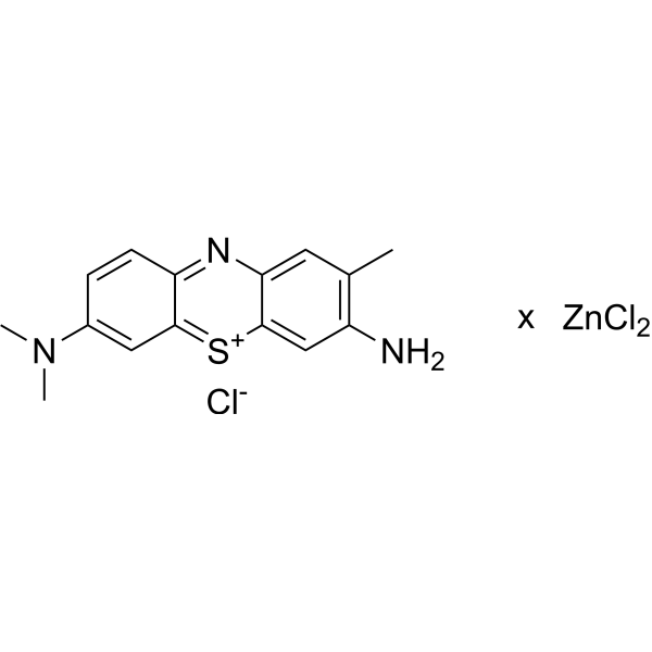
Toluidine blue (ZnCl2) is a basic thiazine dye commonly used as a biological stain for microscopy. It has a deep bluish-purple color and is commonly used to stain nucleic acids such as DNA and RNA, as well as to stain mast cells, cartilage, and other connective tissues. Toluidine blue (ZnCl2) stains the acidic components of these tissues, such as sulfated or carboxylated mucopolysaccharides. It is frequently used in histology, cytology, and pathology applications to aid in the diagnosis of various diseases and conditions. The dye is usually applied to tissue sections prior to microscopic examination and can be differentiated using an acidic alcohol solution. Toluidine blue (ZnCl2) is a relatively simple and inexpensive stain with good reproducibility, making it a popular choice for many laboratories.
Para uso exclusivo en investigación. No vendemos a pacientes.
- Assay : 84.54%
- No. CAS: 6586-04-5
- Fòrmula: C15H16N3S.xCl2Zn.Cl
-
Almacenamiento:
-20°C, protect from light
* In solvent : -80°C, 6 months; -20°C, 1 month (protect from light)

To place orders, for customer services and technical support, please contact: MedChemExpress USA
Tel: 609-228-6898 E-mail: [email protected] [email protected]


-
 Actividad biológica
-
Chemical Information
-
Solvente y solubilidad
- Protocolo
- Pureza y Documentación
-
 Help & FAQs


-
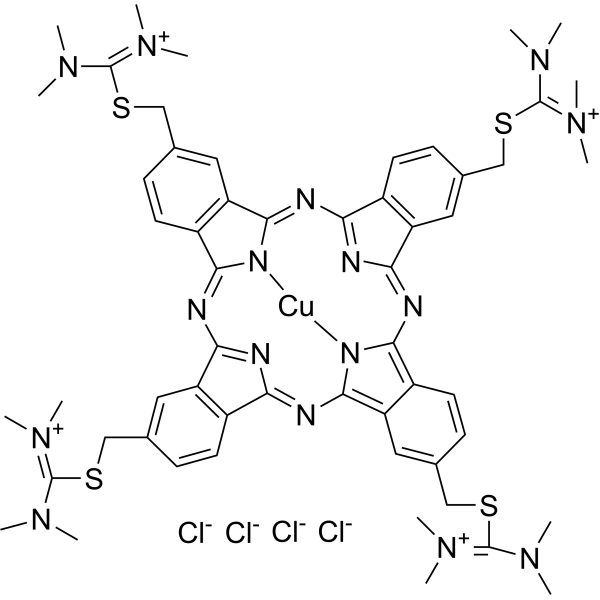
HY-D0001
Alcian Blue 8GXCationic Copper Phthalocyanine Dye -
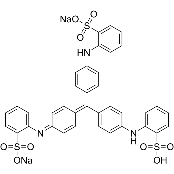
HY-D0003
Methyl BlueAntiseptic Dye -
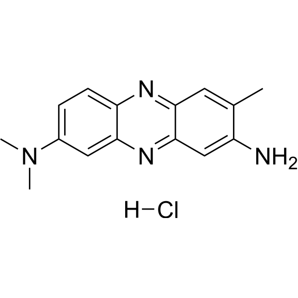
HY-D0166
Neutral RedBiochemical Assay Reagent -
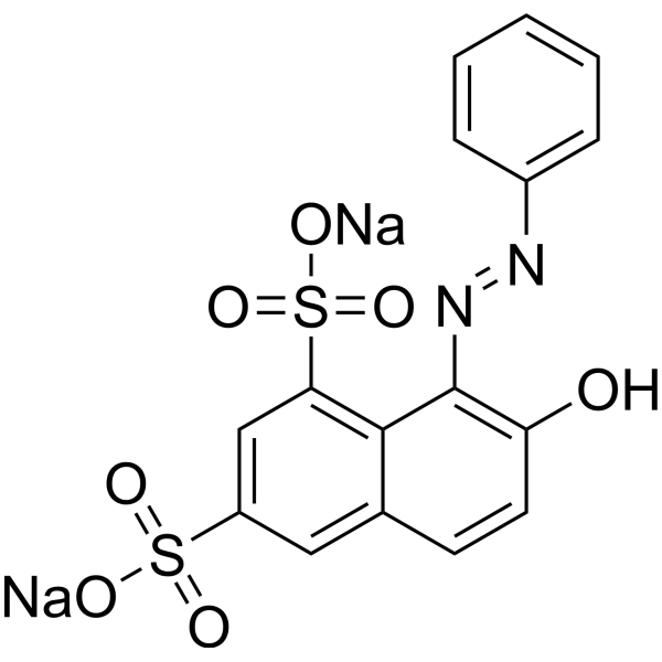
HY-135712
Orange GBiochemical Assay Reagent -
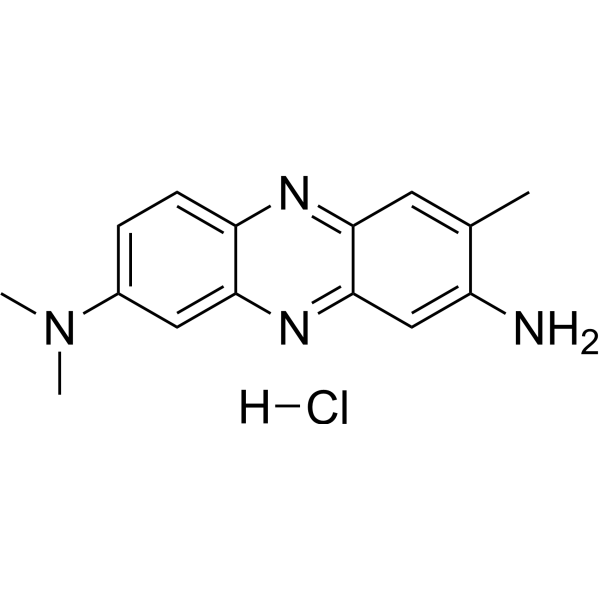
HY-D0166A
Neutral Red (IND)Biochemical Assay Reagent -

HY-D0854
Acid Red sodiumBiochemical Assay Reagent -
HY-D0803
ThymoquinoneNatural Product -
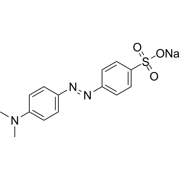
HY-118907
Methyl OrangeAzo Dye -
HY-D0950A
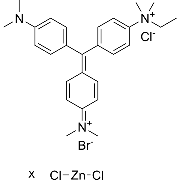
Methyl Green zinc chlorideDNA Fluorescent Dye -

HY-118907R
Methyl Orange (Standard)Azo Dye -

HY-D0803R
Thymoquinone (Standard)Natural Product -
HY-W127718
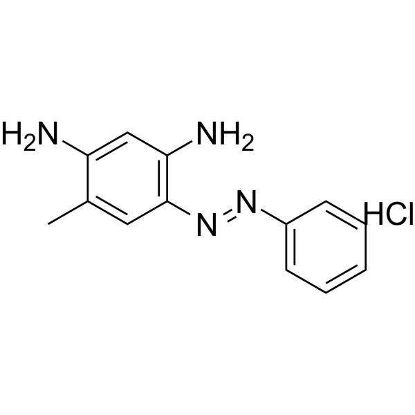
Chrysoidine RBiochemical Assay Reagent


Actividad biológica
Descripciòn
In Vitro
Toluidine blue for microscopy is a biochemical reagent that can be used as a biological material or organic compound for life science related research.
MedChemExpress (MCE) has not independently confirmed the accuracy of these methods. They are for reference only. Further protocols information, click here.


Chemical Information
-
No. CAS 6586-04-5
-
Appearance Solid
-
Fòrmula C15H16N3S.xCl2Zn.Cl
-
Color Brown to black
-
SMILES
NC1=C(C)C=C2N=C3C=CC(N(C)C)=CC3=[S+]C2=C1.Cl[Zn]Cl.[Cl-].[x]
-
Envío
Room temperature in continental US; may vary elsewhere.
-
Almacenamiento
-20°C, protect from light
* In solvent : -80°C, 6 months; -20°C, 1 month (protect from light)


Solvente y solubilidad
In Vitro:
H2O : 1 mg/mL (ultrasonic and warming and heat to 60°C)

Protocolo
-
Paraffin Section
The tissue is embedded in paraffin wax, cut into thin slices by a microtome, stained and sealed and finally made into samples for microscopic observation.
-
Protocol for Hematoxylin-Eosin (H&E) Staining
Hematoxylin-eosin staining is a routine histological method that stains nuclei mainly blue-purple with hemalum and stains cytoplasm, extracellular matrix, and many stromal components pink with eosin, allowing tissue architecture, cell morphology, necrosis, inflammation, fibrosis, tumor growth pattern, and treatment-associated injury to be evaluated by light microscopy. In cancer cells, primary neurons, mouse tumor models, intestinal organoids, inflammatory macrophage preparations, and drug-screening tissues, H&E is a morphology assay rather than a molecular assay; it should be interpreted with complementary molecular or immunostaining assays when the biological question concerns specific proteins, RNA levels, ferroptosis, mitophagy, or immune phenotypes.


Pureza y Documentación
-
Ficha de datos (239 KB)
-
SDS (393 KB)
- English - EN (393 KB)
- Français - FR (393 KB)
- Deutsch - DE (393 KB)
- Norwegian - NO (393 KB)
- Español - ES (393 KB)
- Swedish - SV (393 KB)
- Italian - IT (393 KB)
- Korean - KR (393 KB)
- Portuguese - PT (393 KB)
-
Instrucciones de manejo (2659 KB)

Calculators
Concentration (start) × Volume (start) = Concentration (final) × Volume (final)


Toluidine blue (ZnCl2) Related Classifications
HY-W250143 Related Classifications
 Powered by Bioz