



Trontinemab

Based on 1 Customer Validation
Trontinemab (RG6102) is a brain-penetrant, anti-amyloid, bispecific and humanizedized IgG1-κ antibody, targeting to Aβ plaques and transferrin receptor 1 (TFR1). Trontinemab binds to fibrillar Aβ as well as Aβ plaques triggering plaque clearance by engaging immune cells on Alzheimer disease (AD) brain sections. Trontinemab also shows specific affinity to cynomolgus and human TFR1.
For research use only. We do not sell to patients.
- Purity : 99.9%
- CAS No.: 2568868-35-7
- Molecular Weight:194.25 kDa
-
Storage:
Please store the product under the recommended conditions in the Certificate of Analysis.


To place orders, for customer services and technical support, please contact: MedChemExpress USA
Tel: 609-228-6898 E-mail: [email protected] [email protected]


-
 Biological Activity
-
Chemical Information
- Protocols
- Purity & Documentation
- References
-
 Help & FAQs


-

HY-P99022
GantenerumabAnti-Aβ Monoclonal Antibody -

HY-P99797
Pabinafusp alfaAnti-Transferrin Receptor Antibody -

HY-P99001
Human IgG1 kappa, Isotype ControlIgG1κ Isotype Control -

HY-P991047
Anti-CD71/TfR1 AntibodyAnti TfR1 Antibody -

HY-P990724
Tividenofusp alfaID2S Fusion Protein -

HY-13757A
TamoxifenEstrogen Receptor Modulator -

HY-17394
CisplatinDNA Alkylator -
HY-12216
ResmetiromTHR-β Agonist -

HY-P99575
Tarlatamabanti-DLL3/CD3 BiTE antibody -

HY-15763
ErastinFerroptosis Inducer -

HY-P9908
AdalimumabTNF-α Inhibitor -
HY-134541
SM-102Ionizable Amino Lipid


Biological Activity
Description
Isotype
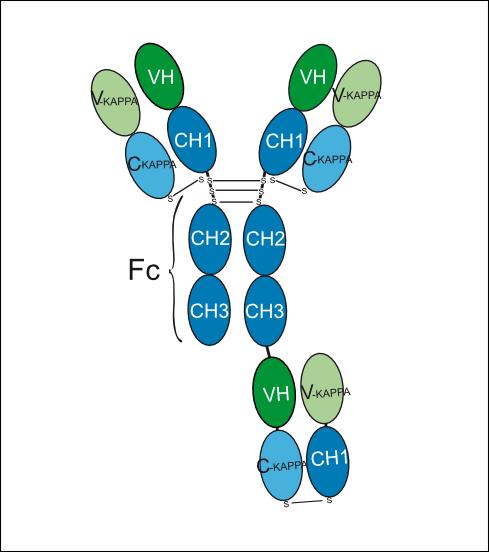
IgG1-kappa-(Fab G1-kappa with domain crossover)
Recommend Isotype Controls
Species Reactivity
Human
IC50 & Target
APP & TFRC
In Vitro
Trontinemab (0-5 μg/mL, 45 min)-decorated Aβ plaques by Fc receptor-mediated phagocytosis is a major mechanism of Aβ plaque clearance in primary human macrophages[3].
MedChemExpress (MCE) has not independently confirmed the accuracy of these methods. They are for reference only. Further protocols information, click here.
-
Cell Line:Primary human macrophages
-
Concentration:0-5 μg/mL
-
Incubation Time:45 min
-
Result:Decreased Aβ plaque load dose-dependently in primary human macrophages.
In Vivo
MedChemExpress (MCE) has not independently confirmed the accuracy of these methods. They are for reference only.
Clinical Trial
| NCT Number | Sponsor | Condition | Start Date |
Phase

|
|---|---|---|---|---|
| NCT01329991 | Plexxikon| | 2011-05 | PHASE1 |
Gene ID
Accession
Conjugated
Unconjugated
Reconsititution
The product can be reconstituted/diluted with sterile PBS or saline.
Format
-
IgG1-kappa-(Fab G1-kappa with domain crossover)
-
IgG1-kappa-(Fab G1-kappa with domain crossover)

Application
ELISA, FACS, Functional assay
Verified Bioactivity
-
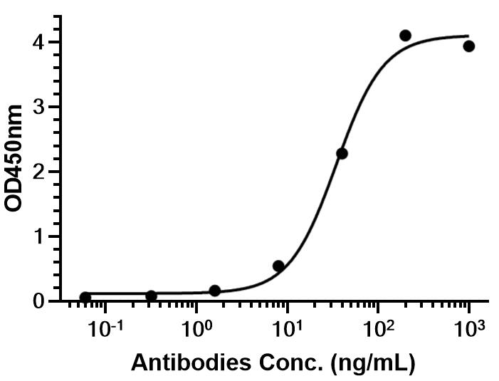 Immobilized Human Beta-amyloid/Beta-APP Protein (hFc Tag) can bind Trontinemab. The EC50 for this effect is 34.64 ng/mL.
 -
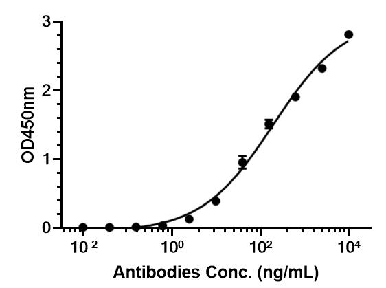 Immobilized HY-P71069 TFRC Protein, Human (HEK293, His) can bind Trontinemab. The EC50 for this effect is 193.5 ng/mL.


Chemical Information
-
CAS No. 2568868-35-7
-
Appearance Liquid
-
Molecular Weight 194.25 kDa
-
Color Colorless to light yellow
-
SMILES
[Trontinemab]
-
Synonyms
RG6102; RO-7126209
-
Shipping
Shipping with dry ice.
-
Formulation
Please refer to the lot-specific COA for specific buffer information.
-
Storage
Please store the product under the recommended conditions in the Certificate of Analysis.

Protocols
-
Protocol for Pharmacokinetic Study
Pharmacokinetic studies quantify how an organism handles a drug over time through absorption, distribution, metabolism, and excretion, and the core experimental readout is the concentration-time profile of parent drug and, when relevant, metabolites in biological matrices such as plasma, whole blood, urine, bile, or tissue. Pharmacokinetic analysis links dose, route, exposure, clearance, half-life, distribution, bioavailability, and systemic exposure to drug efficacy and toxicity hypotheses rather than measuring a signaling pathway directly. The literature links pharmacokinetics to drug-development phenotypes by showing that drug metabolism and pharmacokinetics influence compound progression, exposure-response interpretation, safety margins, dosing strategy, and failure risk during discovery and development. DMPK science contributes to compound optimization by integrating physicochemical properties, in vitro metabolism, transporter behavior, in vivo exposure, and pharmacodynamic contex
-
Amyloid: Congo Red Amyloid Staining
Congo red amyloid staining is a histochemical method used to detect extracellular amyloid deposits in tissue sections based on the affinity of Congo red dye for β-pleated sheet-rich protein aggregates. When bound to amyloid, Congo red produces characteristic apple-green birefringence under polarized light microscopy, which is widely regarded as a diagnostic feature of amyloid deposition in histopathology. The diagnostic principle relies on the combination of dye binding (congophilia) and optical anisotropy under polarized illumination, which distinguishes amyloid from most non-amyloid eosinophilic extracellular deposits in routine histological evaluation. Amyloid identification by Congo red staining remains a cornerstone in diagnostic pathology despite the availability of adjunct methods such as immunohistochemistry and mass spectrometry, particularly because of its ability to localize deposits directly within tissue architecture. The specificity of Congo red-positive deposits is incre
-
Alzheimer’s Disease Modeling
Alzheimer’s Disease (AD) is a neurodegenerative disorder characterized by a progressive decline in cognitive functions and loss of specific types of neurons and synapses. Alzheimer's symptoms can be simulated in mice by injecting drugs (such as Aβ) or genetically modified.


Purity & Documentation
-
Data Sheet (261 KB)
-
SDS (251 KB)
- English - EN (251 KB)
- Français - FR (251 KB)
- Deutsch - DE (251 KB)
- Norwegian - NO (251 KB)
- Español - ES (251 KB)
- Swedish - SV (251 KB)
- Italian - IT (251 KB)
- Korean - KR (251 KB)
- Portuguese - PT (251 KB)
-
Inhibitory Antibodies User Guide (603 KB)
References
[1]. Neațu M, et al. Monoclonal Antibody Therapy in Alzheimer's Disease. Pharmaceutics. 2023 Dec 29;16(1):60. [Content Brief]
[2]. Trontinemab. IMGT/mAb-DB.
[3]. Weber F, et al. Brain Shuttle Antibody for Alzheimer's Disease with Attenuated Peripheral Effector Function due to an Inverted Binding Mode. Cell Rep. 2018 Jan 2;22(1):149-162. [Content Brief]

Calculators
Concentration (start) × Volume (start) = Concentration (final) × Volume (final)


Trontinemab Related Classifications
HY-P9999 Related Classifications
 Powered by Bioz