



TT-012
Based on 2 publication(s) in Google Scholar
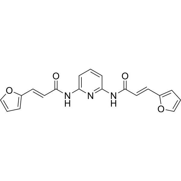
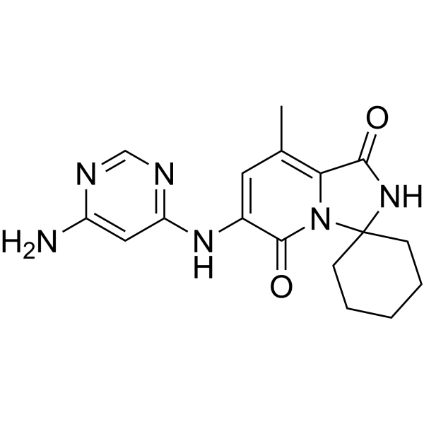
TT-012 is a MITF inhibitor with a human MITF IC50 of 13.1 nM and a human MITF Kd value of 15.5 nM. TT-012 reduces mRNA levels of MITF downstream genes linked to melanosome biogenesis, cell survival, and proliferation, and upregulates cell cycle-inhibiting genes. TT-012 can be used for the research of melanoma[1][2][3].
For research use only. We do not sell to patients.
- Purity : 99.76%
- CAS No.: 1164471-33-3
- Formula: C19H15N3O4
- Molecular Weight:349.34
-
Storage:Powder -20°C, 3 years , 4°C, 2 years ; In solvent -80°C, 6 months , -20°C, 1 month

To place orders, for customer services and technical support, please contact: MedChemExpress USA
Tel: 609-228-6898 E-mail: [email protected] [email protected]


-
 Biological Activity
-
Chemical Information
-
Solvent & Solubility
- Protocols
- Purity & Documentation
- References
-
 Help & FAQs





Publications Citing Use of MedChemExpress (MCE) TT-012
More

Biological Activity
Description
In Vitro
TT-012 inhibits the growth and metastasis of MITF-high melanoma cells by disrupting MITF dimer formation and DNA-binding ability[1].
TT-012 potently and specifically disrupts the dimerization of the human MITF bHLH-LZ domain in a cell-free AlphaScreen assay with an IC50 of 13.1 nM[2].
TT-012 specifically binds the MBP-fused human MITF bHLH-LZ domain in a cell-free SPR assay with a Kd of 15.5 nM[2].
TT-012 (5 μM; 8 h) systematically inhibits the MITF-mediated transcriptional network in B16F10 mouse melanoma cells, down-regulating 33 MITF target genes by more than 2-fold and selectively targeting MITF over other MiT/TFE family members[2].
TT-012 (72 h) potently inhibits the growth of high-MITF B16F10 mouse melanoma cells with an IC50 of 499 nM, with reduced activity in cells overexpressing MITF or the stable MITFΔ3 mutant, and shows selective activity against high-MITF melanoma cell lines[2].
TT-012 (10 μM; 1 h) increases the proportion of monomeric endogenous MITF in B16F10 mouse melanoma cells[3].
TT-012 (1 h) dose-dependently reduces endogenous MITF dimer formation in B16F10 mouse melanoma cells[3].
TT-012 (1.56-50 μM; 8 h) dose-dependently reduces mRNA levels of MITF target genes Tyr and Trpm1 in B16F10 mouse melanoma cells, with IC50 values less than 3.12 μM[3].
MedChemExpress (MCE) has not independently confirmed the accuracy of these methods. They are for reference only.
-
Cell Line:B16F10 mouse melanoma cells
-
Concentration:1.56 μM; 3.12 μM; 6.25 μM; 12.5 μM; 25 μM; 50 μM
-
Incubation Time:8 h
-
Result:Dose-dependently reduced the mRNA levels of MITF target genes Tyr and Trpm1, with IC50 values less than 3.12 μM.
In Vivo
TT-012 (2-5 mg/kg; i.v.; once every 2 days; 18 days) significantly inhibits B16F10 melanoma pulmonary metastasis in C57BL/6 mice[2].
TT-012 (10 mg/kg; i.v.; three times weekly) inhibits tumor growth in a high-MITF melanoma patient-derived xenograft model, with no activity in a low-MITF PDX model[2].
TT-012 (2-5 mg/kg; i.v.; once every 2 days) reduces subcutaneous melanoma tumor weight in female C57BL/6 mice, with tolerable toxicity to liver and immune cells[3].
TT-012 (2-5 mg/kg; i.v.; once every 2 days; 18 days) reduces melanoma pulmonary metastatic burden by ~99% at 5 mg/kg and significantly reduces metastatic niches at 2 mg/kg in female C57BL/6 mice[3].
TT-012 (10 mg/kg; i.v.; three times weekly) inhibits tumor growth in high-MITF melanoma patient-derived xenografts in NPSG mice[3].
MedChemExpress (MCE) has not independently confirmed the accuracy of these methods. They are for reference only.
-
Animal Model:C57BL/6 (female, 6-8 weeks old, subcutaneous implantation of B16F10 melanoma cells)[2]
-
Dosage:2 mg/kg; 5 mg/kg
-
Administration:i.v.; once every 2 days; 5 total doses
-
Result:Potently suppressed tumor growth (P < 0.0001).
Reduced average tumor weight by 79.7% (2 mg/kg) compared to vehicle controls.
Reduced average tumor weight by 93.9% (5 mg/kg) compared to vehicle controls.
Showed no significant difference in body weight compared to vehicle-treated mice.
Caused no significant changes in frequencies of major lymphoid and myeloid subsets across tested tissues.
Caused no significant changes in GPT or ALP levels, indicating no apparent liver damage.
-
Animal Model:C57BL/6 (female, 6-8 weeks old, intravenous tail vein injection of B16F10 melanoma cells)[2]
-
Dosage:2 mg/kg; 5 mg/kg
-
Administration:i.v.; once every 2 days; 18 days
-
Result:Caused a significant decrease in lung metastatic burden (P < 0.0001) at 2 mg/kg compared to vehicle controls.
Reduced metastatic burden by ~99% at 5 mg/kg compared to vehicle controls (P < 0.0001).
-
Animal Model:NPSG (implanted with patient-derived melanoma tissue)[2]
-
Dosage:10 mg/kg
-
Administration:i.v.; three times weekly
-
Result:Attenuated xenograft tumor growth in the high-MITF PDX-case 7 model.
Showed no effect in the low-MITF PDX-case 6 model.
-
Animal Model:C57BL/6 (female, 6-8 weeks old)[3]
-
Dosage:2 mg/kg; 5 mg/kg
-
Administration:i.v.; once every 2 days
-
Result:Suppressed tumor growth (P < 0.0001).
Reduced average tumor weight by 79.7% (2 mg/kg) compared to vehicle-treated mice.
Reduced average tumor weight by 93.9% (5 mg/kg) compared to vehicle-treated mice.
Showed no significant changes in body weight, frequencies of major lymphoid/myeloid immune cell subsets, or liver function markers (GPT, ALP).
-
Animal Model:C57BL/6 (female, 6-8 weeks old)[3]
-
Dosage:2 mg/kg; 5 mg/kg
-
Administration:i.v.; once every 2 days; 18 days
-
Result:Reduced lung metastatic burden significantly (P < 0.0001) at 2 mg/kg compared to vehicle controls.
Reduced metastatic burden by ~99% (P < 0.0001) at 5 mg/kg compared to vehicle controls.
-
Animal Model:NPSG (implanted with patient-derived melanoma tissue)[3]
-
Dosage:10 mg/kg
-
Administration:i.v.; three times weekly
-
Result:Attenuated xenograft tumor growth of PDX-case 7 (high MITF).
Reduced tumor volumes significantly compared to vehicle controls by day 12.


Chemical Information
-
CAS No. 1164471-33-3
-
Appearance Solid
-
Molecular Weight 349.34
-
Formula C19H15N3O4
-
Color Light yellow to brown
-
SMILES
O=C(/C=C/C1=CC=CO1)NC2=CC=CC(NC(/C=C/C3=CC=CO3)=O)=N2
-
Shipping
Room temperature in continental US; may vary elsewhere.
-
Storage
Powder -20°C 3 years 4°C 2 years In solvent -80°C 6 months -20°C 1 month


Publications (2)
-
Journal Impact Factor
-
Most Recent
-
Dev Cell
2025 Feb 19:S1534-5807(25)00063-2. PMID: 40020679 -


Solvent & Solubility
In Vitro:
DMSO : 100 mg/mL (286.25 mM; Need ultrasonic; Hygroscopic DMSO has a significant impact on the solubility of product, please use newly opened DMSO)
Please refer to the solubility information to select the appropriate solvent. Once prepared, please aliquot and store the solution to prevent product inactivation from repeated freeze-thaw cycles.
Storage method and period of stock solution: -80°C, 6 months; -20°C, 1 month. When stored at -80°C, please use it within 6 months. When stored at -20°C, please use it within 1 month.
Please refer to the solubility information to select the appropriate solvent. Once prepared, please aliquot and store the solution to prevent product inactivation from repeated freeze-thaw cycles.
Storage method and period of stock solution: -80°C, 6 months; -20°C, 1 month. When stored at -80°C, please use it within 6 months. When stored at -20°C, please use it within 1 month.
Concentration (start) × Volume (start) = Concentration (final) × Volume (final)
In Vivo:
Select the appropriate dissolution method based on your experimental animal and administration route.
- For the following dissolution methods, please ensure to first prepare a clear stock solution using an In Vitro approach and then sequentially add co-solvents:
- To ensure reliable experimental results, the clarified stock solution can be appropriately stored based on storage conditions. As for the working solution for In Vivo experiments, it is recommended to prepare freshly and use it on the same day.
- The percentages shown for the solvents indicate their volumetric ratio in the final prepared solution. If precipitation or phase separation occurs during preparation, heat and/or sonication can be used to aid dissolution.
Add each solvent one by one: 10% DMSO 40% PEG300 5% Tween-80 45% Saline
Solubility: 2.5 mg/mL (7.16 mM); Clear solution; Need ultrasonic
This protocol yields a clear solution of 2.5 mg/mL.
Taking 1 mL working solution as an example, add 100 μL DMSO stock solution (25.0 mg/mL) to 400 μL PEG300, and mix evenly; then add 50 μL Tween-80 and mix evenly; then add 450 μL Saline to adjust the volume to 1 mL.
Preparation of Saline: Dissolve 0.9 g sodium chloride in ddH₂O and dilute to 100 mL to obtain a clear Saline solution.
In Vivo Dissolution Calculator
Please enter the basic information of animal experiments:
-
-
-
-
Recommended: Prepare an additional quantity of animals to account for potential losses during experiments.
Please enter your animal formula composition:
-
%DMSO +
Recommended: Keep the proportion of DMSO in working solution below 2% if your animal is weak.
-
%+
-
+%Tween-80 + +
-
%Saline +
The co-solvents required include: DMSO, . All of co-solvents are available by MedChemExpress (MCE). , Tween 80. All of co-solvents are available by MedChemExpress (MCE).
Working solution concentration: 0.22 mg/mL
Method for preparing stock solution: mg drug dissolved in μL DMSO. Stock solution concentration: mg/mL.
1. Take μL DMSO stock solution;
2. Add μL .
μL , mix evenly;
3. Then add μL Tween 80, mix evenly;
4. Then add μL
Please ensure that the stock solution in the first step is dissolved to a clear state, and add co-solvents in sequence. You can use ultrasonic heating (ultrasonic cleaner, recommended frequency 20-40 kHz), vortexing, etc. to assist dissolution.


Purity & Documentation
-
Data Sheet (281 KB)
-
SDS (251 KB)
- English - EN (251 KB)
- Français - FR (251 KB)
- Deutsch - DE (251 KB)
- Norwegian - NO (251 KB)
- Español - ES (251 KB)
- Swedish - SV (251 KB)
- Italian - IT (251 KB)
- Korean - KR (251 KB)
- Portuguese - PT (251 KB)
-
Handling Instructions (2659 KB)
References
[1]. Wei C, et al. Delineating the early dissemination mechanisms of acral melanoma by integrating single-cell and spatial transcriptomic analyses. Nat Commun. 2023;14(1):8119. Published 2023 Dec 8. [Content Brief]
[2]. Liu Z, et al. A unique hyperdynamic dimer interface permits small molecule perturbation of the melanoma oncoprotein MITF for melanoma therapy. Cell Res. 2023;33(1):55-70. [Content Brief]


Complete Stock Solution Preparation Table
Please refer to the solubility information to select the appropriate solvent. Once prepared, please aliquot and store the solution to prevent product inactivation from repeated freeze-thaw cycles.
Storage method and period of stock solution: -80°C, 6 months; -20°C, 1 month. When stored at -80°C, please use it within 6 months. When stored at -20°C, please use it within 1 month.
| Optional Solvent | Concentration Solvent Mass | 1 mg | 5 mg | 10 mg | 25 mg |
|---|---|---|---|---|---|
| DMSO | 1 mM | 2.8625 mL | 14.3127 mL | 28.6254 mL | 71.5635 mL |
| 5 mM | 0.5725 mL | 2.8625 mL | 5.7251 mL | 14.3127 mL | |
| 10 mM | 0.2863 mL | 1.4313 mL | 2.8625 mL | 7.1564 mL | |
| 15 mM | 0.1908 mL | 0.9542 mL | 1.9084 mL | 4.7709 mL | |
| 20 mM | 0.1431 mL | 0.7156 mL | 1.4313 mL | 3.5782 mL | |
| 25 mM | 0.1145 mL | 0.5725 mL | 1.1450 mL | 2.8625 mL | |
| 30 mM | 0.0954 mL | 0.4771 mL | 0.9542 mL | 2.3855 mL | |
| 40 mM | 0.0716 mL | 0.3578 mL | 0.7156 mL | 1.7891 mL | |
| 50 mM | 0.0573 mL | 0.2863 mL | 0.5725 mL | 1.4313 mL | |
| 60 mM | 0.0477 mL | 0.2385 mL | 0.4771 mL | 1.1927 mL | |
| 80 mM | 0.0358 mL | 0.1789 mL | 0.3578 mL | 0.8945 mL | |
| 100 mM | 0.0286 mL | 0.1431 mL | 0.2863 mL | 0.7156 mL |


TT-012 Related Classifications
HY-156483 Related Classifications
 Powered by Bioz