



5-PT
Based on 1 publication(s) in Google Scholar
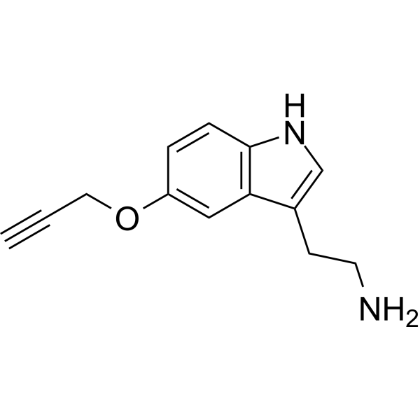
5-PT (5-Propargyltryptamide) is an alkyne-functionalized 5-HT (HY-B1473A) derivative and serotonylation substrate, taken up by living cells. 5-PT conjugated to a biotin enables identification of serotonylated proteins via copper-click chemistry and mass spectrometry. 5-PT labels and isolates serotonylated cells in alveolar epithelial cells for proteomic analysis. 5-PT can be used for the research of bronchopulmonary dysplasia.
For research use only. We do not sell to patients.
- CAS No.: 92085-05-7
- Formula: C13H14N2O
- Molecular Weight:214.26
-
Storage:
Please store the product under the recommended conditions in the Certificate of Analysis.

To place orders, for customer services and technical support, please contact: MedChemExpress USA
Tel: 609-228-6898 E-mail: [email protected] [email protected]


-
 Biological Activity
-
Chemical Information
- Protocols
- Purity & Documentation
- References
-
 Help & FAQs


Publications Citing Use of MedChemExpress (MCE) 5-PT
More

Biological Activity
Description
In Vitro
5-PT (100 μM; 24 h) treatment of MLE-12 alveolar epithelial cells labels 121 unique serotonylated proteins, many of which are associated with cellular processes and cell behavior regulation[1].
MedChemExpress (MCE) has not independently confirmed the accuracy of these methods. They are for reference only.


Chemical Information
-
CAS No. 92085-05-7
-
Molecular Weight 214.26
-
Formula C13H14N2O
-
SMILES
NCCC1=CNC2=C1C=C(OCC#C)C=C2
-
Synonyms
5-Propargyltryptamide
-
Shipping
Room temperature in continental US; may vary elsewhere.
-
Storage
Please store the product under the recommended conditions in the Certificate of Analysis.


Publications (1)
-
Journal Impact Factor
-
Most Recent
-
iScience
2025 Nov 19;28(12):114120. PMID: 41438086


Purity & Documentation
References

Calculators
Concentration (start) × Volume (start) = Concentration (final) × Volume (final)


5-PT Related Classifications
HY-156562 Related Classifications
 Powered by Bioz