



Beta-Sitosterol (purity>80%)
Based on 24 publication(s) in Google Scholar
Beta-Sitosterol (purity≥80%) is orally active. Beta-Sitosterol exhibits multiple activities, including anti-inflammatory, anticancer, antioxidant, antimicrobial, antidiabetic, antioxidant enzyme, and analgesic. Beta-Sitosterol inhibits inflammation and impaired adipogenesis in bovine mammary epithelial cells by reducing levels of ROS, TNF-α, IL-1β, and NF-κB p65 and restoring the activity of the HIF-1α/mTOR signaling pathway. Beta-Sitosterol induces apoptosis in cancer cells through ROS-mediated mitochondrial dysregulation and p53 activation. Beta-Sitosterol exerts its anticancer effects in cancer cells by activating caspase-3, caspase-8, and caspase-9, mediating PARP inactivation, MMP loss, altered Bcl-2-Bax ratio, and cytochrome c release. Beta-Sitosterol modulates macrophage polarization and reduces rheumatoid inflammation in mice. Beta-Sitosterol inhibits tumor growth in multiple mouse cancer models. Beta-Sitosterol can be used in the research of arthritis, lung cancer, breast cancer and other cancers, diabetes, etc.
For research use only. We do not sell to patients.
- Purity : 95.51%
- CAS No.: 83-46-5
- Formula: C29H50O
- Molecular Weight:414.71
-
Storage:
-20°C, protect from light
* In solvent : -80°C, 6 months; -20°C, 1 month (protect from light)

To place orders, for customer services and technical support, please contact: MedChemExpress USA
Tel: 609-228-6898 E-mail: [email protected] [email protected]


-
 Biological Activity
-
Chemical Information
-
Solvent & Solubility
- Protocols
- Purity & Documentation
- References
-
 Help & FAQs


-
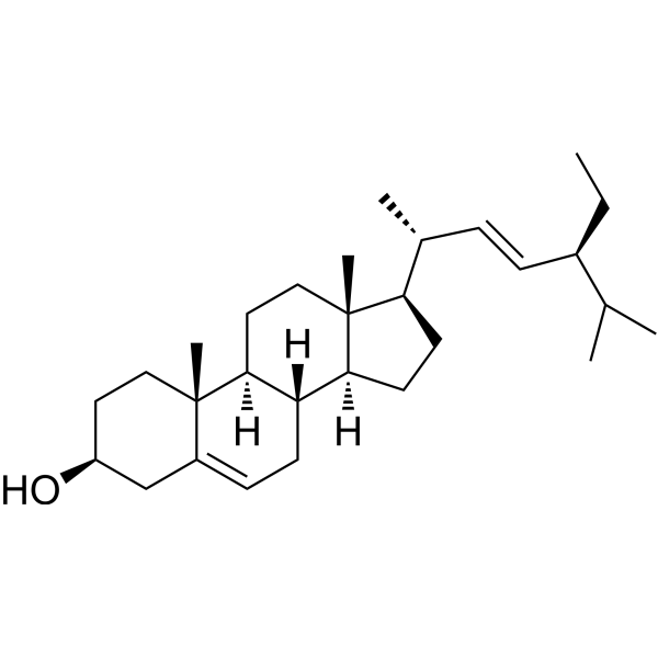
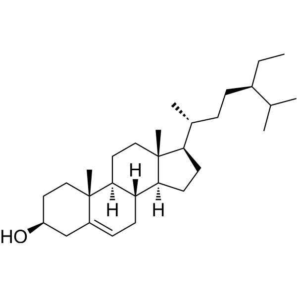
HY-N0131
StigmasterolPlant Sterol -
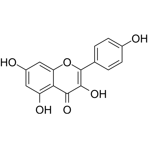
HY-14590
KaempferolEstrogen Receptor α Inhibitor -
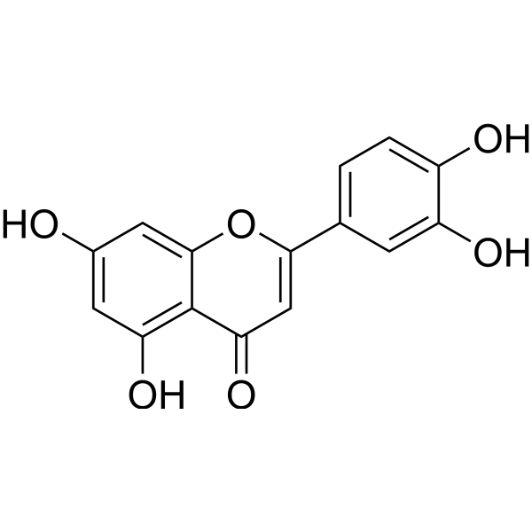
HY-N0162
LuteolinNrf2 Inhibitor -
HY-N0171A
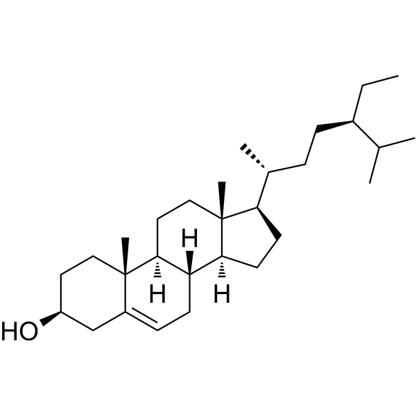
Beta-Sitosterol (purity>98%)Dietary Plant Sterols -
HY-N0171R
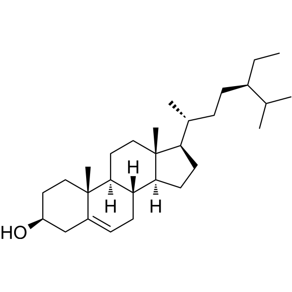
Beta-Sitosterol (Standard) -
HY-18085
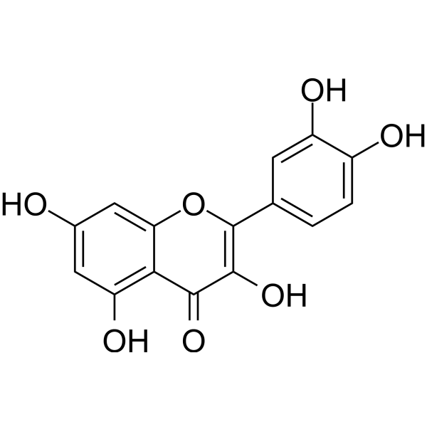
QuercetinFlavonoid Antioxidant -
HY-N1459
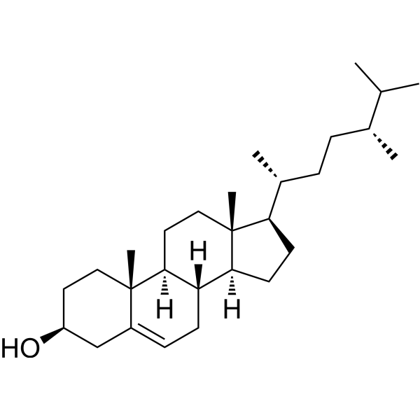
CampesterolEndogenous Metabolite -
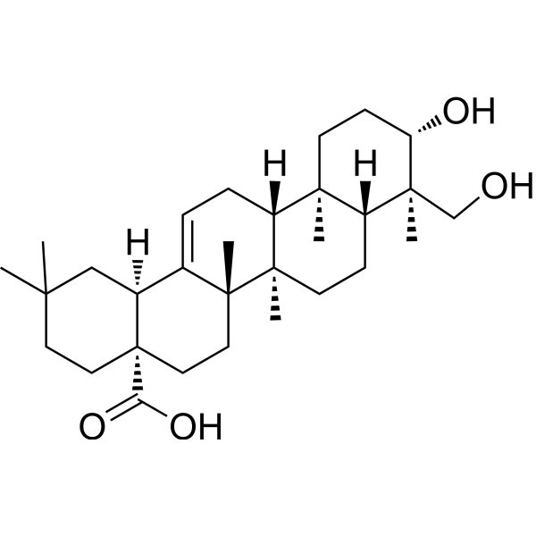
HY-N0256
HederageninCOX Inhibitor -
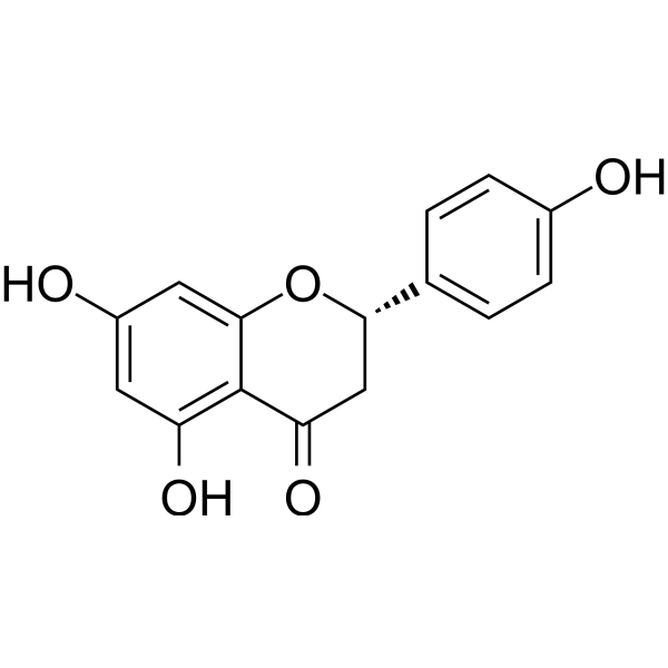
HY-N0100
NaringeninFlavonoid -
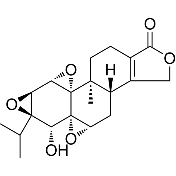
HY-32735
TriptolideImmunosuppressive Agent -
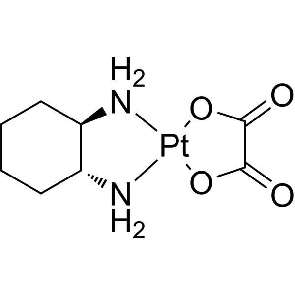
HY-17371
OxaliplatinDNA Synthesis Inhibitor
Publications Citing Use of MedChemExpress (MCE) Beta-Sitosterol (purity>80%)
More- Signal Transduct Target Ther. 2025 Dec 15;10(1):406. [Abstract]
- J Control Release. 2026 Apr 10:392:114691. [Abstract]
- Phytomedicine. 2024 Dec:135:156030. [Abstract]
- Phytother Res. 2026 May;40(5):2491-2513. [Abstract]
- Int J Pharm X. 2026 Apr 13:11:100543. [Abstract]
- J Ethnopharmacol. 2025 Sep 9;355(Pt A):120590. [Abstract]
- Acta Neuropathol Commun. 2020 Apr 22;8(1):56. [Abstract]
- Plant J. 2025 Nov;124(3):e70559. [Abstract]
- Mol Med Rep. 2025 Apr;31(4):95. [Abstract]
- Sci Rep. 2025 Jul 11;15(1):25045. [Abstract]
- Biosci Rep. 2020 Oct 30;40(10):BSR20201349. [Abstract]
- Processes (Basel). 2026 Jan;14(9):1351.
- Front Oncol. 2022 Aug 10;12:882784. [Abstract]
- Int J Environ Health Res. 2026 Feb 17:1-12. [Abstract]
- Int J Clin Pract. 2025 Aug 25.
- Lett Drug Des Discov. 2026 Feb 11.
- bioRxiv. 2026 Jul 6:2026.07.03.736431.
- SSRN. 2026 Jun 16.
- Discover Pharmaceutical Sciences. 2026 Jan 7;2(1):1.
- Res Sq. 2025 May 16.
- SSRN. 2025 May 6.
- Patent. US20240209361A1.
- Research Square Preprint. 2021. Jul.
- Evid Based Complement Alternat Med. 2020 Apr 29:2020:2760979. [Abstract]
 Customer Validation & Images

-
Cell Proliferation/Viability Assay
-
In Vivo Efficacy Study
-
WB
-
Cell Proliferation/Viability Assay
-
ELISA
All Endogenous Metabolite Isoforms
More

Biological Activity
Description
Cellular Effect
|
Cell Line

|
Type | Value | Description | References |
|---|---|---|---|---|
| 1A9 | ED50 |
10.6 μg/mL
Compound: 17
|
Cytotoxicity against human 1A9 cells after 6 days by SRB assay
Cytotoxicity against human 1A9 cells after 6 days by SRB assay
|
[PMID: 14640511] |
| 1A9 | ED50 |
16.8 μg/mL
Compound: 17
|
Cytotoxicity against human 1A9 cells after 3 days by SRB assay
Cytotoxicity against human 1A9 cells after 3 days by SRB assay
|
[PMID: 14640511] |
| 1A9/ptx-10 | ED50 |
20 μg/mL
Compound: 17
|
Cytotoxicity against human 1A9/PTX10 cells after 3 days by SRB assay
Cytotoxicity against human 1A9/PTX10 cells after 3 days by SRB assay
|
[PMID: 14640511] |
| 1A9/ptx-10 | ED50 |
9.5 μg/mL
Compound: 17
|
Cytotoxicity against human 1A9/PTX10 cells after 6 days by SRB assay
Cytotoxicity against human 1A9/PTX10 cells after 6 days by SRB assay
|
[PMID: 14640511] |
| A2780 | IC50 |
>10 μg/mL
Compound: page 1629, R26C1
|
Cytotoxicity against human A2780 cells after 96 hrs by MTT assay
Cytotoxicity against human A2780 cells after 96 hrs by MTT assay
|
[PMID: 17125236] |
| A549 | ED50 |
>20 μg/mL
Compound: 17
|
Cytotoxicity against human A549 cells after 3 days by SRB assay
Cytotoxicity against human A549 cells after 3 days by SRB assay
|
[PMID: 14640511] |
| A549 | IC50 |
>10 μg/mL
Compound: page 1629, R26C1
|
Cytotoxicity against human A549 cells after 96 hrs by MTT assay
Cytotoxicity against human A549 cells after 96 hrs by MTT assay
|
[PMID: 17125236] |
| Bel-7402 | IC50 |
>10 μg/mL
Compound: page 1629, R26C1
|
Cytotoxicity against human Bel-7402 cells after 96 hrs by MTT assay
Cytotoxicity against human Bel-7402 cells after 96 hrs by MTT assay
|
[PMID: 17125236] |
| BGC-823 | IC50 |
>10 μg/mL
Compound: page 1629, R26C1
|
Cytotoxicity against human BGC-823 cells after 96 hrs by MTT assay
Cytotoxicity against human BGC-823 cells after 96 hrs by MTT assay
|
[PMID: 17125236] |
| ECV-304 | IC50 |
472 μM
Compound: beta-sitosterol
|
Membranolytic activity in human ECV304 cells assessed as leakage of intracellular lactate dehydrogenase after 2 hrs by spectrophotometry
Membranolytic activity in human ECV304 cells assessed as leakage of intracellular lactate dehydrogenase after 2 hrs by spectrophotometry
|
[PMID: 22503361] |
| ECV-304 | IC50 |
472 μM
Compound: Beta-sitosterol
|
Membrane toxicity against human ECV304 cells after 2 hrs by LDH release assay
Membrane toxicity against human ECV304 cells after 2 hrs by LDH release assay
|
[PMID: 24084294] |
| ECV-304 | IC50 |
61 μM
Compound: beta-sitosterol
|
Cytotoxicity against human ECV304 cells after 72 hrs by Hoechst 33258 staining based fluorescence assay
Cytotoxicity against human ECV304 cells after 72 hrs by Hoechst 33258 staining based fluorescence assay
|
[PMID: 22503361] |
| ECV-304 | IC50 |
61 μM
Compound: Beta-sitosterol
|
Cytotoxicity against human ECV304 cells after 72 hrs by MTT assay
Cytotoxicity against human ECV304 cells after 72 hrs by MTT assay
|
[PMID: 24084294] |
| HCT-8 | ED50 |
>20 μg/mL
Compound: 17
|
Cytotoxicity against human HCT8 cells after 3 days by SRB assay
Cytotoxicity against human HCT8 cells after 3 days by SRB assay
|
[PMID: 14640511] |
| HCT-8 | IC50 |
>10 μg/mL
Compound: page 1629, R26C1
|
Cytotoxicity against human HCT8 cells after 96 hrs by MTT assay
Cytotoxicity against human HCT8 cells after 96 hrs by MTT assay
|
[PMID: 17125236] |
| HeLa | IC50 |
46.22 μM
Compound: 9
|
Cytotoxicity against human HeLa cells by MTT assay
Cytotoxicity against human HeLa cells by MTT assay
|
[PMID: 19447618] |
| HEp-2 | IC50 |
11.4 μM
Compound: 119
|
Antiproliferative activity against human Hep2 cells by MTT assay
Antiproliferative activity against human Hep2 cells by MTT assay
|
[PMID: 30830783] |
| HT-1080 | IC50 |
2.5 μM
Compound: Angelicin
|
Cytotoxicity against human HT1080 cells assessed as reduction in cell viability
Cytotoxicity against human HT1080 cells assessed as reduction in cell viability
|
[PMID: 30660827] |
| HUVEC | ED50 |
>5 μg/mL
Compound: beta-sitosterol
|
Cytotoxicity against HUVEC
Cytotoxicity against HUVEC
|
[PMID: 15043409] |
| J774 | IC50 |
>241.1 μM
Compound: 11
|
Cytotoxicity against mouse J774 cells by alamar blue assay
Cytotoxicity against mouse J774 cells by alamar blue assay
|
[PMID: 17637068] |
| KB | ED50 |
>20 μg/mL
Compound: 17
|
Cytotoxicity against human KB cells after 3 days by SRB assay
Cytotoxicity against human KB cells after 3 days by SRB assay
|
[PMID: 14640511] |
| KB | IC50 |
>10 μg/mL
Compound: 119
|
Antiproliferative activity against human KB cells assessed as reduction in cell viability
Antiproliferative activity against human KB cells assessed as reduction in cell viability
|
[PMID: 30830783] |
| KB | IC50 |
>241.5 μM
Compound: 119
|
Antiproliferative activity against human KB/HeLa cells assessed as reduction in cell viability
Antiproliferative activity against human KB/HeLa cells assessed as reduction in cell viability
|
[PMID: 30830783] |
| LNCaP | ED50 |
>5 μg/mL
Compound: beta-sitosterol
|
Cytotoxicity against human LNCAP cells
Cytotoxicity against human LNCAP cells
|
[PMID: 15043409] |
| Lu1 | ED50 |
>5 μg/mL
Compound: beta-sitosterol
|
Cytotoxicity against human Lu1 cells
Cytotoxicity against human Lu1 cells
|
[PMID: 15043409] |
| MCF7 | ED50 |
>20 μg/mL
Compound: 17
|
Cytotoxicity against human MCF7 cells after 3 days by SRB assay
Cytotoxicity against human MCF7 cells after 3 days by SRB assay
|
[PMID: 14640511] |
| MCF7 | ED50 |
>5 μg/mL
Compound: beta-sitosterol
|
Cytotoxicity against human MCF7 cells
Cytotoxicity against human MCF7 cells
|
[PMID: 15043409] |
| MCF7 | IC50 |
42.1 μM
Compound: 9
|
Cytotoxicity against human MCF7 cells by MTT assay
Cytotoxicity against human MCF7 cells by MTT assay
|
[PMID: 19447618] |
| PC-3 | ED50 |
>20 μg/mL
Compound: 17
|
Cytotoxicity against human PC3 cells after 3 days by SRB assay
Cytotoxicity against human PC3 cells after 3 days by SRB assay
|
[PMID: 14640511] |
| SK-MEL-1 | IC50 |
>50 μM
Compound: 9
|
Cytotoxicity against human SK-MEL-1 cells by MTT assay
Cytotoxicity against human SK-MEL-1 cells by MTT assay
|
[PMID: 19447618] |
| U-87MG ATCC | ED50 |
>20 μg/mL
Compound: 17
|
Cytotoxicity against human U87MG cells after 3 days by SRB assay
Cytotoxicity against human U87MG cells after 3 days by SRB assay
|
[PMID: 14640511] |
| Vero | IC50 |
>128 μg/mL
Compound: 4
|
Cytotoxicity against african green monkey Vero cells
Cytotoxicity against african green monkey Vero cells
|
[PMID: 18818073] |
In Vitro
Bioactivity-guided isolation afforded three compounds from the hexane fraction of E. indica, namely, Beta-Sitosterol (β-sitosterol), Stigmasterol, and Lutein. Both compounds are found to possess very low PPL inhibition activity, that is, 2.99±0.80% (Beta-Sitosterol) of inhibition at 100 μg/mL (242 μM) and 2.68±0.38% (Stigmasterol) of inhibition at 100 μg/mL (243 μM), respectively. Weak PPL inhibition activity of Beta-Sitosterol and Stigmasterol isolated from Alpinia zerumbet with IC50 value of 99.99±1.86 μg/mL and 125.05±4.76 μg/mL, respectively, in comparison with the inhibition shown by Curcumin (IC50=4.92±0.21 μg/mL) and Quercetin (IC50=18.60±0.86 μg/mL) which are used as positive controls in their study. Beta-Sitosterol and Stigmasterol are recorded with weak PPL inhibitory activity of only 3.0±0.8% and 2.7±0.4% at 100 μg/mL, respectively, (i.e., 242 μM and 243 μM) in contrast (34.5±5.4% at 100 μg/mL), which are comparatively lower than that recorded in literature (i.e., 50% PPL inhibition at 100 μg/mL)[1]. Sitosterol is an important compound extracted from the leaves of Aloe vera. It inhibits the growth of promastigotes of L. donovani, a causative agent for life threatening visceral leishmaniasis disease[2].
MedChemExpress (MCE) has not independently confirmed the accuracy of these methods. They are for reference only. Further protocols information, click here.
In Vivo
MedChemExpress (MCE) has not independently confirmed the accuracy of these methods. They are for reference only.


Chemical Information
-
CAS No. 83-46-5
-
Appearance Solid
-
Molecular Weight 414.71
-
Formula C29H50O
-
Color White to off-white
-
SMILES
CC[C@@H](C(C)C)CC[C@@H](C)[C@H]1CC[C@@]2([H])[C@]3([H])CC=C4C[C@@H](O)CC[C@]4(C)[C@@]3([H])CC[C@]12C
-
Initial Source
-
Shipping
Room temperature in continental US; may vary elsewhere.
-
Storage
-20°C, protect from light
* In solvent : -80°C, 6 months; -20°C, 1 month (protect from light)


Publications (24)
-
Journal Impact Factor
-
Most Recent
-
Signal Transduct Target Ther
Selective depletion of tumor-associated SAMHD1 enhances chemotherapeutic efficacy and antitumor immune responses. [Abstract]2025 Dec 15;10(1):406. PMID: 41392286 -
J Control Release
Single intravitreal injection of lipid nanoparticles delivering circular mRNA of nicotinamide phosphoribosyltransferase protects against dry AMD. [Abstract]2026 Apr 10:392:114691. PMID: 41672305 -
Phytomedicine
β-sitosterol alleviates pulmonary arterial hypertension by altering smooth muscle cell phenotype and DNA damage/cGAS/STING signaling. [Abstract]2024 Dec:135:156030. PMID: 39265206 -
Phytother Res
β-Sitosterol Suppresses Osteoclastogenesis and Alleviates Bone Loss by Targeting Tmed10 to Inhibit RANKL-Induced Hedgehog/MAPK Signaling and ROS. [Abstract]2026 May;40(5):2491-2513. PMID: 41703993 -
Int J Pharm X
Cutaneous delivery of bioactive components from a rice bran oil nanoemulsion and their biodistribution in porcine and human skin. [Abstract]2026 Apr 13:11:100543. PMID: 42028061 -
J Ethnopharmacol
Ethanol extract of Liriodendron tulipifera leaves displays anti-inflammatory activity by suppressing the Syk/Src/NF-κB pathway. [Abstract]2025 Sep 9;355(Pt A):120590. PMID: 40935214 -
Acta Neuropathol Commun
Unilateral intranigral administration of β-sitosterol β-D-glucoside triggers pathological α-synuclein spreading and bilateral nigrostriatal dopaminergic neurodegeneration in the rat. [Abstract]2020 Apr 22;8(1):56. PMID: 32321590 -
Plant J
Unveiling the inequivalent biochemical functions of OsSMO2-1 and OsSMO2-2 in rice phytosterol biosynthesis using a customized sterol standards mixture. [Abstract]2025 Nov;124(3):e70559. PMID: 41194406 -
Mol Med Rep
Mechanism of β‑sitosterol in treating keloids: Network pharmacology, molecular docking and experimental verification. [Abstract]2025 Apr;31(4):95. PMID: 39981895 -
Sci Rep
Beta sitosterol inhibits the proliferation and migration of synoviocytes in rheumatoid arthritis via lactylation of GPI. [Abstract]2025 Jul 11;15(1):25045. PMID: 40646091 -
Biosci Rep
Experimental evidence and network pharmacology-based analysis reveal the molecular mechanism of Tongxinluo capsule administered in coronary heart diseases. [Abstract]2020 Oct 30;40(10):BSR20201349. PMID: 32990315
Beta-Sitosterol (purity>80%) purchased from MedChemExpress. Usage Cited in: Biosci Rep. 2020 Oct 30;40(10):BSR20201349. [Abstract]
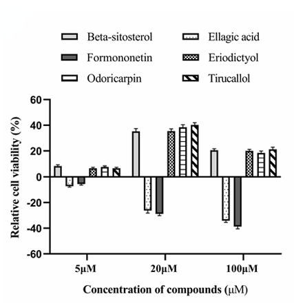
Beta-sitosterol (5–100 μM; 48 h). Different concentrations of active compounds on improvement rate.
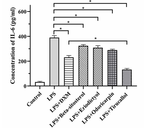
Beta-Sitosterol (purity>80%) purchased from MedChemExpress. Usage Cited in: Biosci Rep. 2020 Oct 30;40(10):BSR20201349. [Abstract]
Beta-Sitosterol (40 μM; 30 min) reduced IL-6 concentration.
-
-
Front Oncol
Salvia chinensis Benth Inhibits Triple-Negative Breast Cancer Progression by Inducing the DNA Damage Pathway. [Abstract]2022 Aug 10;12:882784. PMID: 36033499
Beta-Sitosterol (purity>80%) purchased from MedChemExpress. Usage Cited in: Front Oncol. 2022 Aug 10;12:882784. [Abstract]
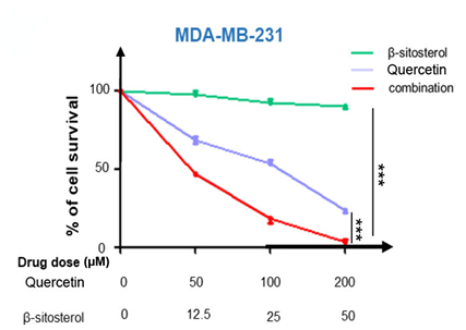
TNBC cells were simultaneously treated with quercetin and/or β-sitosterol for 48 h. Cell viability was measured via CCK-8 assay.
Beta-Sitosterol (purity>80%) purchased from MedChemExpress. Usage Cited in: Front Oncol. 2022 Aug 10;12:882784. [Abstract]
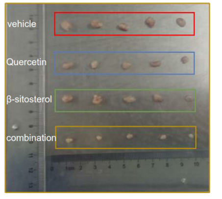
Mice were administered β-sitosterol (75 mg/kg) each day via oral gavage. Nude mice were subcutaneously injected with MDA-MB-231 cells. Representative images showed the dissected tumors in distinct treatment groups as specified at the endpoint.
Beta-Sitosterol (purity>80%) purchased from MedChemExpress. Usage Cited in: Front Oncol. 2022 Aug 10;12:882784. [Abstract]
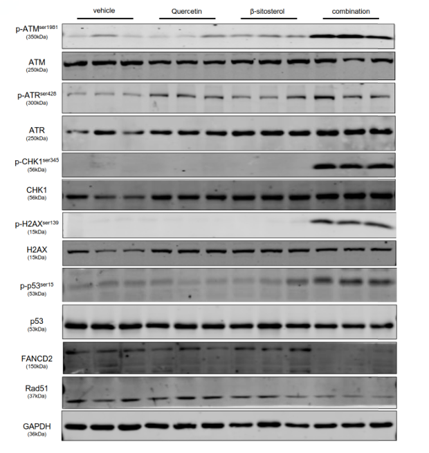
Mice were administered β-sitosterol (75 mg/kg) each day via oral gavage. Tumor lysates were collected, and the protein profile was established by immunoblotting analysis.
-
Int J Environ Health Res
Green extraction and RSM optimization of Peganum harmala shoots for antioxidant and anti-inflammatory properties: modulation of pro-inflammatory cytokines. [Abstract]2026 Feb 17:1-12. PMID: 41700732 -
-
-
-
-
-
-
-
-
-
Evid Based Complement Alternat Med
Ethanol Extract of the Infructescence of Platycarya strobilacea Sieb. et Zucc. Induces Methuosis of Human Nasopharyngeal Carcinoma Cells. [Abstract]2020 Apr 29:2020:2760979. PMID: 32419796


Solvent & Solubility
In Vitro:
Ethanol : 4 mg/mL (9.65 mM; ultrasonic and warming and heat to 60°C)
DMSO : < 1 mg/mL (insoluble or slightly soluble)
H2O : < 0.1 mg/mL (insoluble)
Please refer to the solubility information to select the appropriate solvent. Once prepared, please aliquot and store the solution to prevent product inactivation from repeated freeze-thaw cycles.
Storage method and period of stock solution: -80°C, 6 months; -20°C, 1 month (protect from light). When stored at -80°C, please use it within 6 months. When stored at -20°C, please use it within 1 month.
Please refer to the solubility information to select the appropriate solvent. Once prepared, please aliquot and store the solution to prevent product inactivation from repeated freeze-thaw cycles.
Storage method and period of stock solution: -80°C, 6 months; -20°C, 1 month (protect from light). When stored at -80°C, please use it within 6 months. When stored at -20°C, please use it within 1 month.
Concentration (start) × Volume (start) = Concentration (final) × Volume (final)
In Vivo:
For the following dissolution methods, please prepare the working solution directly:
It is recommended to prepare fresh solutions and use them promptly within a short period of time.
The percentages shown for the solvents indicate their volumetric ratio in the final prepared solution. If precipitation or phase separation occurs during preparation, heat and/or sonication can be used to aid dissolution.
Add each solvent one by one: 0.5% CMC-Na/saline water
Solubility: 20 mg/mL (48.23 mM); Suspended solution; Need ultrasonic

Protocols
-
Research Protocol for Infectious Diseases
Infectious-disease experiments test how pathogens interact with host barriers, innate immune receptors, inflammatory signaling, pathogen replication, and tissue injury; pattern-recognition receptors such as TLRs, RIG-I-like receptors, NOD-like receptors, and inflammasomes detect microbial molecules and activate NF-κB, interferon, and cytokine responses. The central hypothesis is that infection severity reflects the balance between pathogen burden and host response: protective inflammation restricts pathogen growth, whereas excessive or mislocalized inflammation contributes to tissue damage and disease phenotype. Unresolved questions include which host pathways are protective versus pathogenic, why some infection models fail to translate to human disease, and which combined readouts best predict clinically relevant infection outcomes.
-
Mitochondrial membrane-potential fluorescent assay
Mitochondrial membrane potential fluorescent assays estimate ΔΨm in living cells using lipophilic cationic dyes such as TMRM, TMRE, rhodamine 123, and JC-1, which accumulate in mitochondria according to membrane polarization; loss of signal after FCCP or CCCP treatment is interpreted as mitochondrial depolarization. TMRM/TMRE and rhodamine 123 are commonly used for semi-quantitative live-cell microscopy or flow cytometry, while JC-1 can report a shift from red aggregate fluorescence to green monomer fluorescence during depolarization; interpretation requires controls because dye concentration, quenching mode, cell type, dye efflux, and mitochondrial mass can affect fluorescence independently of ΔΨm.
-
Annexin V plus membrane-impermeant dye apoptosis staining
Annexin V-based apoptosis assays rely on the detection of phosphatidylserine (PS) externalization from the inner leaflet of the plasma membrane to the outer leaflet, an early biochemical hallmark of apoptosis. Fluorescently labeled Annexin V binds PS in a calcium-dependent manner, enabling identification of early apoptotic cells by flow cytometry or fluorescence microscopy. When combined with a membrane-impermeant DNA-binding dye (e. g. , propidium iodide), this approach allows discrimination between viable (Annexin V−/dye−), early apoptotic (Annexin V+/dye−), and late apoptotic or necrotic (Annexin V+/dye+) cell populations by assessing membrane integrity and PS exposure.
-
ROS/oxidative-stress fluorescent staining
ROS/oxidative-stress fluorescent staining uses cell-permeant fluorogenic probes that become fluorescent after oxidation inside cells or tissues; commonly used examples include DCFH-DA/DCFDA for broad cellular oxidant detection, DHE for superoxide-related signal detection, MitoSOX for mitochondrial superoxide-related signal detection, and CellROX probes for oxidative-stress-associated fluorescence readouts. The assay detects probe oxidation rather than a single ROS species unless the probe and analysis method have been chemically validated for that species. DCFH-DA enters cells, is deacetylated by intracellular esterases to DCFH, and produces fluorescent DCF after oxidation, so the readout is used as an operational measure of total cellular oxidative stress rather than a species-specific ROS measurement. DHE and MitoSOX can report superoxide-related oxidation, but red fluorescence alone can include non-specific ethidium-like oxidation products; HPLC or optimized spectral approaches are
-
Patient-Derived Orthotopic Xenograft (PDOX)
Patient-derived orthotopic xenograft (PDOX) modeling implants fresh patient tumor tissue or patient-derived tumor cells into the anatomically corresponding organ or tissue site of immunodeficient mice, usually by surgical orthotopic implantation, to preserve patient tumor histology, local microenvironmental context, invasion, metastatic behavior, and treatment-response features better than subcutaneous implantation. PDOX readouts include tumor engraftment, orthotopic tumor growth, local invasion, metastasis, recurrence after resection, histologic similarity to the donor tumor, biomarker retention, molecular concordance, survival, and response or resistance to therapy. PDOX models are used for preclinical drug testing and individualized therapy evaluation, but engraftment success varies by tumor type and specimen quality.
-
Research Protocol for Inflammation-related Diseases
The NLRP3 inflammasome is a cytosolic innate immune signaling platform that integrates priming signals and danger-signal activation to promote caspase-1 activation, maturation of IL-1β and IL-18, and gasdermin D-mediated pyroptotic cell death. The core experimental logic is to determine whether inflammatory disease phenotypes are driven by increased NLRP3 expression, ASC-containing inflammasome assembly, caspase-1 cleavage, GSDMD cleavage, and extracellular release of IL-1β/IL-18 rather than by nonspecific cell injury alone. The pathway is strongly linked to inflammation-related disease phenotypes because monosodium urate crystals activate NALP3/NLRP3 inflammasome signaling in gout-like crystal inflammation, cholesterol crystals activate NLRP3 inflammasomes in atherogenesis models, and DSS-induced intestinal inflammation has been reported to involve NLRP3 inflammasome activity. However, experimental colitis studies also show context-dependent protective effects of NLRP3 inflammasome co
-
Breast Cancer Modeling
Breast cancer is a heterogeneous cancer, and it has been distinguished into four subtypes: luminal A, luminal B, HER2-positive and basal-like. Molecular mutations, epigenetic alterations, hormone exposure and immune microenvironment are related to the progression of breast cancer.
-
Research Protocol for Metabolic Diseases
AMP-activated protein kinase, AMPK, is a conserved cellular energy sensor that responds to reduced cellular energy status and coordinates metabolism by increasing ATP-generating catabolic pathways while suppressing ATP-consuming anabolic processes. In metabolic disease research, the AMPK pathway is experimentally relevant because it regulates hepatic lipid synthesis, fatty acid oxidation, glucose production, skeletal-muscle glucose disposal, mTORC1-linked biosynthesis, autophagy, mitochondrial homeostasis, and whole-body energy balance. The central pathway logic is that energy stress, metformin, exercise-like stimulation, or direct AMPK activators increase AMPKα Thr172 phosphorylation and downstream substrate phosphorylation, including ACC and RAPTOR. Phosphorylation of ACC suppresses lipogenesis and supports fatty acid oxidation, whereas phosphorylation of RAPTOR suppresses mTORC1 signaling and links cellular energy status to growth and protein synthesis control. The pathway is linked
-
3T3-L1 preadipocyte-to-adipocyte differentiation
3T3-L1 preadipocytes are induced to differentiate after growth arrest using adipogenic media containing insulin, dexamethasone, and IBMX; differentiation is assessed by lipid-droplet accumulation, triglyceride increase, Oil Red O staining, and adipocyte-marker induction such as PPARγ and C/EBPα.
-
Collagen-Induced Arthritis
Collagen-induced arthritis (CIA) is an autoimmune murine model of rheumatoid arthritis in which immunization with type II collagen (CII) emulsified in an adjuvant induces a T cell- and autoantibody-driven inflammatory arthritis characterized by synovial hyperplasia, immune cell infiltration, and joint destruction. The model typically relies on genetically susceptible mouse strains (e. g. , DBA/1) and reproduces key features of human rheumatoid arthritis, including anti-collagen immune responses and progressive joint inflammation. Disease onset generally occurs within ~3-4 weeks after immunization, depending on antigen/adjuvant combinations and protocol variation. The immunopathology is driven by adaptive immune activation against CII, leading to systemic and local joint inflammation mediated by pro-inflammatory cytokines and effector immune cells, making CIA a standard preclinical platform for evaluating immunomodulatory and anti-arthritic interventions.
-
Apoptosis
Apoptosis, also called programmed cell death, is generally characterized by distinct morphological characteristics.
-
TUNEL staining for apoptotic DNA fragmentation
TUNEL staining detects DNA strand breaks by using terminal deoxynucleotidyl transferase to add labeled nucleotides to exposed 3′-OH DNA termini, generating either microscopic staining in fixed cells or tissue sections, or fluorescence/cytometric signal in cell suspensions. TUNEL positivity reflects DNA fragmentation but should not be interpreted alone as definitive apoptosis, because TUNEL can also label necrotic, autolytic, mechanically damaged, or DNA-repair-associated DNA breaks.
-
Mitochondrial membrane-potential and mitochondrial mass staining
Mitochondrial membrane potential staining measures the electrochemical polarization across the mitochondrial inner membrane in live cells using lipophilic cationic fluorescent probes; early rhodamine-based work showed that selective mitochondrial dye accumulation is lost when the mitochondrial transmembrane potential is dissipated. JC-1 reports mitochondrial polarization by shifting from green monomer fluorescence to red J-aggregate fluorescence as dye concentration increases within energized mitochondria; therefore, the red/green fluorescence ratio is used as a relative readout of mitochondrial membrane potential. TMRE or TMRM staining provides a single-channel relative readout because these cationic rhodamine esters accumulate in polarized mitochondria, and lower fluorescence indicates reduced mitochondrial polarization when acquisition and dye-loading conditions are controlled. Mitochondrial mass staining is commonly performed with MitoTracker Green FM or related MitoTracker dyes as
-
Patient-Derived Xenograft (PDX)
Patient-derived xenograft (PDX) models are generated by engrafting primary human tumor tissue directly into immunodeficient mice, allowing in vivo propagation of patient tumor biology without initial in vitro adaptation. These models are used to preserve key histopathological and molecular characteristics of the original tumor and enable assessment of tumor growth dynamics and therapeutic response in a living organism. The biological readout is tumor engraftment and subsequent growth in the murine host, which reflects the ability of human tumor cells to survive, vascularize, and expand in an immunocompromised microenvironment.
-
Apoptosis Solutions
Apoptosis is a regulated, generally non-lytic cell-death pathway that removes unwanted, damaged, infected, or abnormal cells through coordinated morphological changes, caspase activation, DNA fragmentation, and membrane remodeling. The intrinsic apoptosis pathway is controlled mainly by mitochondrial outer membrane permeabilization, BCL-2 family proteins, cytochrome c release, apoptosome formation, caspase-9 activation, and downstream executioner caspase-3/7 activation. The extrinsic apoptosis pathway is initiated by death receptors such as Fas, TNFR, and TRAIL receptors, which recruit adaptor proteins and activate caspase-8 before engaging executioner caspases or mitochondrial amplification through BID cleavage. Apoptosis is linked to many phenotypes, including cancer cell killing, tissue homeostasis, immune regulation, neurodegeneration, infection response, and treatment-induced cytotoxicity; unresolved questions include how apoptosis interacts with necroptosis, pyroptosis, ferroptos
-
Primary monocyte-to-macrophage differentiation
Primary human monocytes can be differentiated ex vivo into monocyte-derived macrophages by culturing purified blood monocytes for approximately 5-7 days in macrophage-supporting cytokine conditions; M-CSF commonly yields CD14^high/CD163^high macrophages, while GM-CSF yields a phenotypically distinct macrophage population, so the cytokine condition should be chosen according to the downstream model. The readout of successful differentiation is a combined change in morphology, adherence, surface phenotype, and function: differentiated macrophages become adherent, enlarge, acquire macrophage-associated markers such as CD14, CD68, CD163, CD206, or HLA-DR depending on culture condition, and show increased phagocytic capacity compared with starting monocytes.
-
Subcutaneous Cell-Line-Derived Xenograft
Subcutaneous cell-line-derived xenograft (CDX) models are established by implanting cultured human cancer cell lines into immunodeficient mice, where the injected cells form localized tumors that can be monitored in vivo as a measure of tumorigenic potential, growth kinetics, and treatment response. These models are widely used in oncology research because they allow reproducible tumor formation and enable comparative assessment of tumor growth between different cell lines or genetic manipulations in a controlled in vivo microenvironment. Subcutaneous implantation of cancer cells in immunodeficient mice is a standard approach for evaluating tumor growth behavior and therapeutic response across multiple cancer types, including prostate, esophageal, pancreatic, and colon cancer models.
-
Orthotopic Cell-Line Xenograft
Orthotopic cell-line xenograft models involve implantation of human cancer cell lines into the anatomically corresponding organ of immunodeficient mice to reproduce tumor growth within a native microenvironment, enabling more clinically relevant tumor behavior compared with subcutaneous models. These models are widely used because orthotopic placement better recapitulates tumor progression, including invasion and metastatic spread, which are often underrepresented in heterotopic implantation systems. Compared with conventional xenografts, orthotopic implantation is described as more technically complex but provides improved simulation of tumor-microenvironment interactions and metastatic behavior, making it particularly valuable for translational oncology research. Surgical orthotopic implantation approaches have been emphasized as enabling faithful reproduction of clinical cancer features, including metastasis and disease progression patterns that align with the tumor’s organ of origi
-
Lipid Droplets: Oil Red O/Sudan Dye Lipid Staining
Lipid droplets are intracellular organelles with a neutral-lipid core that stores triacylglycerols and sterol esters, and Oil Red O or Sudan dyes detect these hydrophobic lipid deposits by partitioning into retained lipids in fresh or frozen specimens. Oil Red O stains neutral triglycerides and lipids in frozen tissue sections or air-dried cytologic preparations, while Sudan Black B has also been used as a histochemical fat stain for lipid-rich tissue structures.
-
Pyroptosis Solutions
Pyroptosis is a lytic inflammatory cell-death pathway executed by gasdermin pores, most classically through inflammasome-mediated activation of caspase-1, cleavage of gasdermin D, membrane pore formation, LDH release, and secretion of IL-1β and IL-18. The canonical pathway is commonly modeled by priming cells with an inflammatory signal such as LPS to induce pro-IL-1β and inflammasome components, followed by an activation signal such as ATP or nigericin to activate NLRP3, ASC speck formation, caspase-1 cleavage, GSDMD cleavage, cytokine release, and pyroptotic membrane rupture. The non-canonical pathway is triggered when cytosolic LPS activates mouse caspase-11 or human caspase-4/5, leading to GSDMD cleavage and pyroptosis, and this can secondarily activate NLRP3-dependent IL-1β release. Pyroptosis is linked to inflammatory injury, infection, cancer, liver disease, ocular disease, placental inflammation, and other disease phenotypes, but unresolved questions include which gasdermin fam


Purity & Documentation
-
Data Sheet (293 KB)
-
SDS (393 KB)
- English - EN (393 KB)
- Français - FR (393 KB)
- Deutsch - DE (393 KB)
- Norwegian - NO (393 KB)
- Español - ES (393 KB)
- Swedish - SV (393 KB)
- Italian - IT (393 KB)
- Korean - KR (393 KB)
- Portuguese - PT (393 KB)
-
Handling Instructions (2659 KB)
References
[1]. Bin Sayeed MS, et al. Beta-Sitosterol: A Promising but Orphan Nutraceutical to Fight Against Cancer. Nutr Cancer. 2015;67(8):1214-20. [Content Brief]
[2]. Tariq A, et al. Ethnomedicines and anti-parasitic activities of Pakistani medicinal plants against Plasmodia and Leishmania parasites. Ann Clin Microbiol Antimicrob. 2016 Sep 20;15(1):52. [Content Brief]
[3]. Zhao D, et al. Structural Features and Potent Antidepressant Effects of Total Sterols and β-sitosterol Extracted from Sargassum horneri. Mar Drugs. 2016 Jun 28;14(7). [Content Brief]
[4]. Fan Y, et al. Beta-Sitosterol Suppresses Lipopolysaccharide-Induced Inflammation and Lipogenesis Disorder in Bovine Mammary Epithelial Cells. Int J Mol Sci. 2023 Sep 27;24(19):14644. [Content Brief]
[5]. Rajavel T, et al. Beta-Sitosterol targets Trx/Trx1 reductase to induce apoptosis in A549 cells via ROS mediated mitochondrial dysregulation and p53 activation. Sci Rep. 2018 Feb 1;8(1):2071. [Content Brief]
[6]. Liu R, et al. Beta-Sitosterol modulates macrophage polarization and attenuates rheumatoid inflammation in mice. Pharm Biol. 2019 Dec;57(1):161-168. [Content Brief]
[7]. Villaseñor IM, et al. Bioactivity studies on beta-sitosterol and its glucoside. Phytother Res. 2002 Aug;16(5):417-21. [Content Brief]
[8]. Ju YH, et al. beta-Sitosterol, beta-Sitosterol Glucoside, and a Mixture of beta-Sitosterol and beta-Sitosterol Glucoside Modulate the Growth of Estrogen-Responsive Breast Cancer Cells In Vitro and in Ovariectomized Athymic Mice. J Nutr. 2004 May;134(5):1145-51. [Content Brief]
[9]. Awad AB, et al. beta-Sitosterol activates Fas signaling in human breast cancer cells. Phytomedicine. 2007 Nov;14(11):747-54. [Content Brief]
[10]. Loizou S, et al. Beta-sitosterol exhibits anti-inflammatory activity in human aortic endothelial cells. Mol Nutr Food Res. 2010 Apr;54(4):551-8. [Content Brief]
[11]. Vivancos M, et al. beta-Sitosterol modulates antioxidant enzyme response in RAW 264.7 macrophages. Free Radic Biol Med. 2005 Jul 1;39(1):91-7. [Content Brief]
[12]. Gupta R, et al. Antidiabetic and antioxidant potential of Beta-Sitosterol in streptozotocin-induced experimental hyperglycemia. J Diabetes. 2011 Mar;3(1):29-37. [Content Brief]
[13]. Anwar R, et al. Antimicrobial Activity of Beta-Sitosterol Isolated from Kalanchoe tomentosa Leaves Against Staphylococcus aureus and Klebsiella pneumonia. Pak J Biol Sci. 2022 Jun;25(7):602-607. [Content Brief]


Complete Stock Solution Preparation Table
Please refer to the solubility information to select the appropriate solvent. Once prepared, please aliquot and store the solution to prevent product inactivation from repeated freeze-thaw cycles.
Storage method and period of stock solution: -80°C, 6 months; -20°C, 1 month (protect from light). When stored at -80°C, please use it within 6 months. When stored at -20°C, please use it within 1 month.
| Optional Solvent | Concentration Solvent Mass | 1 mg | 5 mg | 10 mg | 25 mg |
|---|---|---|---|---|---|
| Ethanol | 1 mM | 2.4113 mL | 12.0566 mL | 24.1132 mL | 60.2831 mL |
| 5 mM | 0.4823 mL | 2.4113 mL | 4.8226 mL | 12.0566 mL |


Beta-Sitosterol (purity>80%) Related Classifications
HY-N0171 Related Classifications
 Powered by Bioz