



SM-6586
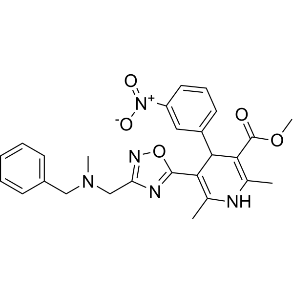
SM-6586 is a calcium channel antagonist and inhibitor of Na+/H+ and Na+/Ca2+ exchange transport, potentially for the treatment of cerebrovasular diseases and hypertension.
For research use only. We do not sell to patients.
- CAS No.: 103898-38-0
- Formula: C26H27N5O5
- Molecular Weight:489.52
-
Storage:
Please store the product under the recommended conditions in the Certificate of Analysis.

To place orders, for customer services and technical support, please contact: MedChemExpress USA
Tel: 609-228-6898 E-mail: [email protected] [email protected]


-
 Biological Activity
-
Chemical Information
- Protocols
- Purity & Documentation
- References
-
 Help & FAQs


-
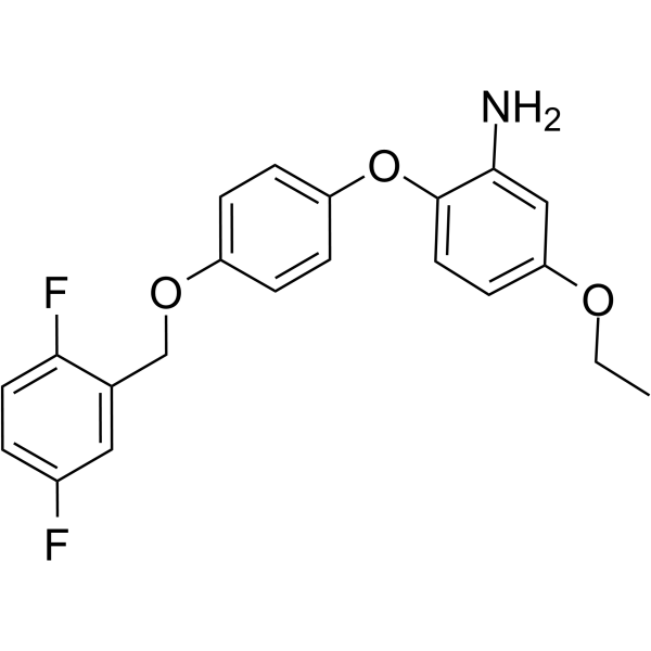
HY-15515
SEA0400Na+/Ca2+ Exchanger Inhibitor -
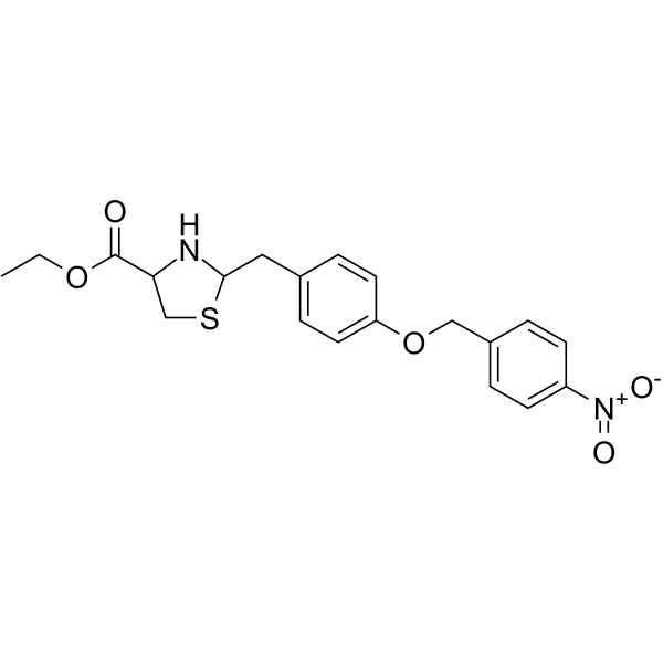
HY-107658
SN 6Na+/Ca2+ Exchanger Inhibitor -
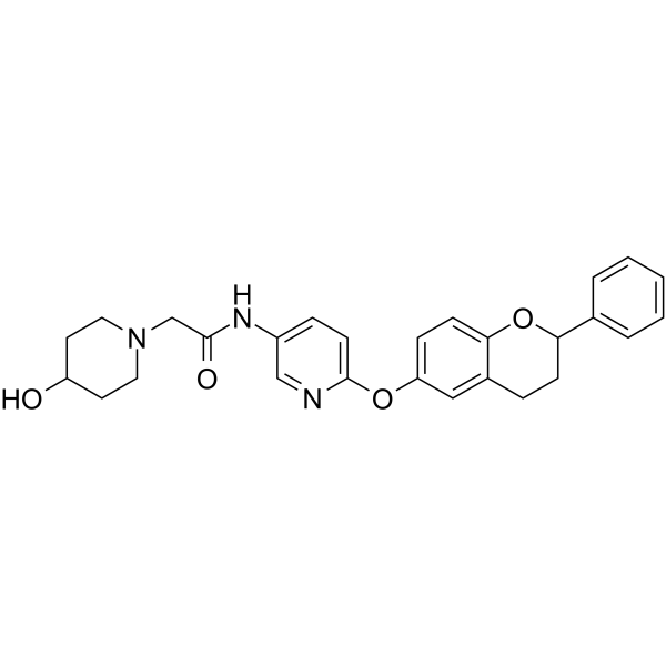
HY-123785
ORM-10962NCX Inhibitor -
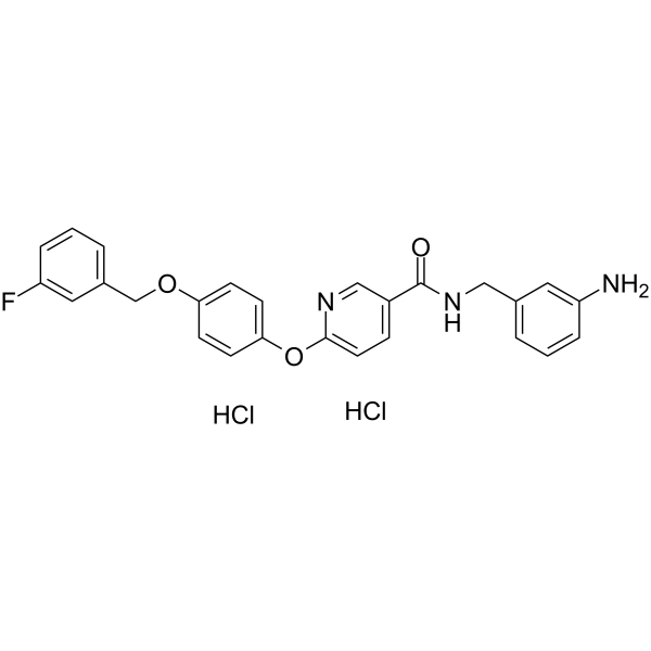
HY-136182
YM-244769 dihydrochlorideNCX Inhibitor -

HY-15515R
SEA0400 (Standard)Na+/Ca2+ Exchanger Inhibitor -
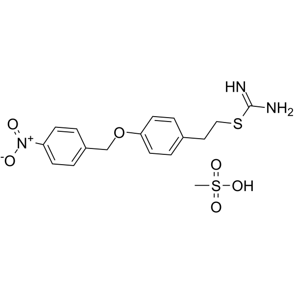
HY-15415
KB-R7943 mesylateNCXrev Inhibitor -

HY-107658R
SN 6 (Standard)Reference Standard -
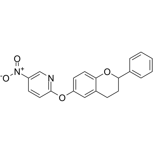
HY-128678
ORM-10103Na+/Ca2+ Exchanger Inhibitor -
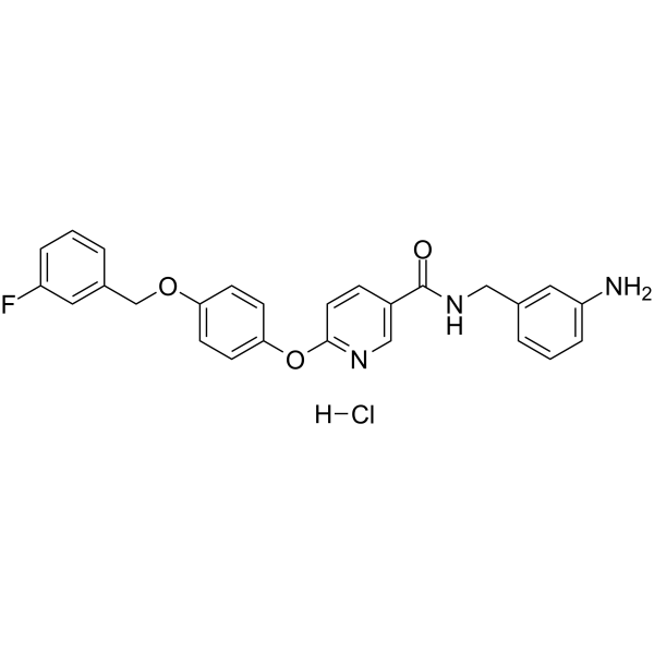
HY-107659
YM-244769 hydrochlorideNCX Inhibitor -
HY-117073
ITH12575mNCX Blocker -

HY-107659R
YM-244769 hydrochloride (Standard)Reference Standard
All Calcium Channel Isoforms
More

Biological Activity
Description
In Vivo
MedChemExpress (MCE) has not independently confirmed the accuracy of these methods. They are for reference only.


Chemical Information
-
CAS No. 103898-38-0
-
Molecular Weight 489.52
-
Formula C26H27N5O5
-
SMILES
O=C(C1=C(C)NC(C)=C(C2=NC(CN(C)CC3=CC=CC=C3)=NO2)C1C4=CC=CC([N+]([O-])=O)=C4)OC
-
Shipping
Room temperature in continental US; may vary elsewhere.
-
Storage
Please store the product under the recommended conditions in the Certificate of Analysis.


Purity & Documentation
References

Calculators
Concentration (start) × Volume (start) = Concentration (final) × Volume (final)


SM-6586 Related Classifications
HY-19062 Related Classifications
 Powered by Bioz