



PR-39 TFA
Based on 2 publication(s) in Google Scholar
PR-39 TFA, a natural proline- and arginine-rich antibacterial peptide, is a noncompetitive, reversible and allosteric proteasome inhibitor. PR-39 TFAreversibly binds to the α7 subunit of the proteasome and blocks degradation of NF-κB inhibitor IκBα by the ubiquitin-proteasome pathway. PR-39 TFA stimulates angiogenesis, inhibits inflammatory responses and significant reduces myocardial infarct size in mice.
For research use only. We do not sell to patients.
- Purity : 99.89%
- Formula: C231H347F3N70O42
- Molecular Weight:4833.76
-
Storage:
Sealed storage, away from moisture.
Powder -80°C, 2 years , -20°C, 1 year* In solvent : -80°C, 6 months; -20°C, 1 month (sealed storage, away from moisture)

To place orders, for customer services and technical support, please contact: MedChemExpress USA
Tel: 609-228-6898 E-mail: [email protected] [email protected]


-
 Biological Activity
-
Chemical Information
-
Solvent & Solubility
- Protocols
- Purity & Documentation
- References
-
 Help & FAQs


Publications Citing Use of MedChemExpress (MCE) PR-39 TFA
More

Biological Activity
Description
In Vitro
PR-39 TFA, shown to selectively affect proteasomemediated protein degradation in vivo, alters the shape of the 20S and 26S cylinder and affects the binding of 19S caps in a reversible manner. PR-39 TFA specifically blocks degradation of IκBα and HIF-1α by the proteasome[1].
PR-39 TFA (100 nM) blocks TNF-α-induced (1 ng/mL; for 20 minutes) activation of VCAM-1 (2 hours) and ICAM-1 (8 hours) expression in human umbilical vein endothelial cells (HUVEC)[2].
PR-39 TFA (10 μM) does not affect the ability to proliferate of ECV304 cell. PR39 is able to inhibit IκBα degradation without significantly affecting overall protein degradation in cells[2].
MedChemExpress (MCE) has not independently confirmed the accuracy of these methods. They are for reference only.
In Vivo
PR-39 TFA (1 μg/kg/day; 7-day intraperitoneal infusion) demonstrates significantly small infarct in C57BL/6 mice[2].
MedChemExpress (MCE) has not independently confirmed the accuracy of these methods. They are for reference only.


Chemical Information
-
Appearance Solid
-
Molecular Weight 4833.76
-
Formula C231H347F3N70O42
-
Color White to off-white
-
Sequence
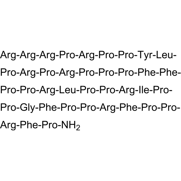
Arg-Arg-Arg-Pro-Arg-Pro-Pro-Tyr-Leu-Pro-Arg-Pro-Arg-Pro-Pro-Pro-Phe-Phe-Pro-Pro-Arg-Leu-Pro-Pro-Arg-Ile-Pro-Pro-Gly-Phe-Pro-Pro-Arg-Phe-Pro-Pro-Arg-Phe-Pro-NH2
-
Sequence Shortening
RRRPRPPYLPRPRPPPFFPPRLPPRIPPGFPPRFPPRFP-NH2
-
Shipping
Room temperature in continental US; may vary elsewhere.
-
Storage
Sealed storage, away from moisture
Powder -80°C 2 years -20°C 1 year * In solvent : -80°C, 6 months; -20°C, 1 month (sealed storage, away from moisture)


Publications (2)
-
Journal Impact Factor
-
Most Recent
-
Int J Pharm
Combined antimicrobial and pro-healing effects of cathelicidins in a polysaccharide-based hydrogel scaffold: treatment of carbapenemase-resistant Pseudomonas aeruginosa wound infections in a porcine model. [Abstract]2026 Jun 25:699:126992. PMID: 42150643 -
Folia Microbiol (Praha)
EDTA enhances antimicrobial activity of PR-39 and Protegrin-1 antimicrobial peptides against carbapenem-resistant Pseudomonas aeruginosa in serum. [Abstract]2026 May 28. PMID: 42207438


Solvent & Solubility
In Vitro:
H2O : 100 mg/mL (20.69 mM; Need ultrasonic)
Please refer to the solubility information to select the appropriate solvent. Once prepared, please aliquot and store the solution to prevent product inactivation from repeated freeze-thaw cycles.
Storage method and period of stock solution: -80°C, 6 months; -20°C, 1 month (sealed storage, away from moisture). When stored at -80°C, please use it within 6 months. When stored at -20°C, please use it within 1 month.
* Note: If you choose water as the stock solution, please dilute it to the working solution, then filter and sterilize it with a 0.22 μm filter before use.
Please refer to the solubility information to select the appropriate solvent. Once prepared, please aliquot and store the solution to prevent product inactivation from repeated freeze-thaw cycles.
Storage method and period of stock solution: -80°C, 6 months; -20°C, 1 month (sealed storage, away from moisture). When stored at -80°C, please use it within 6 months. When stored at -20°C, please use it within 1 month.
* Note: If you choose water as the stock solution, please dilute it to the working solution, then filter and sterilize it with a 0.22 μm filter before use.
Concentration (start) × Volume (start) = Concentration (final) × Volume (final)
In Vivo:
For the following dissolution methods, please prepare the working solution directly:
It is recommended to prepare fresh solutions and use them promptly within a short period of time.
The percentages shown for the solvents indicate their volumetric ratio in the final prepared solution. If precipitation or phase separation occurs during preparation, heat and/or sonication can be used to aid dissolution.
Add each solvent one by one: PBS
Solubility: 50 mg/mL (10.34 mM); Clear solution; Need ultrasonic


Purity & Documentation
-
Data Sheet (288 KB)
-
SDS (252 KB)
- English - EN (252 KB)
- Français - FR (252 KB)
- Deutsch - DE (252 KB)
- Norwegian - NO (252 KB)
- Español - ES (252 KB)
- Swedish - SV (252 KB)
- Italian - IT (252 KB)
- Korean - KR (252 KB)
- Portuguese - PT (252 KB)
-
Handling Instructions (2659 KB)
References
[1]. Maria Gaczynska, et al. Proline- and arginine-rich peptides constitute a novel class of allosteric inhibitors of proteasome activity. Biochemistry. 2003 Jul 29;42(29):8663-70. [Content Brief]
[2]. Y Gao, et al. Inhibition of ubiquitin-proteasome pathway-mediated I kappa B alpha degradation by a naturally occurring antibacterial peptide. J Clin Invest. 2000 Aug;106(3):439-48. [Content Brief]


Complete Stock Solution Preparation Table
Please refer to the solubility information to select the appropriate solvent. Once prepared, please aliquot and store the solution to prevent product inactivation from repeated freeze-thaw cycles.
Storage method and period of stock solution: -80°C, 6 months; -20°C, 1 month (sealed storage, away from moisture). When stored at -80°C, please use it within 6 months. When stored at -20°C, please use it within 1 month.
| Optional Solvent | Concentration Solvent Mass | 1 mg | 5 mg | 10 mg | 25 mg |
|---|---|---|---|---|---|
| H2O | 1 mM | 0.2069 mL | 1.0344 mL | 2.0688 mL | 5.1720 mL |
| 5 mM | 0.0414 mL | 0.2069 mL | 0.4138 mL | 1.0344 mL | |
| 10 mM | 0.0207 mL | 0.1034 mL | 0.2069 mL | 0.5172 mL | |
| 15 mM | 0.0138 mL | 0.0690 mL | 0.1379 mL | 0.3448 mL | |
| 20 mM | 0.0103 mL | 0.0517 mL | 0.1034 mL | 0.2586 mL |
* Note: If you choose water as the stock solution, please dilute it to the working solution, then filter and sterilize it with a 0.22 μm filter before use.


PR-39 TFA Related Classifications
HY-P1259A Related Classifications
 Powered by Bioz