



Canakinumab
Based on 1 publication(s) in Google Scholar
Canakinumab (ACZ885) is a recombinant human anti-IL-1β monoclonal antibody. Canakinumab shows IC50 values of 43.6 and 40.8 pM for human and marmoset IL-1β, respectively. The mode of action of canakinumab is based on the neutralization of IL-1β signaling, resulting in suppression of inflammation related to disorders of autoimmune origin.
For research use only. We do not sell to patients.
- Purity : 98.84%
- CAS No.: 914613-48-2
- Molecular Weight:145.5 kDa
-
Storage:
Please store the product under the recommended conditions in the Certificate of Analysis.


To place orders, for customer services and technical support, please contact: MedChemExpress USA
Tel: 609-228-6898 E-mail: [email protected] [email protected]


-
 Biological Activity
-
Chemical Information
- Protocols
- Purity & Documentation
- References
-
 Help & FAQs


-

HY-P9956
SiltuximabAnti-IL-6 mAb -

HY-108841
RaleukinIL-1R Antagonist -

HY-P99171
GevokizumabIL-1β Modulator -
HY-P99396
SpesolimabAnti-IL-36R Antibody -

HY-P9927
SecukinumabIL-17A Antagonist -

HY-P9917
TocilizumabIL-6R Neutralizing Antibody -

HY-P99721
LutikizumabAnti-IL-1α/β Immunoglobulin -

HY-P99379
Nidanilimab (Nadunolimab)Anti-IL1RAP Antibody -

HY-P99012
ClazakizumabIL-6 Monoclonal Antibody Inhibitor -

HY-P99505
ZiltivekimabIL-6 Ligand Inhibitor -

HY-P9970
InfliximabTNF-α Inhibitor -

HY-P9908
AdalimumabTNF-α Inhibitor
Publications Citing Use of MedChemExpress (MCE) Canakinumab
More

Biological Activity
Description
Isotype
Human IgG1(K214R) kappa
Recommend Isotype Controls
Species Reactivity
Human
IC50 & Target
IL-1b
In Vitro
Canakinumab (0-7 nM) dose dependently represses IL-6 production in marmoset peripheral blood mononuclear cells with IC50s of 43.6 and 40.8 pM for human and marmoset IL-1β[2]. Canakinumab effectively competes with IL-1RI and IL-1RII for binding to IL-1b[2].
MedChemExpress (MCE) has not independently confirmed the accuracy of these methods. They are for reference only. Further protocols information, click here.
Clinical Trial
| NCT Number | Sponsor | Condition | Start Date |
Phase

|
|---|---|---|---|---|
| NCT01329991 | Plexxikon| | 2011-05 | PHASE1 |
Gene ID
Accession
Conjugated
Unconjugated
Reconsititution
The product can be reconstituted/diluted with sterile PBS or saline.
Format
-
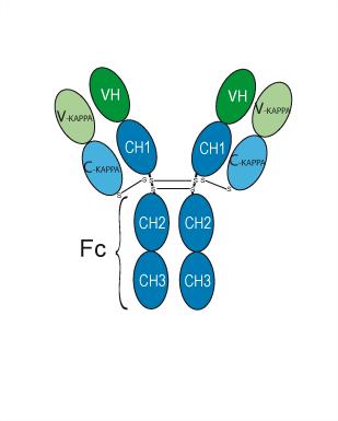
Human IgG1 kappa
Application
ELISA, FACS, Functional assay
Verified Bioactivity
-
 Immobilized IL-1 beta Protein, Human (HY-P7028) can bind Canakinumab. The EC50 for this effect is 6.151 ng/mL.
 -
 Flow cytometric analysis of 1.5X10⁶ RAW264.7 cells treated with 50 nM phorbol-12-myristate-13-acetate (PMA, HY-18739) and 5 μg/ml lipopolysaccharide (LPS, HY-D1056) for 24 hours with Canakinumab (HY-108810, red). Cells were fixed with 4% paraformaldehyde. Then stained with the primary antibody at 1/200 dilution for an hour at 4℃. Goat Anti-Human IgG H&L (AF488) (HY-P83776) was used as the secondary antibody at 1/1,000 dilution for 30 minutes at 4℃, Human IgG1 kappa, Isotype Control (HY-P99001, blue) was used as the isotype control.


Chemical Information
-
CAS No. 914613-48-2
-
Appearance Liquid
-
Molecular Weight 145.5 kDa
-
Color Colorless to light yellow
-
SMILES
[Canakinumab]
-
Synonyms
Ilaris; ACZ 885
-
Shipping
Shipping with dry ice.
-
Formulation
Please refer to the lot-specific COA for specific buffer information.
-
Storage
Please store the product under the recommended conditions in the Certificate of Analysis.


Publications (1)
-
Journal Impact Factor
-
Most Recent
-
Biochem Biophys Res Commun
Allograft inflammatory factor-1 in macrophages promotes renal tubular cell epithelial-mesenchymal transition via the NF-κB/IL-1β axis in diabetic kidney disease. [Abstract]2026 Jun 23:829:154192. PMID: 42349122

Protocols
-
Cell Cytotoxicity Assay
Cytotoxicity assays are usually based on the assessment of cell membrane damage, which can also be indirectly detected by measuring cell viability. Detection methods include MTT assay, CKK-8 assay, LDH assay and ATP assay, etc.
-
Research Protocol for Inflammation-related Diseases
The NLRP3 inflammasome is a cytosolic innate immune signaling platform that integrates priming signals and danger-signal activation to promote caspase-1 activation, maturation of IL-1β and IL-18, and gasdermin D-mediated pyroptotic cell death. The core experimental logic is to determine whether inflammatory disease phenotypes are driven by increased NLRP3 expression, ASC-containing inflammasome assembly, caspase-1 cleavage, GSDMD cleavage, and extracellular release of IL-1β/IL-18 rather than by nonspecific cell injury alone. The pathway is strongly linked to inflammation-related disease phenotypes because monosodium urate crystals activate NALP3/NLRP3 inflammasome signaling in gout-like crystal inflammation, cholesterol crystals activate NLRP3 inflammasomes in atherogenesis models, and DSS-induced intestinal inflammation has been reported to involve NLRP3 inflammasome activity. However, experimental colitis studies also show context-dependent protective effects of NLRP3 inflammasome co
-
Pyroptosis Solutions
Pyroptosis is a lytic inflammatory cell-death pathway executed by gasdermin pores, most classically through inflammasome-mediated activation of caspase-1, cleavage of gasdermin D, membrane pore formation, LDH release, and secretion of IL-1β and IL-18. The canonical pathway is commonly modeled by priming cells with an inflammatory signal such as LPS to induce pro-IL-1β and inflammasome components, followed by an activation signal such as ATP or nigericin to activate NLRP3, ASC speck formation, caspase-1 cleavage, GSDMD cleavage, cytokine release, and pyroptotic membrane rupture. The non-canonical pathway is triggered when cytosolic LPS activates mouse caspase-11 or human caspase-4/5, leading to GSDMD cleavage and pyroptosis, and this can secondarily activate NLRP3-dependent IL-1β release. Pyroptosis is linked to inflammatory injury, infection, cancer, liver disease, ocular disease, placental inflammation, and other disease phenotypes, but unresolved questions include which gasdermin fam


Purity & Documentation
-
Data Sheet (257 KB)
-
SDS (251 KB)
- English - EN (251 KB)
- Français - FR (251 KB)
- Deutsch - DE (251 KB)
- Norwegian - NO (251 KB)
- Español - ES (251 KB)
- Swedish - SV (251 KB)
- Italian - IT (251 KB)
- Korean - KR (251 KB)
- Portuguese - PT (251 KB)
-
Inhibitory Antibodies User Guide (603 KB)
References

Calculators
Concentration (start) × Volume (start) = Concentration (final) × Volume (final)


Canakinumab Related Classifications
HY-108810 Related Classifications
 Powered by Bioz