



Withanolide A

Based on 1 Customer Validation
Withanolide A is an orally active extract from the Indian herb Ashwagandha. Withanolide A can induce apoptosis. Withanolide A has anti-inflammatory and antitumor activity. Withanolide A can be used in the study of neurodegenerative diseases.
商品は「研究用試薬」です。人や動物の医療用・臨床診断用・食品用の製品ではありません。
研究用途以外に使用した場合、当社は一切の責任を負いかねます。
- 純度 : 99.98%
- CAS 番号: 32911-62-9
- 分子式: C28H38O6
- 分子量:470.60
-
保管条件:
-20°C, protect from light
* In solvent : -80°C, 6 months; -20°C, 1 month (protect from light)

To place orders, for customer services and technical support, please contact: MedChemExpress USA
Tel: 609-228-6898 E-mail: [email protected] [email protected]


-
 生物活性
生物活性
-
化学情報
-
溶剤 & 溶解度
- プロトコル
- 純度とドキュメンテーション
- 参考文献
-
 Help & FAQs


-
HY-N2065
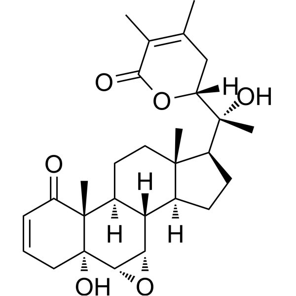
Withaferin ANF-κB Inhibitor -
HY-N3014
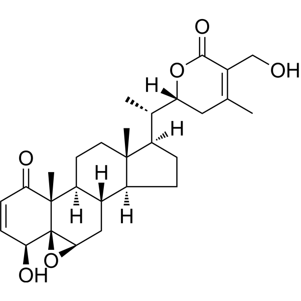
Bruceine DApoptosis Inducer -
HY-16561
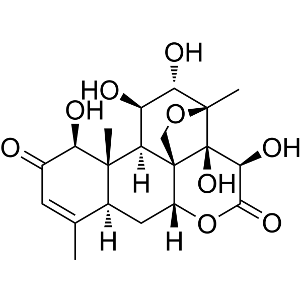
ResveratrolPolyphenolic Phytoalexin -

HY-18739
Phorbol 12-myristate 13-acetatePKC Activator -
HY-N0529
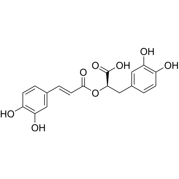
Rosmarinic acidAnti-Inflammatory Agent -
HY-129566
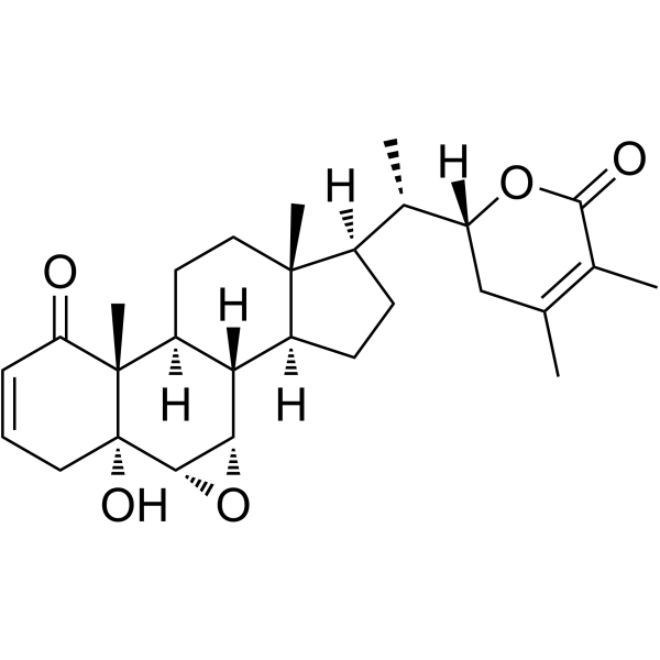
Withanolide BNeuroprotective Agent -

HY-N7028R
Withanolide A (Standard)NeuroprotectIve Agent -
HY-12449
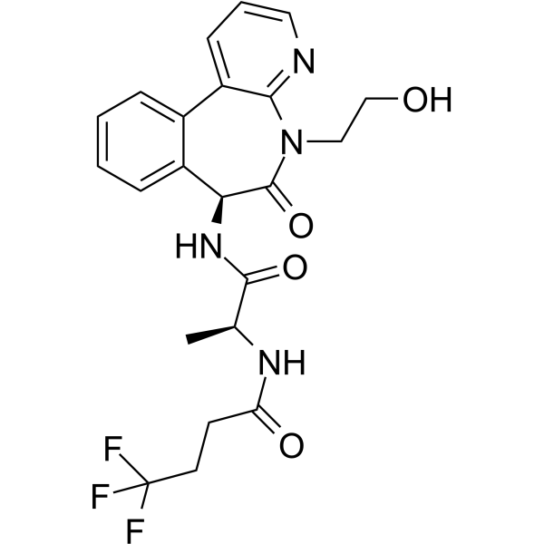
CrenigacestatNotch/γ-secretase Inhibitor -
HY-100431
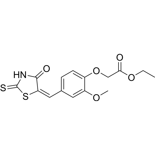
IMR-1Notch Inhibitor -
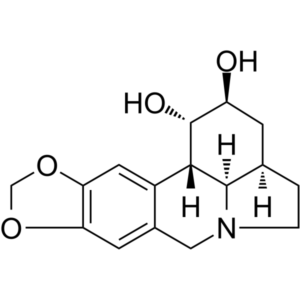
HY-N2403
DihydrolycorineProtein Synthesis Inhibitor -
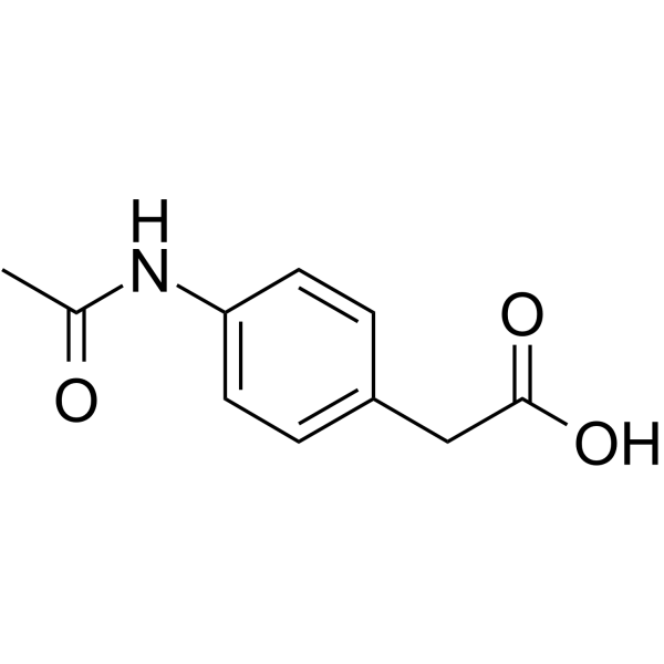
HY-76938
ActaritCAII Inhibitor -
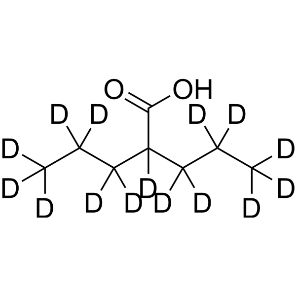
HY-10585S2
Valproic acid-d15Stable Isotope


生物活性
製品説明
Cellular Effect
|
Cell Line

|
Type | Value | Description | References |
|---|---|---|---|---|
| NCI-H929 | IC50 |
>10 μM
Compound: 8
|
Cytotoxicity against human NCI-H929 cells after 3 days by Alamar blue assay
Cytotoxicity against human NCI-H929 cells after 3 days by Alamar blue assay
|
[PMID: 24625088] |
体外実験
Withanolide A (1 μM, 4 days) can induce presynaptic and postsynaptic reconstruction of rat neurons and induce significant regeneration of axons and dendrites[1].
Withanolide A (0.5-2 μM, 24-48 h) inhibits growth of CaOV3 and SKOV3 ovarian cancer cells by targeting Notch1 and Notch3[2].
Withanolide A (0.5-50 μg/mL, 3 h) has the potential of vascular relaxation by promoting the production of NO in endothelial cells[3].
MedChemExpress (MCE) has not independently confirmed the accuracy of these methods. They are for reference only. Further protocols information, click here.
-
Cell Line:CaOV3、SKOV3、OVCAR3、TOV112D and TOV21G
-
Concentration:0, 0.078, 0.156, 0.313, 0.625, 1.25, 2.5, 5, 10 μM
-
Incubation Time:72 h
-
Result:Decreased the cell viability and with an IC50 concentrations at 520 nM, 627 nM, 452 nM, 262 nM, 243 nM, respectively.
-
Cell Line:CaOV3, SKOV3
-
Concentration:0.5, 1, 2 μM
-
Incubation Time:24, 48 h
-
Result:Induced apoptosis in a dose-dependent manner.
-
Cell Line:CaOV3, SKOV3
-
Concentration:0.5, 1, 2 μM
-
Incubation Time:24, 48 h
-
Result:Induced G2/M phase cell cycle arrest.
-
Cell Line:CaOV3, SKOV3
-
Concentration:0.5, 1, 2 μM
-
Incubation Time:24, 48 h
-
Result:Decreased cdc25C protein. Cyclin B1 stayed unchanged at 24 h but decreased by 48 h in a dose-dependent manner in CaOV3 cells, transiently increased in SKOV3 cells. Reduced the levels of phospho-Akt (ser 473) and Bcl-2. Reduced Notch 3 protein levels in CaOV3 cells and Notch1 protein levels in SKOV3 cells.
体内実験
MedChemExpress (MCE) has not independently confirmed the accuracy of these methods. They are for reference only.
-
Animal Model:Adult male Sprague Dawley rats weighing 240–250 g[4]
-
Dosage:10 μmol/kg
-
Administration:p.o. (dissolved in 0.5% gum arabic solution)
-
Result:Decreased the level of reactive oxygen species generation and lipid peroxidation. Increased the level of GSH and the activity of glutathione reductase, glutathione s transferase and superoxide dismutase. Increased the ATP, NADPH and GCLC activity. Decreased corticosterone level and glucocorticoid receptor.


化学情報
-
CAS 番号 32911-62-9
-
性状 Solid
-
分子量 470.60
-
分子式 C28H38O6
-
Color White to off-white
-
SMILES
O=C1O[C@@]([H])([C@@](C)([C@H]2CC[C@]3([C@@]4([C@H]5[C@@H]([C@@](O)(CC=CC6=O)[C@]6([C@]4(CC[C@@]32C)[H])C)O5)[H])[H])O)CC(C)=C1C
-
別名
ウィザノリドA
-
Structure Classification
-
Initial Source
-
輸送条件
Room temperature in continental US; may vary elsewhere.
-
保管条件
-20°C, protect from light
* In solvent : -80°C, 6 months; -20°C, 1 month (protect from light)


溶剤 & 溶解度
体外:
DMSO : 10 mg/mL (21.25 mM; Need ultrasonic; Hygroscopic DMSO has a significant impact on the solubility of product, please use newly opened DMSO)
Please refer to the solubility information to select the appropriate solvent. Once prepared, please aliquot and store the solution to prevent product inactivation from repeated freeze-thaw cycles.
Storage method and period of stock solution: -80°C, 6 months; -20°C, 1 month (protect from light). When stored at -80°C, please use it within 6 months. When stored at -20°C, please use it within 1 month.
Please refer to the solubility information to select the appropriate solvent. Once prepared, please aliquot and store the solution to prevent product inactivation from repeated freeze-thaw cycles.
Storage method and period of stock solution: -80°C, 6 months; -20°C, 1 month (protect from light). When stored at -80°C, please use it within 6 months. When stored at -20°C, please use it within 1 month.
濃度 (開始) × 体積 (開始) = 濃度 (終了) × 体積 (終了)
体内:
Select the appropriate dissolution method based on your experimental animal and administration route.
- For the following dissolution methods, please ensure to first prepare a clear stock solution using an In Vitro approach and then sequentially add co-solvents:
- To ensure reliable experimental results, the clarified stock solution can be appropriately stored based on storage conditions. As for the working solution for In Vivo experiments, it is recommended to prepare freshly and use it on the same day.
- The percentages shown for the solvents indicate their volumetric ratio in the final prepared solution. If precipitation or phase separation occurs during preparation, heat and/or sonication can be used to aid dissolution.
In Vivo Dissolution Calculator
Please enter the basic information of animal experiments:
-
-
-
-
Recommended: Prepare an additional quantity of animals to account for potential losses during experiments.
Please enter your animal formula composition:
-
%DMSO +
Recommended: Keep the proportion of DMSO in working solution below 2% if your animal is weak.
-
%+
-
+%Tween-80 + +
-
%Saline +
The co-solvents required include: DMSO, . All of co-solvents are available by MedChemExpress (MCE). , Tween 80. All of co-solvents are available by MedChemExpress (MCE).
Working solution concentration: 0.22 mg/mL
Method for preparing stock solution: mg drug dissolved in μL DMSO. Stock solution concentration: mg/mL. * In solvent : -80°C, 6 months; -20°C, 1 month (protect from light)
1. Take μL DMSO stock solution;
2. Add μL .
μL , mix evenly;
3. Then add μL Tween 80, mix evenly;
4. Then add μL
Please ensure that the stock solution in the first step is dissolved to a clear state, and add co-solvents in sequence. You can use ultrasonic heating (ultrasonic cleaner, recommended frequency 20-40 kHz), vortexing, etc. to assist dissolution.

プロトコル
-
Apoptosis
Apoptosis, also called programmed cell death, is generally characterized by distinct morphological characteristics.
-
TUNEL staining for apoptotic DNA fragmentation
TUNEL staining detects DNA strand breaks by using terminal deoxynucleotidyl transferase to add labeled nucleotides to exposed 3′-OH DNA termini, generating either microscopic staining in fixed cells or tissue sections, or fluorescence/cytometric signal in cell suspensions. TUNEL positivity reflects DNA fragmentation but should not be interpreted alone as definitive apoptosis, because TUNEL can also label necrotic, autolytic, mechanically damaged, or DNA-repair-associated DNA breaks.
-
Annexin V plus membrane-impermeant dye apoptosis staining
Annexin V-based apoptosis assays rely on the detection of phosphatidylserine (PS) externalization from the inner leaflet of the plasma membrane to the outer leaflet, an early biochemical hallmark of apoptosis. Fluorescently labeled Annexin V binds PS in a calcium-dependent manner, enabling identification of early apoptotic cells by flow cytometry or fluorescence microscopy. When combined with a membrane-impermeant DNA-binding dye (e. g. , propidium iodide), this approach allows discrimination between viable (Annexin V−/dye−), early apoptotic (Annexin V+/dye−), and late apoptotic or necrotic (Annexin V+/dye+) cell populations by assessing membrane integrity and PS exposure.
-
Apoptosis Solutions
Apoptosis is a regulated, generally non-lytic cell-death pathway that removes unwanted, damaged, infected, or abnormal cells through coordinated morphological changes, caspase activation, DNA fragmentation, and membrane remodeling. The intrinsic apoptosis pathway is controlled mainly by mitochondrial outer membrane permeabilization, BCL-2 family proteins, cytochrome c release, apoptosome formation, caspase-9 activation, and downstream executioner caspase-3/7 activation. The extrinsic apoptosis pathway is initiated by death receptors such as Fas, TNFR, and TRAIL receptors, which recruit adaptor proteins and activate caspase-8 before engaging executioner caspases or mitochondrial amplification through BID cleavage. Apoptosis is linked to many phenotypes, including cancer cell killing, tissue homeostasis, immune regulation, neurodegeneration, infection response, and treatment-induced cytotoxicity; unresolved questions include how apoptosis interacts with necroptosis, pyroptosis, ferroptos
-
Research Protocol for Inflammation-related Diseases
The NLRP3 inflammasome is a cytosolic innate immune signaling platform that integrates priming signals and danger-signal activation to promote caspase-1 activation, maturation of IL-1β and IL-18, and gasdermin D-mediated pyroptotic cell death. The core experimental logic is to determine whether inflammatory disease phenotypes are driven by increased NLRP3 expression, ASC-containing inflammasome assembly, caspase-1 cleavage, GSDMD cleavage, and extracellular release of IL-1β/IL-18 rather than by nonspecific cell injury alone. The pathway is strongly linked to inflammation-related disease phenotypes because monosodium urate crystals activate NALP3/NLRP3 inflammasome signaling in gout-like crystal inflammation, cholesterol crystals activate NLRP3 inflammasomes in atherogenesis models, and DSS-induced intestinal inflammation has been reported to involve NLRP3 inflammasome activity. However, experimental colitis studies also show context-dependent protective effects of NLRP3 inflammasome co
-
Subcutaneous Cell-Line-Derived Xenograft
Subcutaneous cell-line-derived xenograft (CDX) models are established by implanting cultured human cancer cell lines into immunodeficient mice, where the injected cells form localized tumors that can be monitored in vivo as a measure of tumorigenic potential, growth kinetics, and treatment response. These models are widely used in oncology research because they allow reproducible tumor formation and enable comparative assessment of tumor growth between different cell lines or genetic manipulations in a controlled in vivo microenvironment. Subcutaneous implantation of cancer cells in immunodeficient mice is a standard approach for evaluating tumor growth behavior and therapeutic response across multiple cancer types, including prostate, esophageal, pancreatic, and colon cancer models.


純度とドキュメンテーション
-
データシート (287 KB)
-
SDS (252 KB)
- English - EN (252 KB)
- Français - FR (252 KB)
- Deutsch - DE (252 KB)
- Norwegian - NO (252 KB)
- Español - ES (252 KB)
- Swedish - SV (252 KB)
- Italian - IT (252 KB)
- Korean - KR (252 KB)
- Portuguese - PT (252 KB)
-
取扱説明書 (2659 KB)
参考文献
[1]. Kuboyama T, et al. Neuritic regeneration and synaptic reconstruction induced by withanolide A. Br J Pharmacol. 2005 Apr;144(7):961-71.. [Content Brief]
[2]. Zhang X, et al. Inhibition of cell growth and induction of apoptosis in ovarian carcinoma cell lines CaOV3 and SKOV3 by natural withanolide Withaferin A. Gynecol Oncol. 2012 Mar;124(3):606-12. [Content Brief]
[3]. Pathak P, et al. Standardized root extract of Withania somnifera and Withanolide A exert moderate vasorelaxant effect in the rat aortic rings by enhancing nitric oxide generation. J Ethnopharmacol. 2021 Oct 5;278:114296. [Content Brief]
[4]. Baitharu I, et al. Withanolide A prevents neurodegeneration by modulating hippocampal glutathione biosynthesis during hypoxia. PLoS One. 2014 Oct 13;9(10):e105311. [Content Brief]


Complete Stock Solution Preparation Table
Please refer to the solubility information to select the appropriate solvent. Once prepared, please aliquot and store the solution to prevent product inactivation from repeated freeze-thaw cycles.
Storage method and period of stock solution: -80°C, 6 months; -20°C, 1 month (protect from light). When stored at -80°C, please use it within 6 months. When stored at -20°C, please use it within 1 month.
| Optional Solvent | Concentration Solvent Mass | 1 mg | 5 mg | 10 mg | 25 mg |
|---|---|---|---|---|---|
| DMSO | 1 mM | 2.1249 mL | 10.6247 mL | 21.2495 mL | 53.1237 mL |
| 5 mM | 0.4250 mL | 2.1249 mL | 4.2499 mL | 10.6247 mL | |
| 10 mM | 0.2125 mL | 1.0625 mL | 2.1249 mL | 5.3124 mL | |
| 15 mM | 0.1417 mL | 0.7083 mL | 1.4166 mL | 3.5416 mL | |
| 20 mM | 0.1062 mL | 0.5312 mL | 1.0625 mL | 2.6562 mL |


Withanolide A 関連分類
HY-N7028 関連分類
 Powered by Bioz