



Xaluritamig

Based on 1 Customer Validation
Xaluritamig (AMG-509) is a bispecific T cell engager and cytolytic agent with a Kd of 27.6 nM for human CD3ε. Xaluritamig binds to CD3ε via an anti-CD3 single-chain variable fragment (scFv) domain, and to STEAP1 via a bispecific anti-STEAP1 antigen-binding fragment (Fab) domain, thereby recruiting and activating T cells and forming a bridge between T cells and STEAP1-expressing cancer cells. Xaluritamig induces T cell-mediated redirected cytotoxicity, tumor cell lysis, cytokine release, CD8+ T cell activation and expansion, as well as tumor stasis or regression. Xaluritamig contains an Fc domain with no effector function, which prolongs serum half-life, exhibits only minimal activity against cells with low STEAP1 expression and normal cells, and shows extremely low target-related off-tumor toxicity in cynomolgus monkeys. Xaluritamig is used in STEAP1×CD3 XmAb 2+1 immunotherapy and in research on metastatic castration-resistant prostate cancer and Ewing sarcoma.
For research use only. We do not sell to patients.
- Purity : 99.44%
- CAS No.: 2559056-68-5
- Molecular Weight:173.555 kDa
-
Storage:
Please store the product under the recommended conditions in the Certificate of Analysis.


To place orders, for customer services and technical support, please contact: MedChemExpress USA
Tel: 609-228-6898 E-mail: [email protected] [email protected]


-
 Biological Activity
-
Chemical Information
- Protocols
- Purity & Documentation
- References
-
 Help & FAQs


-

HY-P99575
Tarlatamabanti-DLL3/CD3 BiTE antibody -

HY-P991015
PasritamigKLK2-CD3R Binder -

HY-P99418
AcapatamabAnti-CD3E/FOLH1 Antibody -

HY-P9908
AdalimumabTNF-α Inhibitor -

HY-P99001
Human IgG1 kappa, Isotype ControlIgG1κ Isotype Control -

HY-P991503
Anti-STEAP2 AntibodySTEAP2 Antibody -

HY-P99517
VoxalatamabPSMA/CD3 Dual Antibody -

HY-P9963
BlinatumomabAntineoplastic Agent -

HY-P990673
VandortuzumabSTEAP1 ADC Antibody -

HY-P99539
UbamatamabAnti-MUC16/CD3 Antibody -

HY-P9999
TrontinemabAβ/TFRC Antibody -
HY-17393
CarboplatinDNA Synthesis Inhibitor


Biological Activity
Description
Isotype
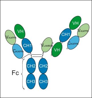
half-IgG1-kappa/VH-CH1-h-(scFv-heavy-lambda)-h-CH2-CH3
Species Reactivity
Human
IC50 & Target
CD3E & STEAP1
In Vitro
Xaluritamig (AMG-509) binds specifically to 293T cells transfected with human STEAP1, with an EC50 of 3.8 nM[2].
Xaluritamig potently induces T cell-mediated lysis of STEAP1-positive human cancer cells, with a median EC50 of 37 pM across 19 cell lines; it shows no activity against STEAP1-knockout prostate cancer cells[2].\n
Xaluritamig redirects prostate cancer cell lysis with higher potency than the anti-STEAP1 Fab XmAb molecule alone; it preferentially kills cancer cells with high STEAP1 expression and exhibits extremely low activity against normal cells[2].
Xaluritamig induces only extremely low levels of cytotoxicity in STEAP1-low-expressing human normal bronchial smooth muscle cells and aortic endothelial cells cultured in vitro[3].
MedChemExpress (MCE) has not independently confirmed the accuracy of these methods. They are for reference only. Further protocols information, click here.
In Vivo
Xaluritamig (0.3 mg/kg; intravenous injection; administered twice at a 7-day interval; total duration of 17 days) significantly inhibits tumor growth in the tibial bone metastasis model of Myc-CaP-LucSTEAP1 in FVB/N huCD3ε knock-in mice, but its combination with anti-PD-1 antibody does not enhance anti-tumor activity[4].
Xaluritamig (10 μg/kg, or an additional 40 μg/kg; intravenous injection; for 28 consecutive days) is well tolerated in male and female Mauritian-origin cynomolgus monkeys. Serum exposure is nearly dose-proportional, with no serious target-related off-tumor toxicity[4].
MedChemExpress (MCE) has not independently confirmed the accuracy of these methods. They are for reference only.
-
Animal Model:Male NSG mice (NOD.Cg-Prkdcscid Il2rgtm1N/j/SzJ), no specified weight and age + 22Rv1-LucSTEAP1 subcutaneous xenograft model[4]
-
Dosage:0.01 mg/kg, 0.1 mg/kg, 1 mg/kg
-
Administration:Intravenous injection; twice, 7 days apart; 16 days.
-
Result:Exerted significant antitumor activity.
The 0.01 mg/kg dose achieved 99.7% tumor growth inhibition with complete tumor stasis, while the 0.1 mg/kg and 1 mg/kg doses induced 61.6% and 66.2% tumor regression, respectively, after two administrations on days 9 and 16.
-
Animal Model:Male and female cynomolgus monkeys (Mauritian origin), normal toxicology evaluation model[4]
-
Dosage:10 μg/kg flat dose, 10 μg/kg initial dose followed by 40 μg/kg weekly
-
Administration:Intravenous injection; single-dose group: once; step-dose group: four times; 28 days.
-
Result:Was well tolerated during the 28-day study, with nearly dose-proportional and no gender differences in serum exposure.
Transient changes related to acute phase response were observed, including increased C-reactive protein, decreased absolute counts of total T cells, cytotoxic T cells, helper T cells and natural killer cells, and mild increases in neutrophils and globulins.
No severe target-related off-tumor toxicity was detected.
Clinical Trial
| NCT Number | Sponsor | Condition | Start Date |
Phase

|
|---|---|---|---|---|
| NCT01329991 | Plexxikon| | 2011-05 | PHASE1 |
Gene ID
Accession
Conjugated
Unconjugated
Reconsititution
The product can be reconstituted/diluted with sterile PBS or saline.
Format
-
half-IGG1-kappa/VH-CH1-h-(scFv-heavy-lambda)-h-CH2-CH3
Application
ELISA, FACS, Functional assay
Verified Bioactivity
-
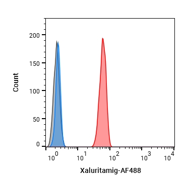 Flow cytometric analysis of 1X10⁶ Jurkat cells with Xaluritamig (HY-P990688, red). Cells were fixed with 4% paraformaldehyde. Then stained with the primary antibody at 1/200 dilution for an hour at 4℃. Alexa Fluor 488-conjugated AffiniPure Goat Anti-Human IgG H&L (HY-P83776) was used as the secondary antibody at 1/1,000 dilution for 30 minutes at 4℃. Human IgG1 kappa (HY-P99001, blue) was used as the isotype control, cells without incubation with primary antibody were used as the unlabeled control (black).
 -
 Flow cytometric analysis of 1X10⁶ FaDu cells with Xaluritamig (HY-P990688, red). Cells were fixed with 4% paraformaldehyde. Then stained with the primary antibody at 1/200 dilution for an hour at 4℃. AF 488-conjugated AffiniPure Goat Anti-Human IgG H&L (HY-P83776) was used as the secondary antibody at 1/1,000 dilution for 30 minutes at 4℃. Human IgG1 kappa (HY-P99001, blue) was used as the isotype control.


Chemical Information
-
CAS No. 2559056-68-5
-
Appearance Liquid
-
Molecular Weight 173.555 kDa
-
Color Colorless to light yellow
-
Synonyms
AMG-509
-
Shipping
Shipping with dry ice.
-
Formulation
Please refer to the lot-specific COA for specific buffer information.
-
Storage
Please store the product under the recommended conditions in the Certificate of Analysis.

Protocols
-
RNA extraction experimental
By lysing cells, releasing RNA, and removing impurities such as proteins and DNA, high-purity RNA products are finally obtained. The commonly used traditional method is the guanidine isothiocyanate/phenol/chloroform method (Trizol), which is suitable for a variety of animal materials including animal tissues, microorganisms, cultured cells, etc., and most plant materials.
-
Cell Cytotoxicity Assay
Cytotoxicity assays are usually based on the assessment of cell membrane damage, which can also be indirectly detected by measuring cell viability. Detection methods include MTT assay, CKK-8 assay, LDH assay and ATP assay, etc.
-
Mammalian live/dead viability and cytotoxicity staining
Live/dead viability and cytotoxicity staining assays are based on the simultaneous detection of intracellular esterase activity in metabolically active (viable) cells and membrane integrity loss in non-viable cells. In commonly used dual-staining approaches, membrane-permeant fluorogenic substrates are converted by intracellular esterases into fluorescent products in live cells, while impermeant DNA-binding dyes selectively enter cells with compromised plasma membranes and label nucleic acids in dead or dying cells, enabling discrimination between viable and non-viable populations by fluorescence microscopy or flow cytometry.
-
Protocol for Pharmacokinetic Study
Pharmacokinetic studies quantify how an organism handles a drug over time through absorption, distribution, metabolism, and excretion, and the core experimental readout is the concentration-time profile of parent drug and, when relevant, metabolites in biological matrices such as plasma, whole blood, urine, bile, or tissue. Pharmacokinetic analysis links dose, route, exposure, clearance, half-life, distribution, bioavailability, and systemic exposure to drug efficacy and toxicity hypotheses rather than measuring a signaling pathway directly. The literature links pharmacokinetics to drug-development phenotypes by showing that drug metabolism and pharmacokinetics influence compound progression, exposure-response interpretation, safety margins, dosing strategy, and failure risk during discovery and development. DMPK science contributes to compound optimization by integrating physicochemical properties, in vitro metabolism, transporter behavior, in vivo exposure, and pharmacodynamic contex
-
Protein Extraction
Protein extraction uses physical, chemical or biological methods, such as ultrasonic disruption, salting out, cell lysis, electrophoresis, etc., to destroy the cell membrane structure and to separate the proteins from different components according to their characteristics.
-
Research Protocol for Inflammation-related Diseases
The NLRP3 inflammasome is a cytosolic innate immune signaling platform that integrates priming signals and danger-signal activation to promote caspase-1 activation, maturation of IL-1β and IL-18, and gasdermin D-mediated pyroptotic cell death. The core experimental logic is to determine whether inflammatory disease phenotypes are driven by increased NLRP3 expression, ASC-containing inflammasome assembly, caspase-1 cleavage, GSDMD cleavage, and extracellular release of IL-1β/IL-18 rather than by nonspecific cell injury alone. The pathway is strongly linked to inflammation-related disease phenotypes because monosodium urate crystals activate NALP3/NLRP3 inflammasome signaling in gout-like crystal inflammation, cholesterol crystals activate NLRP3 inflammasomes in atherogenesis models, and DSS-induced intestinal inflammation has been reported to involve NLRP3 inflammasome activity. However, experimental colitis studies also show context-dependent protective effects of NLRP3 inflammasome co
-
Research Protocol for Cancer Immunology
Cancer immunology studies how the immune system recognizes, suppresses, edits, or fails to eliminate malignant cells through tumor antigen release, antigen presentation, T-cell priming, immune trafficking, tumor-cell killing, and feedback inhibition in the tumor microenvironment. The cancer-immunity cycle links tumor antigenicity, dendritic-cell priming, CD8+ T-cell infiltration, cytotoxic function, and immune-checkpoint regulation to tumor rejection or immune escape. Immune-checkpoint pathways such as PD-1/PD-L1 and CTLA-4 suppress antitumor T-cell activity and can be therapeutically blocked, but many tumors remain resistant because of poor antigen presentation, weak T-cell infiltration, suppressive myeloid cells, regulatory T cells, and tumor-intrinsic immune-exclusion programs. Unresolved questions include which immune-cell states predict response, how tumor-intrinsic pathways exclude immune cells, how myeloid suppression limits checkpoint blockade, and which combination strategies
-
Cell Viability Determination by MTT Colorimetric Assay
The following protocol uses the MTT colorimetric assay as a classic literature-established method for assessing cell viability/metabolic activity in cultured mammalian cells. MTT[3-(4,5-dimethylthiazol-2-yl)-2,5-diphenyltetrazolium bromide] is reduced by metabolically active cells to a colored formazan product; the amount of formazan is quantified spectrophotometrically and provides an indirect measure of metabolically active viable cells. Importantly, MTT reduction reflects cellular oxidoreductase/metabolic activity rather than an absolute direct count of living cells, so changes in cellular metabolism can alter the signal independently of cell number.


Purity & Documentation
-
Data Sheet (264 KB)
-
SDS (251 KB)
- English - EN (251 KB)
- Français - FR (251 KB)
- Deutsch - DE (251 KB)
- Norwegian - NO (251 KB)
- Español - ES (251 KB)
- Swedish - SV (251 KB)
- Italian - IT (251 KB)
- Korean - KR (251 KB)
- Portuguese - PT (251 KB)
-
Inhibitory Antibodies User Guide (603 KB)
References
[1]. Kelly WK, et al. Xaluritamig, a STEAP1 × CD3 XmAb 2+1 Immune Therapy for Metastatic Castration-Resistant Prostate Cancer: Results from Dose Exploration in a First-in-Human Study. Cancer Discov. 2024 Jan 12;14(1):76-89. [Content Brief]
[4]. Nolan-Stevaux O, et al. AMG 509 (Xaluritamig), an Anti-STEAP1 XmAb 2+1 T-cell Redirecting Immune Therapy with Avidity-Dependent Activity against Prostate Cancer. Cancer Discov. 2024 Jan 12;14(1):90-103. [Content Brief]

Calculators
Concentration (start) × Volume (start) = Concentration (final) × Volume (final)


Xaluritamig Related Classifications
HY-P990688 Related Classifications
 Powered by Bioz