



ZLDI-8
Based on 1 publication(s) in Google Scholar
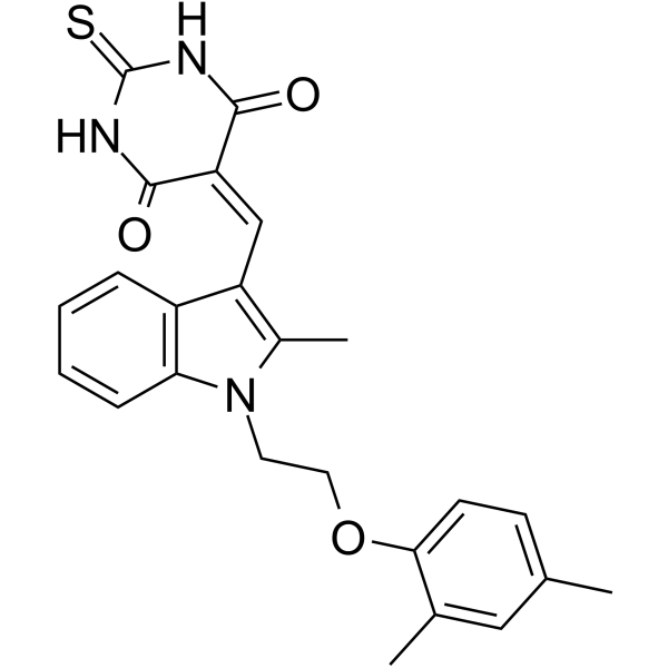
ZLDI-8 is a Notch activating/cleaving enzyme ADAM-17 inhibitor and inhibits the cleavage of Notch protein. ZLDI-8 decreases the expression of pro-survival/anti-apoptosis and epithelial-mesenchymal transition (EMT) related proteins. ZLDI-8 is also a competitive and irreversible tyrosine phosphatase (Lyp) inhibitor with an IC50 of 31.6 μM and a Ki of 26.22 μM. ZLDI-8 inhibits the growth of MHCC97-H cells with an IC50 of 5.32 μM.
For research use only. We do not sell to patients.
- Purity : 98.47%
- CAS No.: 667880-38-8
- Formula: C24H23N3O3S
- Molecular Weight:433.52
-
Storage:Powder -20°C, 3 years , 4°C, 2 years ; In solvent -80°C, 2 years , -20°C, 1 year

To place orders, for customer services and technical support, please contact: MedChemExpress USA
Tel: 609-228-6898 E-mail: [email protected] [email protected]


-
 Biological Activity
-
Chemical Information
-
Solvent & Solubility
- Protocols
- Purity & Documentation
- References
-
 Help & FAQs


-
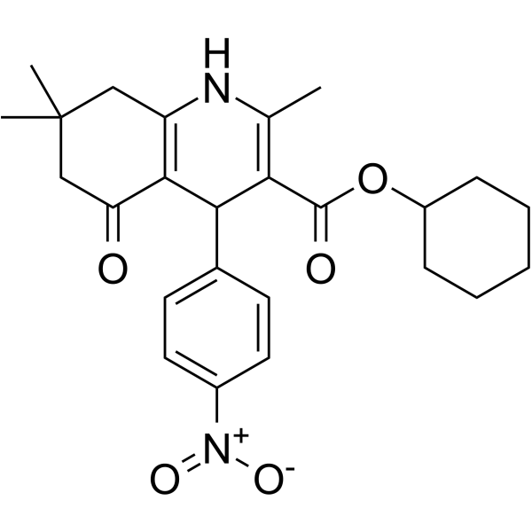
HY-15860
FLI-06Notch Inhibitor -
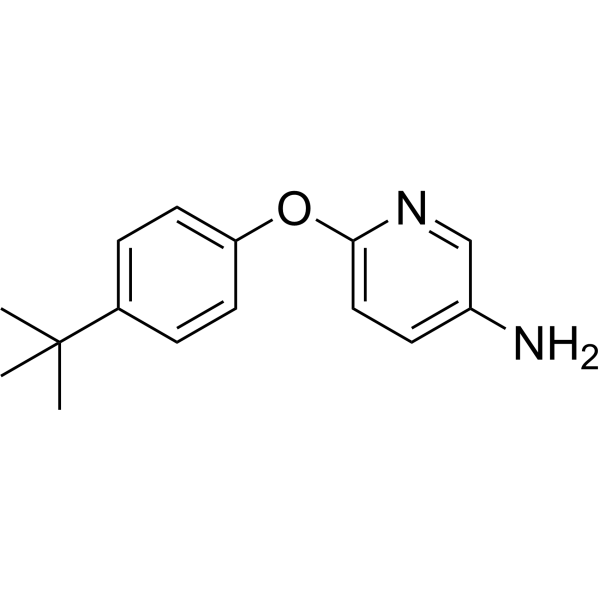
HY-135145
LimantrafinNotch Inhibitor -
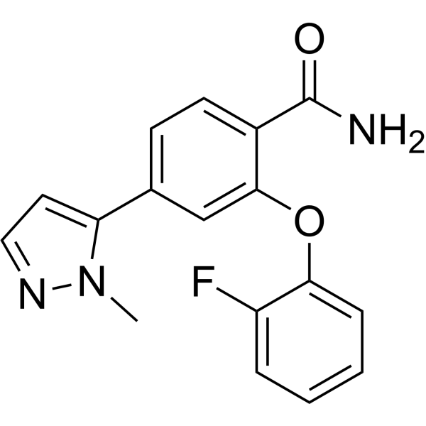
HY-137471
RBPJ Inhibitor-1RBPJ Inhibitor -
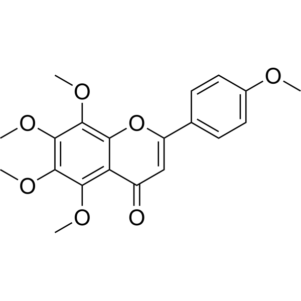
HY-N0133
TangeretinApoptosis Inducer -
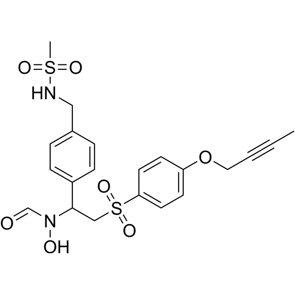
HY-110397
KP-457 -
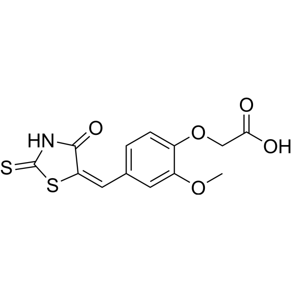
HY-100431A
IMR-1ANotch Inhibitor -
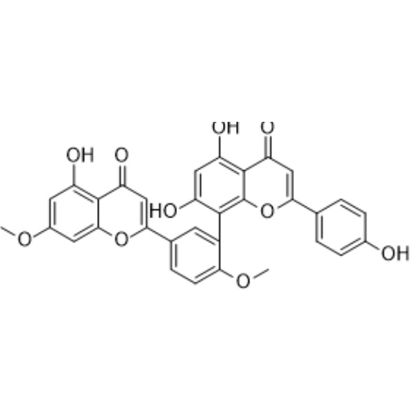
HY-N0889
GinkgetinAutophagy Inducer -

HY-N0133R
Tangeretin (Standard)Apoptosis Inducer -
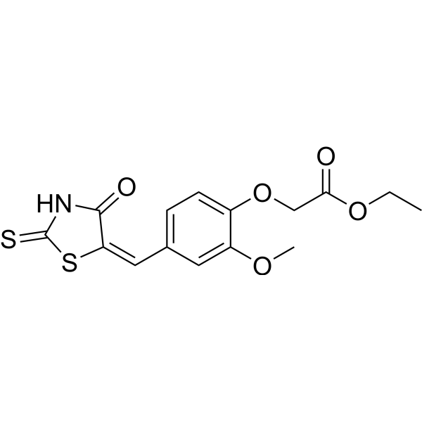
HY-100431
IMR-1Notch Inhibitor -
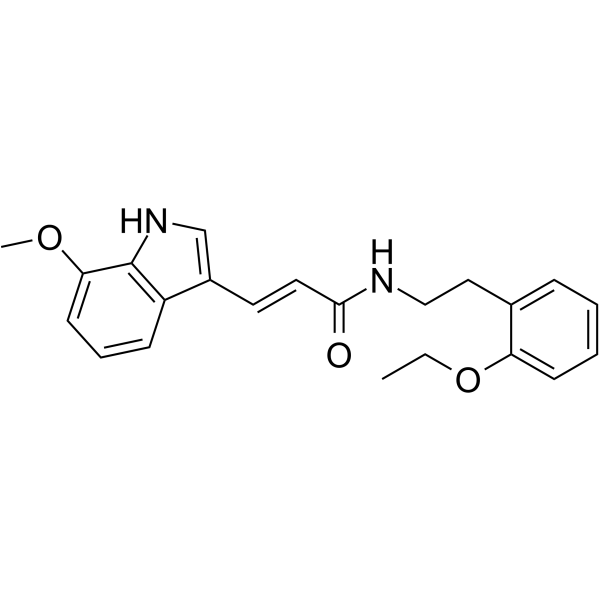
HY-117113
JI051Hes1-PHB2 Interaction Stabilizer -

HY-100431AR
IMR-1A (Standard)Notch Inhibitor -
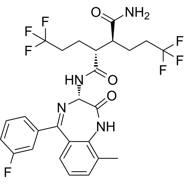
HY-12860
BMS-986115Notch Inhibitor
Publications Citing Use of MedChemExpress (MCE) ZLDI-8
More

Biological Activity
Description
IC50 & Target
ADAM-17[1]
IC50: 31.6 μM (Tyrosine phosphatase)[1]
Ki: 26.22 μM (Tyrosine phosphatase)[1]
In Vitro
ZLDI-8 (0.03-30 μM; 6-72 hours; MHCC97-H cells) treatment reduces cell viability in a time- and dose-dependent manner[1].
ZLDI-8 (1-10 μM; 6-72 hours; MHCC97-H cells) significantly decreases the level of NICD and the accumulation of NICD in the nucleus. ZLDI-8 could also reduce the expression of pro-survival/anti-apoptosis regulators, Survivin and cIAP1/2. And also increases the expression of epithelial marker E-Cadherin and reduced mesenchymal markers N-Cadherin and Vimentin[1].
ZLDI-8 enhances chemotherapy effects on tumor cell proliferation blockage, induction of apoptosis and cell-cycle arrest by inhibiting Notch pathway and blocking chemical resistance[1].
MedChemExpress (MCE) has not independently confirmed the accuracy of these methods. They are for reference only. Further protocols information, click here.
-
Cell Line:MHCC97-H cells
-
Concentration:0.03 μM, 0.1 μM, 0.3 μM, 1 μM, 3 μM, 10 μM, 30 μM
-
Incubation Time:6 hours, 12 hours, 24 hours, 48 hours, 72 hours
-
Result:Emerged cytotoxic effect on MHCC97-H cells in a time- and dose-dependent manner.
-
Cell Line:MHCC97-H cells
-
Concentration:1 μM, 3 μM, 10 μM
-
Incubation Time:6 hours, 12 hours, 24 hours, 48 hours, 72 hours
-
Result:Significantly decreased the level of NICD and the accumulation of NICD in the nucleus. Also reduced the expression of pro-survival/anti-apoptosis regulators, Survivin and cIAP1/2
In Vivo
MedChemExpress (MCE) has not independently confirmed the accuracy of these methods. They are for reference only.
-
Animal Model:Nude mice with MHCC-97H cells[1]
-
Dosage:2 mg/kg, 1 mg/kg, 500 μg/kg, or 200 μg/kg
-
Administration:Intraperitoneal injection; every two days; for 20 days
-
Result:Inhibited tumor growth in nude HCC-bearing mice model.


Chemical Information
-
CAS No. 667880-38-8
-
Appearance Solid
-
Molecular Weight 433.52
-
Formula C24H23N3O3S
-
Color Yellow to orange
-
SMILES
O=C(/C(C(N1)=O)=C/C2=C(C)N(CCOC3=CC=C(C)C=C3C)C4=C2C=CC=C4)NC1=S
-
Shipping
Room temperature in continental US; may vary elsewhere.
-
Storage
Powder -20°C 3 years 4°C 2 years In solvent -80°C 2 years -20°C 1 year


Publications (1)
-
Journal Impact Factor
-
Most Recent


Solvent & Solubility
In Vitro:
DMSO : 62.5 mg/mL (144.17 mM; Need ultrasonic; Hygroscopic DMSO has a significant impact on the solubility of product, please use newly opened DMSO)
Please refer to the solubility information to select the appropriate solvent. Once prepared, please aliquot and store the solution to prevent product inactivation from repeated freeze-thaw cycles.
Storage method and period of stock solution: -80°C, 2 years; -20°C, 1 year. When stored at -80°C, please use it within 2 years. When stored at -20°C, please use it within 1 year.
Please refer to the solubility information to select the appropriate solvent. Once prepared, please aliquot and store the solution to prevent product inactivation from repeated freeze-thaw cycles.
Storage method and period of stock solution: -80°C, 2 years; -20°C, 1 year. When stored at -80°C, please use it within 2 years. When stored at -20°C, please use it within 1 year.
Concentration (start) × Volume (start) = Concentration (final) × Volume (final)

Protocols
-
RNA extraction experimental
By lysing cells, releasing RNA, and removing impurities such as proteins and DNA, high-purity RNA products are finally obtained. The commonly used traditional method is the guanidine isothiocyanate/phenol/chloroform method (Trizol), which is suitable for a variety of animal materials including animal tissues, microorganisms, cultured cells, etc., and most plant materials.
-
Apoptosis
Apoptosis, also called programmed cell death, is generally characterized by distinct morphological characteristics.
-
TUNEL staining for apoptotic DNA fragmentation
TUNEL staining detects DNA strand breaks by using terminal deoxynucleotidyl transferase to add labeled nucleotides to exposed 3′-OH DNA termini, generating either microscopic staining in fixed cells or tissue sections, or fluorescence/cytometric signal in cell suspensions. TUNEL positivity reflects DNA fragmentation but should not be interpreted alone as definitive apoptosis, because TUNEL can also label necrotic, autolytic, mechanically damaged, or DNA-repair-associated DNA breaks.
-
Cell Cytotoxicity Assay
Cytotoxicity assays are usually based on the assessment of cell membrane damage, which can also be indirectly detected by measuring cell viability. Detection methods include MTT assay, CKK-8 assay, LDH assay and ATP assay, etc.
-
Annexin V plus membrane-impermeant dye apoptosis staining
Annexin V-based apoptosis assays rely on the detection of phosphatidylserine (PS) externalization from the inner leaflet of the plasma membrane to the outer leaflet, an early biochemical hallmark of apoptosis. Fluorescently labeled Annexin V binds PS in a calcium-dependent manner, enabling identification of early apoptotic cells by flow cytometry or fluorescence microscopy. When combined with a membrane-impermeant DNA-binding dye (e. g. , propidium iodide), this approach allows discrimination between viable (Annexin V−/dye−), early apoptotic (Annexin V+/dye−), and late apoptotic or necrotic (Annexin V+/dye+) cell populations by assessing membrane integrity and PS exposure.
-
Apoptosis Solutions
Apoptosis is a regulated, generally non-lytic cell-death pathway that removes unwanted, damaged, infected, or abnormal cells through coordinated morphological changes, caspase activation, DNA fragmentation, and membrane remodeling. The intrinsic apoptosis pathway is controlled mainly by mitochondrial outer membrane permeabilization, BCL-2 family proteins, cytochrome c release, apoptosome formation, caspase-9 activation, and downstream executioner caspase-3/7 activation. The extrinsic apoptosis pathway is initiated by death receptors such as Fas, TNFR, and TRAIL receptors, which recruit adaptor proteins and activate caspase-8 before engaging executioner caspases or mitochondrial amplification through BID cleavage. Apoptosis is linked to many phenotypes, including cancer cell killing, tissue homeostasis, immune regulation, neurodegeneration, infection response, and treatment-induced cytotoxicity; unresolved questions include how apoptosis interacts with necroptosis, pyroptosis, ferroptos
-
Notch Pathway Solutions
The Notch pathway is a contact-dependent signaling pathway that controls cell-fate decisions, differentiation, proliferation, and tissue patterning through interactions between membrane-bound Notch receptors and membrane-bound ligands on neighboring cells. Canonical Notch signaling is activated when ligand engagement triggers proteolytic release of the Notch intracellular domain, which enters the nucleus and regulates transcription together with DNA-binding transcriptional complexes. In the canonical mechanism, ligand-dependent Notch activation leads to release of the intracellular Notch domain, and presenilin-dependent γ-secretase activity is required for production of the active intracellular signaling fragment. The released intracellular domain functions as a nuclear signal that converts Notch receptor activation at the membrane into transcriptional regulation of target programs such as HES/HEY-family genes and other context-dependent downstream targets. The literature links Notch p


Purity & Documentation
-
Data Sheet (279 KB)
-
SDS (393 KB)
- English - EN (393 KB)
- Français - FR (393 KB)
- Deutsch - DE (393 KB)
- Norwegian - NO (393 KB)
- Español - ES (393 KB)
- Swedish - SV (393 KB)
- Italian - IT (393 KB)
- Korean - KR (393 KB)
- Portuguese - PT (393 KB)
-
Handling Instructions (2659 KB)
References
[1]. Zhang Y, et al. Novel ADAM-17 inhibitor ZLDI-8 enhances the in vitro and in vivo chemotherapeutic effects of Sorafenib on hepatocellular carcinoma cells. Cell Death Dis. 2018 Jul 3;9(7):743. [Content Brief]
[2]. Hou X, et al. Fast identification of novel lymphoid tyrosine phosphatase inhibitors using target-ligand interaction-based virtual screening. J Med Chem. 2014 Nov 26;57(22):9309-22. [Content Brief]


Complete Stock Solution Preparation Table
Please refer to the solubility information to select the appropriate solvent. Once prepared, please aliquot and store the solution to prevent product inactivation from repeated freeze-thaw cycles.
Storage method and period of stock solution: -80°C, 2 years; -20°C, 1 year. When stored at -80°C, please use it within 2 years. When stored at -20°C, please use it within 1 year.
| Optional Solvent | Concentration Solvent Mass | 1 mg | 5 mg | 10 mg | 25 mg |
|---|---|---|---|---|---|
| DMSO | 1 mM | 2.3067 mL | 11.5335 mL | 23.0670 mL | 57.6675 mL |
| 5 mM | 0.4613 mL | 2.3067 mL | 4.6134 mL | 11.5335 mL | |
| 10 mM | 0.2307 mL | 1.1533 mL | 2.3067 mL | 5.7667 mL | |
| 15 mM | 0.1538 mL | 0.7689 mL | 1.5378 mL | 3.8445 mL | |
| 20 mM | 0.1153 mL | 0.5767 mL | 1.1533 mL | 2.8834 mL | |
| 25 mM | 0.0923 mL | 0.4613 mL | 0.9227 mL | 2.3067 mL | |
| 30 mM | 0.0769 mL | 0.3844 mL | 0.7689 mL | 1.9222 mL | |
| 40 mM | 0.0577 mL | 0.2883 mL | 0.5767 mL | 1.4417 mL | |
| 50 mM | 0.0461 mL | 0.2307 mL | 0.4613 mL | 1.1533 mL | |
| 60 mM | 0.0384 mL | 0.1922 mL | 0.3844 mL | 0.9611 mL | |
| 80 mM | 0.0288 mL | 0.1442 mL | 0.2883 mL | 0.7208 mL | |
| 100 mM | 0.0231 mL | 0.1153 mL | 0.2307 mL | 0.5767 mL |


ZLDI-8 Related Classifications
HY-123931 Related Classifications
 Powered by Bioz