



Zoptarelin doxorubicin

Based on 1 Customer Validation
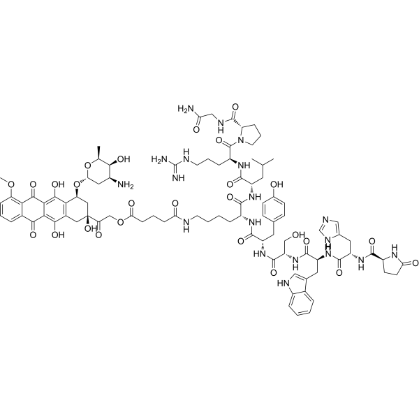
Zoptarelin doxorubicin (AEZS-108; AN-152) is a hybrid anticancer agent, containing Zoptarelin and Doxorubicin. Zoptarelin doxorubicin has been used to research targeting tumors expressing LHRH receptors. Zoptarelin doxorubicin abolishes tumor progression and induces remarkable apoptosis in vitro.
For research use only. We do not sell to patients.
- CAS No.: 139570-93-7
- Formula: C91H117N19O26
- Molecular Weight:1893.01
-
Storage:
Sealed storage, away from moisture and light.
Powder -80°C, 2 years , -20°C, 1 year* In solvent : -80°C, 6 months; -20°C, 1 month (sealed storage, away from moisture and light)

To place orders, for customer services and technical support, please contact: MedChemExpress USA
Tel: 609-228-6898 E-mail: [email protected] [email protected]


-
 Biological Activity
-
Chemical Information
-
Solvent & Solubility
- Protocols
- Purity & Documentation
- References
-
 Help & FAQs


-
HY-16474
RelugolixGnRH Antagonist -
HY-14789
(R)-ElagolixGnRH Receptor Antagonist -
HY-13534
AbarelixGnRH Antagonist -

HY-16168
Degarelix acetateGnRH Anagonist -
HY-14369
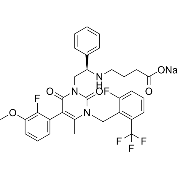
Elagolix sodiumGnRH Receptor Antagonist -
HY-P0013
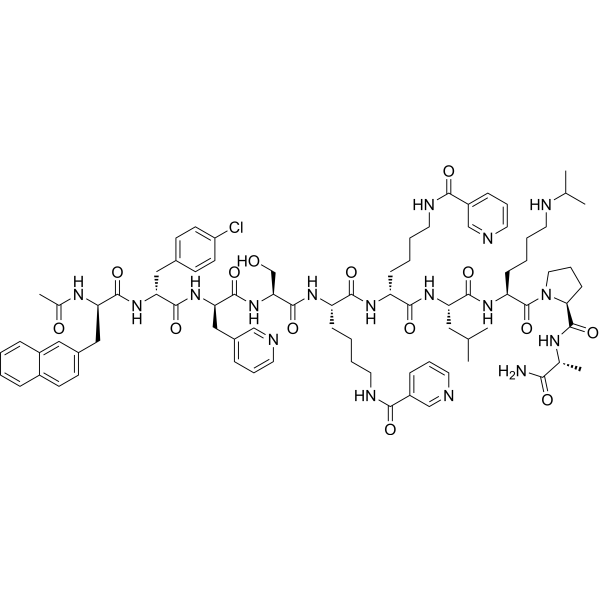
AntideLHRH Antagonist -

HY-16474R
Relugolix (Standard)GnRH Antagonist -
HY-16168A
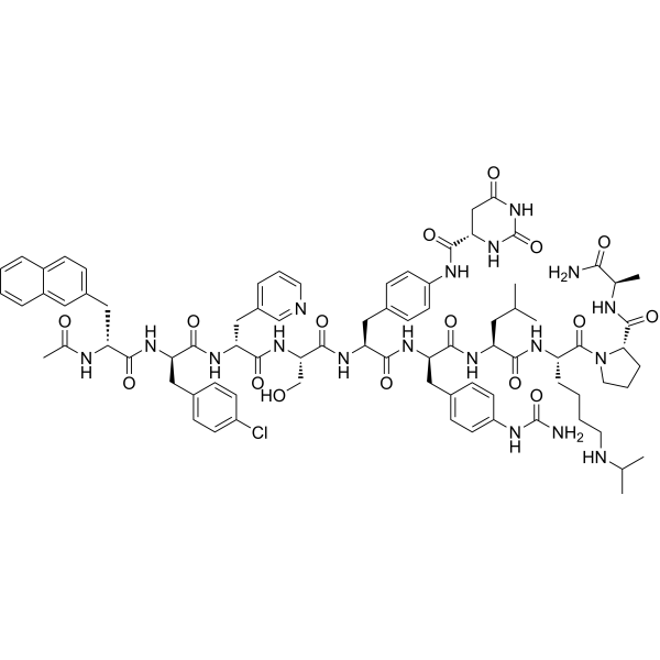
DegarelixGnRHR Antagonist -

HY-14789R
(R)-Elagolix (Standard)GnRH Receptor Antagonist -
HY-13699
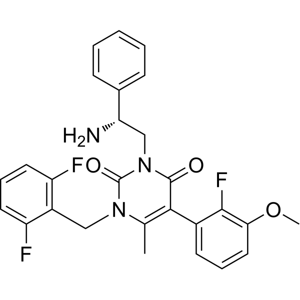
NBI-42902GnRH receptor Antagonist -
HY-P0009A
Cetrorelix acetateGnRH Receptor Antagonist -
HY-P1808
LGnRH-III, lampreyGnRH Agonist


Biological Activity
Description
IC50 & Target
Apoptosis, LHRH receptors[1]
In Vitro
Zoptarelin doxorubicin (AN-152) inhibits almost 70 % of glioblastoma cell growth, increases almost 250% apoptosis and causes a greater increase in calcein retention[1].
Zoptarelin doxorubicin (AN-152) up-regulates the tumor suppressor and pro-apoptotic p53, and inhibits the expression of the primordial, neuroectodermal stem cell marker, nestin[1].
MedChemExpress (MCE) has not independently confirmed the accuracy of these methods. They are for reference only. Further protocols information, click here.
-
Cell Line:U-87 MG
-
Concentration:100 nM
-
Incubation Time:48 h
-
Result:Brought about an almost 70 % inhibition of tumor cell growth.
-
Cell Line:U-87 MG
-
Concentration:100 nM
-
Incubation Time:48 h
-
Result:Increased almost 250 % apoptosis.
-
Cell Line:U-87 MG
-
Concentration:100 nM
-
Incubation Time:24 h
-
Result:Did not induce any down-regulation of LHRH-R.
Inhibited the expression of the primordial, neuroectodermal stem cell marker, nestin.
Up-regulated the tumor suppressor and pro-apoptotic p53.
In Vivo
MedChemExpress (MCE) has not independently confirmed the accuracy of these methods. They are for reference only.
-
Animal Model:Female nude mice (injected in the flanks with 1×10⁶ glioblastoma U-87 MG cells)[1]
-
Dosage:413 nM/20g
-
Administration:i.v.; once weekly; for 6 weeks
-
Result:Significantly inhibited tumor growth compared to the control.
Clinical Trial
| NCT Number | Sponsor | Condition | Start Date |
Phase

|
|---|---|---|---|---|
| NCT01329991 | Plexxikon| | 2011-05 | PHASE1 |


Chemical Information
-
CAS No. 139570-93-7
-
Appearance Solid
-
Molecular Weight 1893.01
-
Formula C91H117N19O26
-
Color Orange to red
-
Synonyms
AEZS-108; AN-152
-
Shipping
Room temperature in continental US; may vary elsewhere.
-
Storage
Sealed storage, away from moisture and light
Powder -80°C 2 years -20°C 1 year * In solvent : -80°C, 6 months; -20°C, 1 month (sealed storage, away from moisture and light)


Solvent & Solubility
In Vitro:
H2O : 50 mg/mL (26.41 mM; Need ultrasonic)
Please refer to the solubility information to select the appropriate solvent. Once prepared, please aliquot and store the solution to prevent product inactivation from repeated freeze-thaw cycles.
Storage method and period of stock solution: -80°C, 6 months; -20°C, 1 month (sealed storage, away from moisture and light). When stored at -80°C, please use it within 6 months. When stored at -20°C, please use it within 1 month.
* Note: If you choose water as the stock solution, please dilute it to the working solution, then filter and sterilize it with a 0.22 μm filter before use.
Please refer to the solubility information to select the appropriate solvent. Once prepared, please aliquot and store the solution to prevent product inactivation from repeated freeze-thaw cycles.
Storage method and period of stock solution: -80°C, 6 months; -20°C, 1 month (sealed storage, away from moisture and light). When stored at -80°C, please use it within 6 months. When stored at -20°C, please use it within 1 month.
* Note: If you choose water as the stock solution, please dilute it to the working solution, then filter and sterilize it with a 0.22 μm filter before use.
Concentration (start) × Volume (start) = Concentration (final) × Volume (final)

Protocols
-
Apoptosis
Apoptosis, also called programmed cell death, is generally characterized by distinct morphological characteristics.
-
TUNEL staining for apoptotic DNA fragmentation
TUNEL staining detects DNA strand breaks by using terminal deoxynucleotidyl transferase to add labeled nucleotides to exposed 3′-OH DNA termini, generating either microscopic staining in fixed cells or tissue sections, or fluorescence/cytometric signal in cell suspensions. TUNEL positivity reflects DNA fragmentation but should not be interpreted alone as definitive apoptosis, because TUNEL can also label necrotic, autolytic, mechanically damaged, or DNA-repair-associated DNA breaks.
-
Annexin V plus membrane-impermeant dye apoptosis staining
Annexin V-based apoptosis assays rely on the detection of phosphatidylserine (PS) externalization from the inner leaflet of the plasma membrane to the outer leaflet, an early biochemical hallmark of apoptosis. Fluorescently labeled Annexin V binds PS in a calcium-dependent manner, enabling identification of early apoptotic cells by flow cytometry or fluorescence microscopy. When combined with a membrane-impermeant DNA-binding dye (e. g. , propidium iodide), this approach allows discrimination between viable (Annexin V−/dye−), early apoptotic (Annexin V+/dye−), and late apoptotic or necrotic (Annexin V+/dye+) cell populations by assessing membrane integrity and PS exposure.
-
Apoptosis Solutions
Apoptosis is a regulated, generally non-lytic cell-death pathway that removes unwanted, damaged, infected, or abnormal cells through coordinated morphological changes, caspase activation, DNA fragmentation, and membrane remodeling. The intrinsic apoptosis pathway is controlled mainly by mitochondrial outer membrane permeabilization, BCL-2 family proteins, cytochrome c release, apoptosome formation, caspase-9 activation, and downstream executioner caspase-3/7 activation. The extrinsic apoptosis pathway is initiated by death receptors such as Fas, TNFR, and TRAIL receptors, which recruit adaptor proteins and activate caspase-8 before engaging executioner caspases or mitochondrial amplification through BID cleavage. Apoptosis is linked to many phenotypes, including cancer cell killing, tissue homeostasis, immune regulation, neurodegeneration, infection response, and treatment-induced cytotoxicity; unresolved questions include how apoptosis interacts with necroptosis, pyroptosis, ferroptos


Purity & Documentation
-
Data Sheet (277 KB)
-
SDS (252 KB)
- Français - FR (252 KB)
- Deutsch - DE (252 KB)
- Norwegian - NO (252 KB)
- Español - ES (252 KB)
- Swedish - SV (252 KB)
- Italian - IT (252 KB)
- Korean - KR (252 KB)
- Portuguese - PT (252 KB)
-
Handling Instructions (2659 KB)
References


Complete Stock Solution Preparation Table
Please refer to the solubility information to select the appropriate solvent. Once prepared, please aliquot and store the solution to prevent product inactivation from repeated freeze-thaw cycles.
Storage method and period of stock solution: -80°C, 6 months; -20°C, 1 month (sealed storage, away from moisture and light). When stored at -80°C, please use it within 6 months. When stored at -20°C, please use it within 1 month.
| Optional Solvent | Concentration Solvent Mass | 1 mg | 5 mg | 10 mg | 25 mg |
|---|---|---|---|---|---|
| H2O | 1 mM | 0.5283 mL | 2.6413 mL | 5.2826 mL | 13.2065 mL |
| 5 mM | 0.1057 mL | 0.5283 mL | 1.0565 mL | 2.6413 mL | |
| 10 mM | 0.0528 mL | 0.2641 mL | 0.5283 mL | 1.3206 mL | |
| 15 mM | 0.0352 mL | 0.1761 mL | 0.3522 mL | 0.8804 mL | |
| 20 mM | 0.0264 mL | 0.1321 mL | 0.2641 mL | 0.6603 mL | |
| 25 mM | 0.0211 mL | 0.1057 mL | 0.2113 mL | 0.5283 mL |
* Note: If you choose water as the stock solution, please dilute it to the working solution, then filter and sterilize it with a 0.22 μm filter before use.


Zoptarelin doxorubicin Related Classifications
HY-16532 Related Classifications
 Powered by Bioz