



1,1,1,1-Kestohexaose

Based on 1 Customer Validation
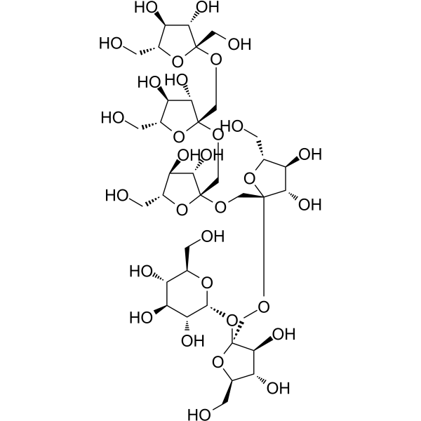
1,1,1,1-Kestohexaose (1,1,1,1-Kestohexose) is an insulin-type fructooligosaccharide hexasaccharide. 1,1,1,1-Kestohexaose exhibits neuroprotective and anti-depressant-like activities. 1,1,1,1-Kestohexaose can alleviate Corticosteron (HY-B1618)-induced decrease in cell viability and NGF expression in vitro.
For research use only. We do not sell to patients.
- Purity : 98.0%
- CAS No.: 62512-19-0
- Formula: C36H62O31
- Molecular Weight:990.86
-
Storage:
4°C, protect from light
* In solvent : -80°C, 6 months; -20°C, 1 month (protect from light)

To place orders, for customer services and technical support, please contact: MedChemExpress USA
Tel: 609-228-6898 E-mail: [email protected] [email protected]


-
 Biological Activity
-
Chemical Information
-
Solvent & Solubility
- Protocols
- Purity & Documentation
- References
-
 Help & FAQs


-
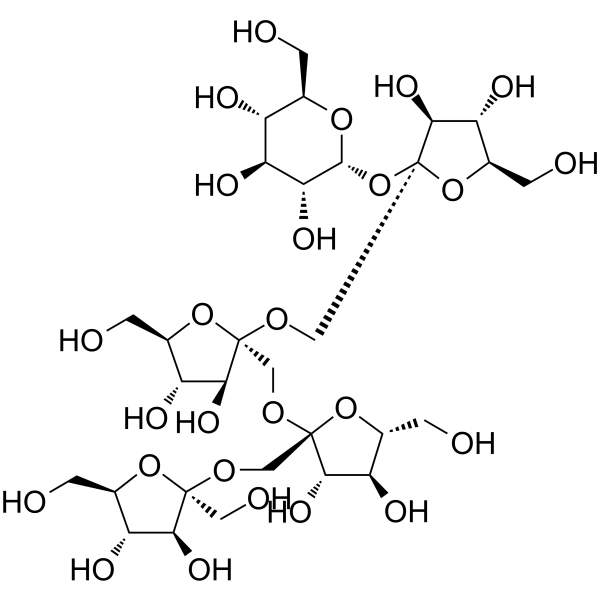
HY-N2577
1F-FructofuranosylnystoseFructo-oligose -
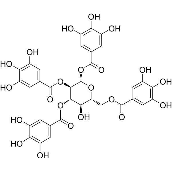
HY-111832
1,2,3,6-TetragalloylglucoseUGT1A1 Inhibitor -
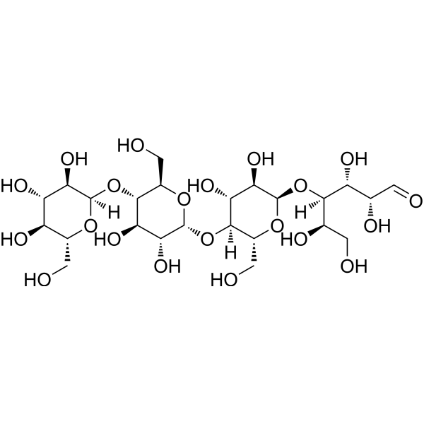
HY-N2464
MaltotetraoseAmylase Substrate -
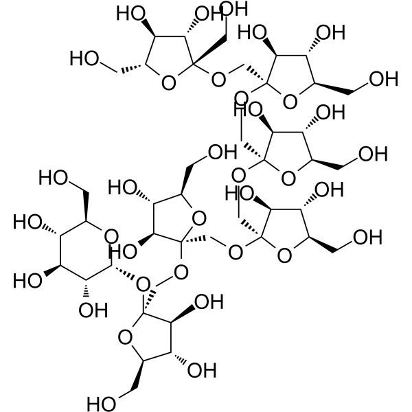
HY-N6837
Fructo-oligosaccharide DP7/GF6 -
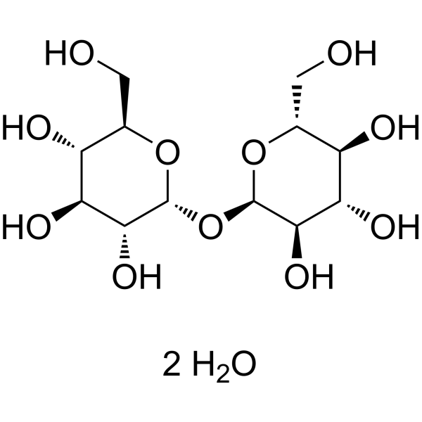
HY-N1132A
D-(+)-Trehalose dihydrateEndogenous Metabolite -
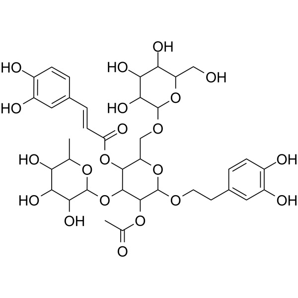
HY-N2155
Tubuloside AHepatoprotective Agent -
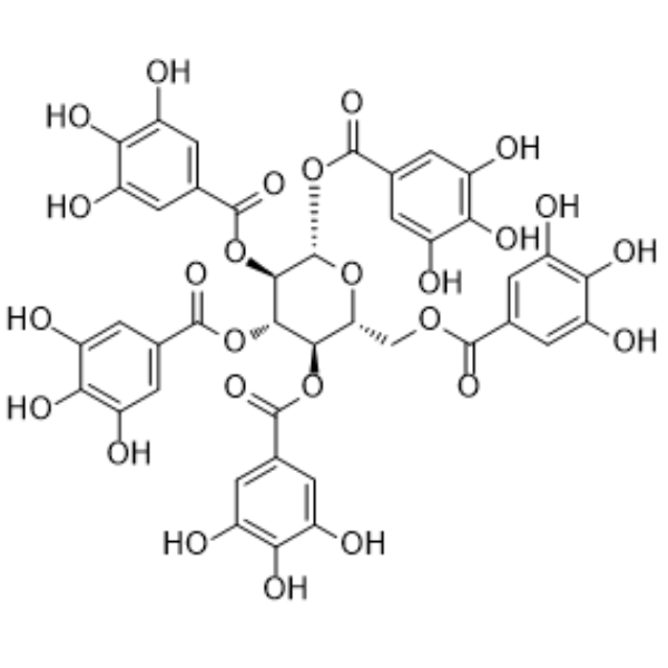
HY-N0527
PentagalloylglucoseAntioxidant -
HY-N0148
RutinCBR1 Inhibitor -
HY-N6839
1,4-b-D-Xylopentaoseα-L-arabinofuranosidase Substrate -
HY-135072
N,N',N''-TriacetylchitotrioseLysozyme Inhibitor -

HY-N1132AR
D-(+)-Trehalose dihydrate (Standard)Endogenous Metabolite -
HY-N3519
Platycodin D3Triterpenoid Saponin


Biological Activity
Description
In Vitro
1,1,1,1-Kestohexaose (Compound IHS) (0.625-1.25 μM, 48 h) protects PC12 cells from the lesion induced by Corticosteron (Cort)[2].
1,1,1,1-Kestohexaose (2.5-10 μM, 48 h) attenuates the Cort-induced [Ca2+] overloading in PC12 cells[2].
1,1,1,1-Kestohexaose (5-10 μM, 48 h) reverses the decrease in NGF mRNA level induced by Cort in PC12 cells[2].
MedChemExpress (MCE) has not independently confirmed the accuracy of these methods. They are for reference only. Further protocols information, click here.


Chemical Information
-
CAS No. 62512-19-0
-
Appearance Solid
-
Molecular Weight 990.86
-
Formula C36H62O31
-
Color White to off-white
-
SMILES
OC[C@]1(O[C@H](CO)[C@@H](O)[C@@H]1O)OC[C@]2(O[C@H](CO)[C@@H](O)[C@@H]2O)OC[C@]3(O[C@H](CO)[C@@H](O)[C@@H]3O)OC[C@]4(O[C@H](CO)[C@@H](O)[C@@H]4O)OC[C@]5(O[C@H](CO)[C@@H](O)[C@@H]5O)O[C@H]6O[C@@H]([C@@H](O)[C@H](O)[C@H]6O)CO
-
Synonyms
1,1,1,1-Kestohexose
-
Structure Classification
-
Initial Source
-
Shipping
Room temperature in continental US; may vary elsewhere.
-
Storage
4°C, protect from light
* In solvent : -80°C, 6 months; -20°C, 1 month (protect from light)


Solvent & Solubility
In Vitro:
H2O : 250 mg/mL (252.31 mM; Need ultrasonic)
DMSO : 100 mg/mL (100.92 mM; Need ultrasonic; Hygroscopic DMSO has a significant impact on the solubility of product, please use newly opened DMSO)
Please refer to the solubility information to select the appropriate solvent. Once prepared, please aliquot and store the solution to prevent product inactivation from repeated freeze-thaw cycles.
Storage method and period of stock solution: -80°C, 6 months; -20°C, 1 month (protect from light). When stored at -80°C, please use it within 6 months. When stored at -20°C, please use it within 1 month.
* Note: If you choose water as the stock solution, please dilute it to the working solution, then filter and sterilize it with a 0.22 μm filter before use.
Please refer to the solubility information to select the appropriate solvent. Once prepared, please aliquot and store the solution to prevent product inactivation from repeated freeze-thaw cycles.
Storage method and period of stock solution: -80°C, 6 months; -20°C, 1 month (protect from light). When stored at -80°C, please use it within 6 months. When stored at -20°C, please use it within 1 month.
* Note: If you choose water as the stock solution, please dilute it to the working solution, then filter and sterilize it with a 0.22 μm filter before use.
Concentration (start) × Volume (start) = Concentration (final) × Volume (final)
In Vivo:
For the following dissolution methods, please prepare the working solution directly:
It is recommended to prepare fresh solutions and use them promptly within a short period of time.
The percentages shown for the solvents indicate their volumetric ratio in the final prepared solution. If precipitation or phase separation occurs during preparation, heat and/or sonication can be used to aid dissolution.
Add each solvent one by one: PBS
Solubility: 100 mg/mL (100.92 mM); Clear solution; Need ultrasonic

Protocols
-
PC12 NGF-Induced Neuronal Differentiation Culture
PC12 cells are a rat adrenal pheochromocytoma clonal line that responds to NGF by stopping proliferation and extending branching neurite-like processes; after longer NGF exposure, cells develop long processes and neuronal-like ultrastructural and functional features. NGF-induced differentiation is read out mainly by neurite outgrowth, reduced proliferation, microtubule assembly, and neuronal differentiation-associated proteins such as MAPs, tau, GAP-43, and synapsin-1.
-
RNA extraction experimental
By lysing cells, releasing RNA, and removing impurities such as proteins and DNA, high-purity RNA products are finally obtained. The commonly used traditional method is the guanidine isothiocyanate/phenol/chloroform method (Trizol), which is suitable for a variety of animal materials including animal tissues, microorganisms, cultured cells, etc., and most plant materials.
-
Cell Cytotoxicity Assay
Cytotoxicity assays are usually based on the assessment of cell membrane damage, which can also be indirectly detected by measuring cell viability. Detection methods include MTT assay, CKK-8 assay, LDH assay and ATP assay, etc.
-
Mammalian live/dead viability and cytotoxicity staining
Live/dead viability and cytotoxicity staining assays are based on the simultaneous detection of intracellular esterase activity in metabolically active (viable) cells and membrane integrity loss in non-viable cells. In commonly used dual-staining approaches, membrane-permeant fluorogenic substrates are converted by intracellular esterases into fluorescent products in live cells, while impermeant DNA-binding dyes selectively enter cells with compromised plasma membranes and label nucleic acids in dead or dying cells, enabling discrimination between viable and non-viable populations by fluorescence microscopy or flow cytometry.
-
Research Protocol for Endocrine Diseases
Endocrine diseases often arise from disrupted hormone production, hormone signaling, or target-tissue responsiveness; for diabetes-focused endocrine disease models, insulin signaling regulates glucose uptake, hepatic glucose output, lipid metabolism, and β-cell compensation. Type 2 diabetes develops through interacting defects in insulin resistance, β-cell dysfunction, adipose inflammation, hepatic glucose overproduction, altered incretin signaling, and ectopic lipid metabolism. A major unresolved question is whether endocrine dysfunction is driven primarily by target-tissue insulin resistance, intrinsic β-cell failure, immune/inflammatory stress, or combined multi-organ failure that differs by disease stage.
-
CCK-8/WST-8 Cell Proliferation Assay
The CCK-8/WST-8 assay is based on the reduction of the water-soluble tetrazolium salt WST-8 to a water-soluble formazan product by cellular dehydrogenases in metabolically active cells, where the generated formazan amount is proportional to the number of living cells and is quantified by measuring absorbance in the visible range, providing a colorimetric readout for cell viability and proliferation assessment. This class of tetrazolium-based assays improves upon earlier MTT-based systems by producing a water-soluble formazan, eliminating the need for organic solubilization steps and enabling direct spectrophotometric measurement in culture medium.
-
PC12 NGF-induced neuronal-like differentiation
PC12 cells are a rat adrenal pheochromocytoma-derived clonal cell line that responds to nerve growth factor by stopping proliferation and extending neurites, producing a sympathetic neuron-like phenotype used to study neuronal differentiation and neurite outgrowth. NGF acts through TrkA-dependent signaling, and neurite outgrowth is associated with ERK/Akt signaling, microtubule organization, neuronal-marker expression, and increased electrophysiological neuronal features such as sodium-channel density. The main assay readout is morphological differentiation, usually measured as the percentage of neurite-bearing cells, neurite length, neurite number, or total neurite length per cell. Additional readouts include GAP-43, tyrosine hydroxylase, βIII-tubulin, neurofilament, synapsin I, synaptophysin, ERK phosphorylation, Akt phosphorylation, and sodium-channel current density.
-
MTT Cell Proliferation Assay
The MTT assay is a colorimetric endpoint assay for estimating viable cell number, cell growth, cytotoxicity, or cell activation in cultured mammalian cells. Living cells reduce the yellow tetrazolium salt MTT into purple/blue formazan, while dead cells do not generate the same signal; the resulting color can be quantified with a multiwell spectrophotometer. MTT reduction is commonly interpreted as a readout of metabolic activity that often correlates with viable cell number, but it should not be treated as a direct cell-counting method unless the assay is optimized for the cell type and experimental condition. Studies show that MTT reduction can involve mitochondrial and non-mitochondrial reducing systems, and formazan may accumulate in intracellular lipid droplets rather than simply marking mitochondria.
-
Directly Induced Neuron Culture
Directly induced neuron culture converts somatic cells, most commonly fibroblasts, into induced neurons without passing through a pluripotent or neural progenitor stage; classic evidence shows that mouse fibroblasts can be converted by Ascl1, Brn2/Pou3f2, and Myt1l, human fibroblasts can be converted by defined neuronal transcription factors, and human fibroblasts can also be converted by miR-9/9-124 with neurogenic or subtype-specifying transcription factors. The readout is acquisition of neuronal identity and function, assessed by neuronal morphology, neuronal markers such as Tuj1/βIII-tubulin, MAP2, synapsin, and subtype markers when relevant, together with functional assays such as action-potential firing, synaptic activity, and electrophysiology.
-
Cell Viability Determination by MTT Colorimetric Assay
The following protocol uses the MTT colorimetric assay as a classic literature-established method for assessing cell viability/metabolic activity in cultured mammalian cells. MTT[3-(4,5-dimethylthiazol-2-yl)-2,5-diphenyltetrazolium bromide] is reduced by metabolically active cells to a colored formazan product; the amount of formazan is quantified spectrophotometrically and provides an indirect measure of metabolically active viable cells. Importantly, MTT reduction reflects cellular oxidoreductase/metabolic activity rather than an absolute direct count of living cells, so changes in cellular metabolism can alter the signal independently of cell number.


Purity & Documentation
-
Data Sheet (279 KB)
-
SDS (252 KB)
- English - EN (252 KB)
- Français - FR (252 KB)
- Deutsch - DE (252 KB)
- Norwegian - NO (252 KB)
- Español - ES (252 KB)
- Swedish - SV (252 KB)
- Italian - IT (252 KB)
- Korean - KR (252 KB)
- Portuguese - PT (252 KB)
-
Handling Instructions (2659 KB)
References


Complete Stock Solution Preparation Table
Please refer to the solubility information to select the appropriate solvent. Once prepared, please aliquot and store the solution to prevent product inactivation from repeated freeze-thaw cycles.
Storage method and period of stock solution: -80°C, 6 months; -20°C, 1 month (protect from light). When stored at -80°C, please use it within 6 months. When stored at -20°C, please use it within 1 month.
| Optional Solvent | Concentration Solvent Mass | 1 mg | 5 mg | 10 mg | 25 mg |
|---|---|---|---|---|---|
| DMSO / H2O | 1 mM | 1.0092 mL | 5.0461 mL | 10.0922 mL | 25.2306 mL |
| 5 mM | 0.2018 mL | 1.0092 mL | 2.0184 mL | 5.0461 mL | |
| 10 mM | 0.1009 mL | 0.5046 mL | 1.0092 mL | 2.5231 mL | |
| 15 mM | 0.0673 mL | 0.3364 mL | 0.6728 mL | 1.6820 mL | |
| 20 mM | 0.0505 mL | 0.2523 mL | 0.5046 mL | 1.2615 mL | |
| 25 mM | 0.0404 mL | 0.2018 mL | 0.4037 mL | 1.0092 mL | |
| 30 mM | 0.0336 mL | 0.1682 mL | 0.3364 mL | 0.8410 mL | |
| 40 mM | 0.0252 mL | 0.1262 mL | 0.2523 mL | 0.6308 mL | |
| 50 mM | 0.0202 mL | 0.1009 mL | 0.2018 mL | 0.5046 mL | |
| 60 mM | 0.0168 mL | 0.0841 mL | 0.1682 mL | 0.4205 mL | |
| 80 mM | 0.0126 mL | 0.0631 mL | 0.1262 mL | 0.3154 mL | |
| 100 mM | 0.0101 mL | 0.0505 mL | 0.1009 mL | 0.2523 mL |
* Note: If you choose water as the stock solution, please dilute it to the working solution, then filter and sterilize it with a 0.22 μm filter before use.


1,1,1,1-Kestohexaose Related Classifications
HY-N6838 Related Classifications
 Powered by Bioz