



1-Myristoyl-2-hydroxy-sn-glycero-3-phosphoethanolamine

Based on 1 Customer Validation
1-Myristoyl-2-hydroxy-sn-glycero-3-phosphoethanolamine (14:0 Lyso PE) is a lysophospholipid with a phosphoethanolamine head and a myristoyl tail. The free amine group can conjugate with NHS active ester or coupled with carboxylic acid in the presence of a coupling agent. It also induces transient increases in intracellular calcium in PC12 cells . Serum levels of 1-Myristoyl-2-hydroxy-sn-glycero-3-phosphoethanolamine are elevated in patients with malignant breast cancer compared to healthy controls.
For research use only. We do not sell to patients.
- Purity : 99.1%
- CAS No.: 123060-40-2
- Formula: C19H40NO7P
- Molecular Weight:425.50
-
Storage:Powder -20°C, 3 years ; In solvent -80°C, 6 months , -20°C, 1 month


To place orders, for customer services and technical support, please contact: MedChemExpress USA
Tel: 609-228-6898 E-mail: [email protected] [email protected]


-
 Biological Activity
-
Chemical Information
- Protocols
- Purity & Documentation
- References
-
 Help & FAQs


-
HY-W127499
1,2-Dierucoyl-sn-glycero-3-phosphocholineLiposome Membrane Composition -

HY-131652
1,2-Di-O-hexadecyl-sn-glycero-3-phosphocholinePhospholipids -
HY-112005
DOPEFerroptosis Inhibitor -
HY-144004
DSPE-PEG2000-Mal ammoniumLipid Carrier -
HY-139533
Phosphatidylinositols, soya, sodium saltsPhosphatidylinositols Mixture -
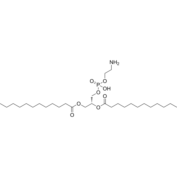
HY-W322225
1,2-Dilauroyl-sn-glycero-3-phosphoethanolamineAnionic Phospholipid -
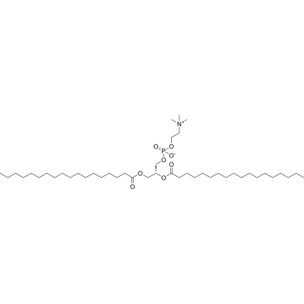
HY-W040193
DSPCLiposome Compound -
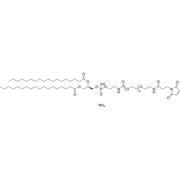
HY-144004A
DSPE-PEG5000-Mal ammoniumLipid Carrier -

HY-139414
LysophosphatidylcholinesHemolytic Agent -
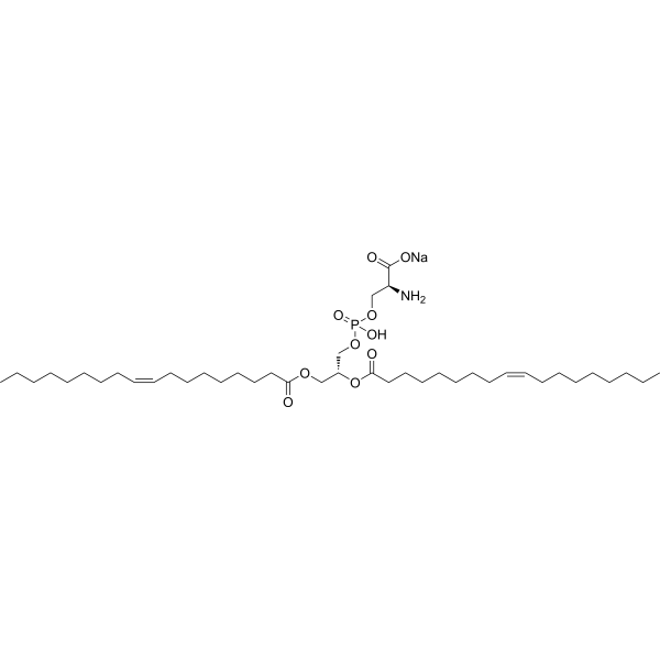
HY-141613
1,2-Dioleoyl-sn-glycero-3-phospho-L-serine sodiumPhosphoserine/Phosphatidylserine Ubstitute -
HY-W355700
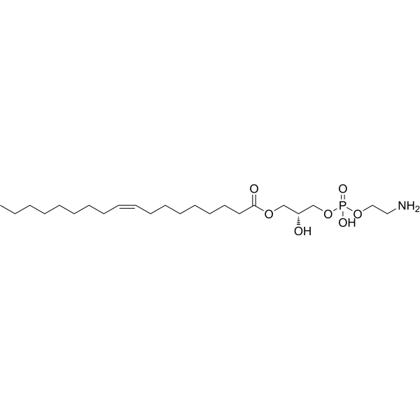
1-Oleoyl-2-hydroxy-sn-glycero-3-phosphoethanolamineDeltamethrin Metabolite -

HY-109541
Colfosceril miristatePhospholipid Compound


Biological Activity
Description


Chemical Information
-
CAS No. 123060-40-2
-
Appearance Solid
-
Molecular Weight 425.50
-
Formula C19H40NO7P
-
Color White to off-white
-
SMILES
CCCCCCCCCCCCCC(OC[C@@H](O)COP(OCCN)(O)=O)=O
-
Synonyms
14:0 Lyso PE; 1-Myristoyl-2-hydroxy-sn-glycero-3-PE; 1-Tetradecanoyl-sn-glycero-3-phosphoethanolamine
-
Shipping
Room temperature in continental US; may vary elsewhere.
-
Storage
Powder -20°C 3 years In solvent -80°C 6 months -20°C 1 month

Protocols
-
Calcium Spark Assay
Calcium sparks are localized, transient increases in intracellular calcium concentration ([Ca2+]i) that occur in cardiac myocytes and represent elementary events underlying excitation-contraction coupling. These events are generated by the coordinated opening of clusters of ryanodine receptors (RyRs) on the sarcoplasmic reticulum membrane, leading to a brief release of Ca2+ into the cytosol. The detection and analysis of calcium sparks provide insights into the mechanisms of calcium handling and signaling in cardiac cells. Imaging techniques using fluorescent calcium indicators such as Fluo-3 are employed to visualize these subcellular calcium transients with high spatial and temporal resolution. The protocol is based on established methodologies described in primary literature for both experimental measurement and automated analysis of calcium sparks.
-
Ca2+ Staining Technique
Ca2+ staining is an experimental technique that utilizes specific fluorescent probes (such as Fluo-4 AM, Fura-2, etc.) to qualitatively or quantitatively detect dynamic changes in intracellular Ca2+ concentrations; this is achieved by monitoring the changes in fluorescent signals generated when these probes bind to free intracellular calcium ions. The underlying principle relies primarily on the presence of chelating groups within the probe's molecular structure that possess high affinity for calcium ions.
-
Breast Cancer Modeling
Breast cancer is a heterogeneous cancer, and it has been distinguished into four subtypes: luminal A, luminal B, HER2-positive and basal-like. Molecular mutations, epigenetic alterations, hormone exposure and immune microenvironment are related to the progression of breast cancer.


Purity & Documentation
-
Data Sheet (266 KB)
-
SDS (254 KB)
- English - EN (254 KB)
- Français - FR (254 KB)
- Deutsch - DE (254 KB)
- Norwegian - NO (254 KB)
- Español - ES (254 KB)
- Swedish - SV (254 KB)
- Italian - IT (254 KB)
- Korean - KR (254 KB)
- Portuguese - PT (254 KB)
-
Handling Instructions (2659 KB)
References

Calculators
Concentration (start) × Volume (start) = Concentration (final) × Volume (final)


1-Myristoyl-2-hydroxy-sn-glycero-3-phosphoethanolamine Related Classifications
HY-145547 Related Classifications
 Powered by Bioz