



11-cis-Retinal
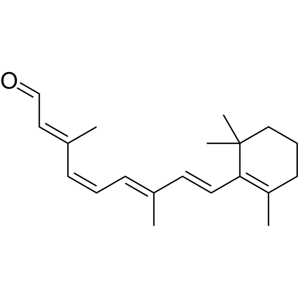
11-cis-Retinal (11-cis Retinaldehyde; 11-cis Vitamin A aldehyde), oxidized form of 11-cis-Retinol (HY-W587807), is a naturally occurring visual component. 11-cis-Retinal binds to opsin in the mammalian visual system as an inverse agonist forming the inactive conformation of rhodopsin. 11-cis-Retinal plays a crucial role in vision, growth and development. 11-cis-Retinal can restore visual function in moths with visual impairments. 11-cis-Retinal can restore retinal function in an early-age LCA2 retinal degeneration 12 mouse model.
For research use only. We do not sell to patients.
- CAS No.: 564-87-4
- Formula: C20H28O
- Molecular Weight:284.44
-
Storage:
-20°C, stored under nitrogen
* In solvent : -80°C, 6 months; -20°C, 1 month (stored under nitrogen)

To place orders, for customer services and technical support, please contact: MedChemExpress USA
Tel: 609-228-6898 E-mail: [email protected] [email protected]


-
 Biological Activity
-
Chemical Information
- Protocols
- Purity & Documentation
- References
-
 Help & FAQs


-

HY-138233
KUS121VCP Modulator -

HY-121600
GAT229CB1 Positive Allosteric Modulator (PAM) -
HY-B1719A
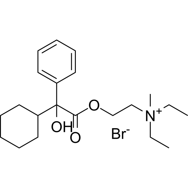
Oxyphenonium bromidemAChR Antagonist -

HY-112076A
Atropine methyl nitratemAChR Antagonist -
HY-106007
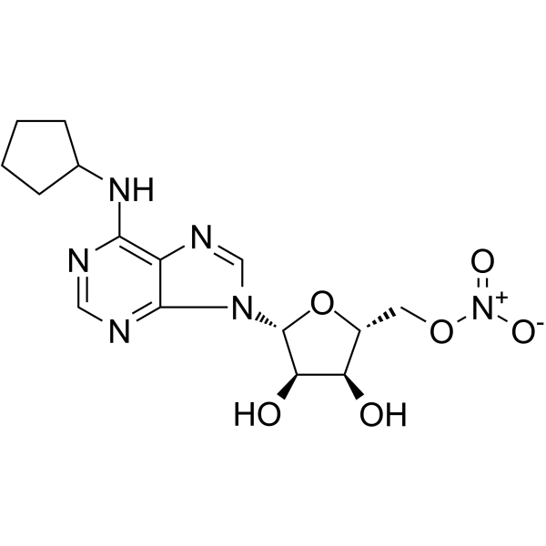
TrabodenosonAdenosine A1 Receptor Agonist -
HY-110014
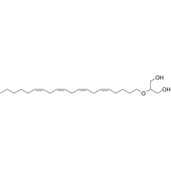
Noladin etherCB1 Agonist -

HY-B1719AR
Oxyphenonium bromide (Standard)Reference Standard -
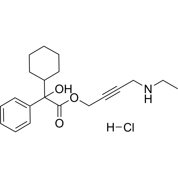
HY-W739521
N-Desethyloxybutynin hydrochlorideMetabolite of Oxybutynin -
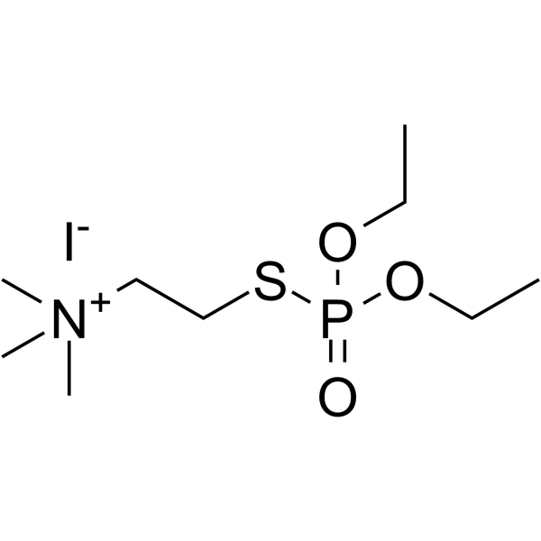
HY-16183
Echothiopate iodideAChE Inhibitor -

HY-W587807
11-cis-RetinolA Retinol Form -
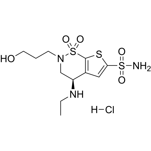
HY-W424792A
O-Desmethyl Brinzolamide hydrochlorideCarbonic Anhydrase Inhibitor -
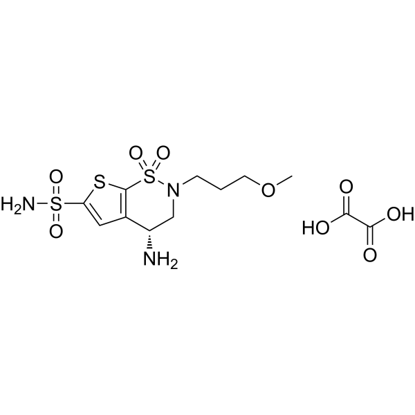
HY-137966A
N-Desethyl Brinzolamide oxalateCarbonic anhydrase Inhibitor
All Endogenous Metabolite Isoforms
More

Biological Activity
Description
IC50 & Target
|
Human Endogenous Metabolite |
Cellular Effect
|
Cell Line

|
Type | Value | Description | References |
|---|---|---|---|---|
| HEK293 | CC50 |
23 μM
Compound: 11-cis-retinal
|
Cytotoxicity against HEK293 cells by WST-1 assay
Cytotoxicity against HEK293 cells by WST-1 assay
|
[PMID: 20805032] |
In Vivo
11-cis-Retinal (i.p., from postnatal day 14 to 21) restores retinal function in an early-age LCA2 retinal degeneration 12 mouse model[3].
MedChemExpress (MCE) has not independently confirmed the accuracy of these methods. They are for reference only.


Chemical Information
-
CAS No. 564-87-4
-
Appearance Liquid (Density: 1.07 g/cm3)
-
Molecular Weight 284.44
-
Formula C20H28O
-
Color Yellow to brown
-
SMILES
CC(/C=C\C=C(\C=C\C1=C(CCCC(C)1C)C)C)=C\C=O
-
Synonyms
11-cis Retinaldehyde; 11-cis Vitamin A aldehyde
-
Structure Classification
-
Initial Source
-
Shipping
Room temperature in continental US; may vary elsewhere.
-
Storage
-20°C, stored under nitrogen
* In solvent : -80°C, 6 months; -20°C, 1 month (stored under nitrogen)

Protocols
-
How to Choose the Right Model Animal
Choosing the right model animal is a validity-driven decision in which the species, strain, sex, age, genetic background, disease-induction method, outcome measures, and welfare burden must match the scientific question rather than laboratory tradition or convenience. A model should be selected by judging face validity, construct validity, and predictive validity: whether it resembles the human phenotype, whether it reproduces relevant mechanisms, and whether results are likely to predict human biology or treatment response. Animal studies often fail to translate because of species differences, weak disease resemblance, poor experimental design, inadequate reporting, publication bias, and underuse of randomization, blinding, and sample-size justification. Unresolved questions include how to rank competing models objectively, how much human-disease complexity must be reproduced for a given objective, and when non-animal systems such as organoids, ex vivo tissue, or computational models


Purity & Documentation
-
Data Sheet (267 KB)
-
SDS (557 KB)
- English - EN (557 KB)
- Français - FR (557 KB)
- Deutsch - DE (557 KB)
- Norwegian - NO (557 KB)
- Español - ES (557 KB)
- Swedish - SV (557 KB)
- Italian - IT (557 KB)
- Korean - KR (557 KB)
- Portuguese - PT (557 KB)
-
Handling Instructions (2659 KB)
References
[1]. Chen P, et al. Interaction of 11-cis-retinol dehydrogenase with the chromophore of retinal g protein-coupled receptor opsin. J Biol Chem. 2001 Jun 15;276(24):21098-104. [Content Brief]
[2]. Bennett RR, et al. 11-cis retinal restores visual function in vitamin A-deficient Manduca. Vis Neurosci. 1991 May;6(5):473-9. [Content Brief]
[3]. Dai X, et al. Intraperitoneal chromophore injections delay early-onset and rapid retinal cone degeneration in a mouse model of Leber congenital amaurosis. Exp Eye Res. 2021 Nov;212:108776. [Content Brief]

Calculators
Concentration (start) × Volume (start) = Concentration (final) × Volume (final)


11-cis-Retinal Related Classifications
HY-116711 Related Classifications
 Powered by Bioz