



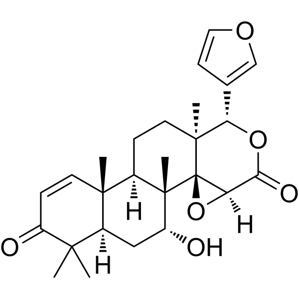
7-Deacetylgedunin
7-Deacetylgedunin is an activator of Keap1/Nrf2/HO-1. 7-Deacetylgedunin alleviates mice mortality induced by LPS. 7-Deacetylgedunin inhibits Keap1 expression and suppresses macrophage proliferation. 7-Deacetylgedunin suppresses inflammation in vivo and in vitro.
For research use only. We do not sell to patients.
- CAS No.: 10314-90-6
- Formula: C26H32O6
- Molecular Weight:440.53
-
Storage:
Please store the product under the recommended conditions in the Certificate of Analysis.

To place orders, for customer services and technical support, please contact: MedChemExpress USA
Tel: 609-228-6898 E-mail: [email protected] [email protected]


-
 Biological Activity
-
Chemical Information
- Protocols
- Purity & Documentation
- References
-
 Help & FAQs




Biological Activity
Description
Cellular Effect
|
Cell Line

|
Type | Value | Description | References |
|---|---|---|---|---|
| A549 | IC50 |
>20 μM
Compound: 17
|
Cytotoxicity against human A549 cells assessed as cell viability after 48 hrs by MTT assay
Cytotoxicity against human A549 cells assessed as cell viability after 48 hrs by MTT assay
|
[PMID: 21381696] |
| AZ-521 cell line | IC50 |
16.9 μM
Compound: 17
|
Cytotoxicity against human AZ-521 cells assessed as cell viability after 48 hrs by MTT assay
Cytotoxicity against human AZ-521 cells assessed as cell viability after 48 hrs by MTT assay
|
[PMID: 21381696] |
| HepG2 | IC50 |
10.26 μM
Compound: 12
|
Cytotoxicity against human HepG2 cells after 3 days by MTT assay
Cytotoxicity against human HepG2 cells after 3 days by MTT assay
|
[PMID: 19908853] |
| HL-60 | IC50 |
>20 μM
Compound: 17
|
Cytotoxicity against human HL60 cells assessed as cell viability after 48 hrs by MTT assay
Cytotoxicity against human HL60 cells assessed as cell viability after 48 hrs by MTT assay
|
[PMID: 21381696] |
| Huh-7 | CC50 |
105 μM
Compound: 9
|
Cytotoxicity against human HuH7 cells after 72 hrs by MTS assay
Cytotoxicity against human HuH7 cells after 72 hrs by MTS assay
|
[PMID: 25330401] |
| Huh-7 | EC50 |
12.5 μM
Compound: 9
|
Antiviral activity against dengue virus 2 infected in human HuH7 cells after 72 hrs by Western blot and ECL detection based assay
Antiviral activity against dengue virus 2 infected in human HuH7 cells after 72 hrs by Western blot and ECL detection based assay
|
[PMID: 25330401] |
| L6 | IC50 |
13.3 μg/mL
Compound: 4
|
Cytotoxicity against rat L6 cells
Cytotoxicity against rat L6 cells
|
[PMID: 17253841] |
| L6 | IC50 |
5.2 μg/mL
Compound: 4
|
Antiprotozoal activity against Trypanosoma cruzi Tulahuen C2C4 trypomastigotes in L6 cells
Antiprotozoal activity against Trypanosoma cruzi Tulahuen C2C4 trypomastigotes in L6 cells
|
[PMID: 17253841] |
| MCF7 | IC50 |
28.96 μM
Compound: 2
|
Antiproliferative activity against human MCF7 cells after 72 hrs
Antiproliferative activity against human MCF7 cells after 72 hrs
|
[PMID: 18816111] |
| P388 | IC50 |
4.5 μg/mL
Compound: 6
|
Cytotoxicity against mouse P388 cells
Cytotoxicity against mouse P388 cells
|
[PMID: 16989525] |
| RAW264.7 | IC50 |
<10 μM
Compound: 7
|
Inhibition of LPS-induced nitric oxide production in mouse RAW264.7 cells assessed as nitrite level after 24 hrs by Griess method
Inhibition of LPS-induced nitric oxide production in mouse RAW264.7 cells assessed as nitrite level after 24 hrs by Griess method
|
[PMID: 21733687] |
| RAW264.7 | IC50 |
37.58 μM
Compound: 7
|
Cytotoxicity against mouse RAW264.7 cells after 24 hrs by MTT assay
Cytotoxicity against mouse RAW264.7 cells after 24 hrs by MTT assay
|
[PMID: 21733687] |
| RAW264.7 | IC50 |
9.191 μM
Compound: B; DAG
|
Antiinflammatory activity in mouse RAW264.7 assessed as inhibition of LPS-induced NO production incubated for 1 hrs followed by LPS stimulation for 18 hrs by Griess reagent based assay
Antiinflammatory activity in mouse RAW264.7 assessed as inhibition of LPS-induced NO production incubated for 1 hrs followed by LPS stimulation for 18 hrs by Griess reagent based assay
|
[PMID: 35427124] |
| SK-BR-3 | IC50 |
12 μM
Compound: 17
|
Cytotoxicity against human SK-BR-3 cells assessed as cell viability after 48 hrs by MTT assay
Cytotoxicity against human SK-BR-3 cells assessed as cell viability after 48 hrs by MTT assay
|
[PMID: 21381696] |
| SK-BR-3 | IC50 |
21.09 μM
Compound: 2
|
Antiproliferative activity against human SKBr3 cells after 72 hrs
Antiproliferative activity against human SKBr3 cells after 72 hrs
|
[PMID: 18816111] |


Chemical Information
-
CAS No. 10314-90-6
-
Molecular Weight 440.53
-
Formula C26H32O6
-
SMILES
C[C@]12[C@]34[C@]([C@H](C5=COC=C5)OC([C@@]3([H])O4)=O)(CC[C@]1([H])[C@@]6([C@@](C(C)(C(C=C6)=O)C)([H])C[C@H]2O)C)C
-
Structure Classification
-
Initial Source
-
Shipping
Room temperature in continental US; may vary elsewhere.
-
Storage
Please store the product under the recommended conditions in the Certificate of Analysis.

Protocols
-
RNA extraction experimental
By lysing cells, releasing RNA, and removing impurities such as proteins and DNA, high-purity RNA products are finally obtained. The commonly used traditional method is the guanidine isothiocyanate/phenol/chloroform method (Trizol), which is suitable for a variety of animal materials including animal tissues, microorganisms, cultured cells, etc., and most plant materials.
-
Research Protocol for Inflammation-related Diseases
The NLRP3 inflammasome is a cytosolic innate immune signaling platform that integrates priming signals and danger-signal activation to promote caspase-1 activation, maturation of IL-1β and IL-18, and gasdermin D-mediated pyroptotic cell death. The core experimental logic is to determine whether inflammatory disease phenotypes are driven by increased NLRP3 expression, ASC-containing inflammasome assembly, caspase-1 cleavage, GSDMD cleavage, and extracellular release of IL-1β/IL-18 rather than by nonspecific cell injury alone. The pathway is strongly linked to inflammation-related disease phenotypes because monosodium urate crystals activate NALP3/NLRP3 inflammasome signaling in gout-like crystal inflammation, cholesterol crystals activate NLRP3 inflammasomes in atherogenesis models, and DSS-induced intestinal inflammation has been reported to involve NLRP3 inflammasome activity. However, experimental colitis studies also show context-dependent protective effects of NLRP3 inflammasome co
-
Cell Viability Determination by MTT Colorimetric Assay
The following protocol uses the MTT colorimetric assay as a classic literature-established method for assessing cell viability/metabolic activity in cultured mammalian cells. MTT[3-(4,5-dimethylthiazol-2-yl)-2,5-diphenyltetrazolium bromide] is reduced by metabolically active cells to a colored formazan product; the amount of formazan is quantified spectrophotometrically and provides an indirect measure of metabolically active viable cells. Importantly, MTT reduction reflects cellular oxidoreductase/metabolic activity rather than an absolute direct count of living cells, so changes in cellular metabolism can alter the signal independently of cell number.


Purity & Documentation
References

Calculators
Concentration (start) × Volume (start) = Concentration (final) × Volume (final)


7-Deacetylgedunin Related Classifications
HY-124941 Related Classifications
 Powered by Bioz