



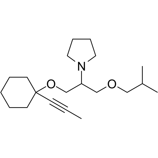
Dopropidil
Dopropidil is a novel anti-anginal calcium ion modulating agent, possessing intracellular calcium antagonist activity and anti-ischemic effects in several predictive animal models.
For research use only. We do not sell to patients.
- CAS No.: 79700-61-1
- Formula: C20H35NO2
- Molecular Weight:321.50
-
Storage:
Please store the product under the recommended conditions in the Certificate of Analysis.

To place orders, for customer services and technical support, please contact: MedChemExpress USA
Tel: 609-228-6898 E-mail: [email protected] [email protected]


-
 Biological Activity
-
Chemical Information
- Protocols
- Purity & Documentation
- References
-
 Help & FAQs


All Calcium Channel Isoforms
More

Biological Activity
Description
IC50 & Target
Calcium[1]
In Vitro
Dopropidil is able to inhibit caffeine-induced contractions of rabbit renal arteries in a calcium-free medium (IC50=30.0 uM). Dopropidil inhibits norepinephnne (NE)-induced responses with IC50s of 2.7 and 29.8 uM, respectively. At 3 and 10 μM, Dopropidil significantly reduces the maximum increase in diastolic tension evoked by veratrine (IC50=2.8 μM)[1].
MedChemExpress (MCE) has not independently confirmed the accuracy of these methods. They are for reference only.
In Vivo
MedChemExpress (MCE) has not independently confirmed the accuracy of these methods. They are for reference only.


Chemical Information
-
CAS No. 79700-61-1
-
Molecular Weight 321.50
-
Formula C20H35NO2
-
SMILES
CC#CC1(OCC(N2CCCC2)COCC(C)C)CCCCC1
-
Shipping
Room temperature in continental US; may vary elsewhere.
-
Storage
Please store the product under the recommended conditions in the Certificate of Analysis.


Purity & Documentation
References

Calculators
Concentration (start) × Volume (start) = Concentration (final) × Volume (final)


Dopropidil Related Classifications
HY-U00151 Related Classifications
 Powered by Bioz