



GNE-617
Based on 7 publication(s) in Google Scholar
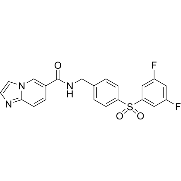
GNE-617 is a specific NAMPT inhibitor that inhibits the biochemical activity of NAMPT with an IC50 of 5 nM and exhibits efficacy in xenograft models of cancer.
Nur für Forschungszwecke. Wir verkaufen nicht an Patienten.
- Reinheit : 99.62%
- CAS. Nr.: 1362154-70-8
- Formel: C21H15F2N3O3S
- Molecular Weight:427.42
-
Speicherung:Powder -20°C, 3 years , 4°C, 2 years ; In solvent -80°C, 2 years , -20°C, 1 year

To place orders, for customer services and technical support, please contact: MedChemExpress USA
Tel: 609-228-6898 E-mail: [email protected] [email protected]


-
 Biologische Aktivität
-
Chemical Information
-
Lösungsmittel & Löslichkeit
- Protokoll
- Reinheit & Dokumentation
- Verweise
-
 Help & FAQs


-
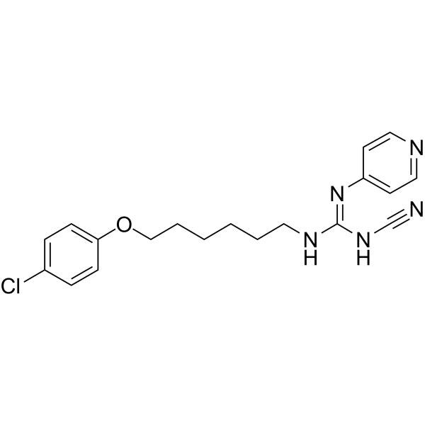
HY-10079
CHS-828NAMPT Inhibitor -
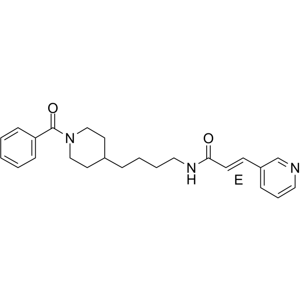
HY-50876
DaporinadNMPRT Inhibitor -
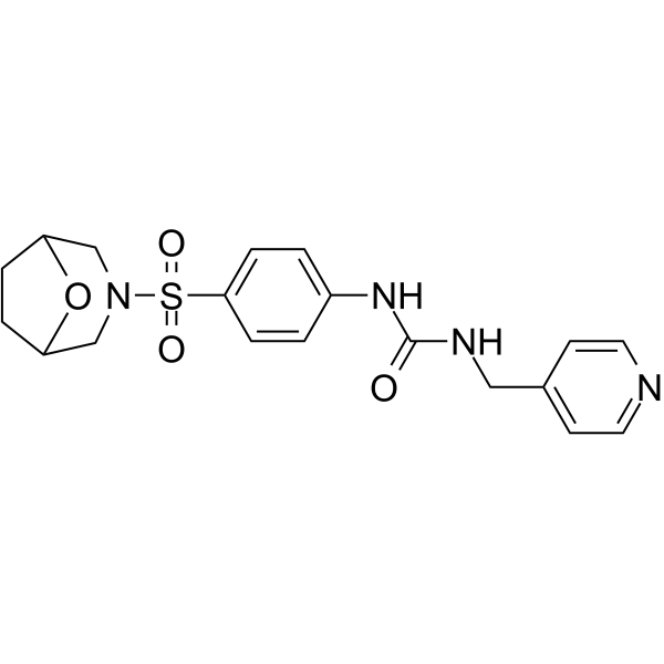
HY-126255
SBI-797812NAMPT Activator -
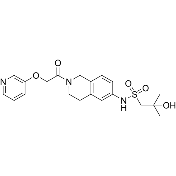
HY-12971
Nampt-IN-1NAMPT Inhibitor -
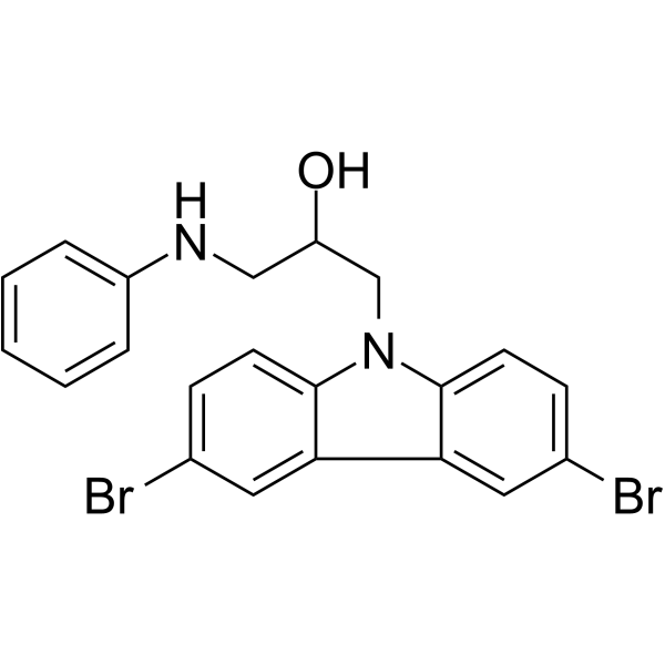
HY-15976
P7C3Neuroprotective Compound -
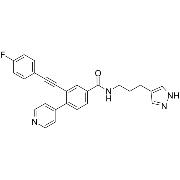
HY-136241
OT-82Nampt Inhibitor -
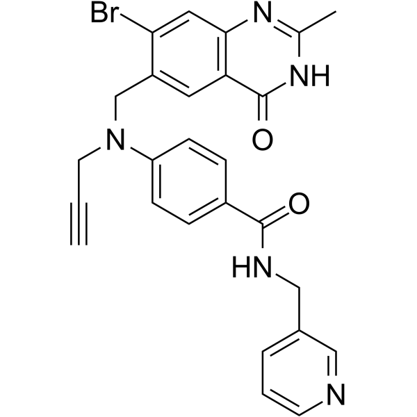
HY-14373
CB30865Nampt Inhibitor -
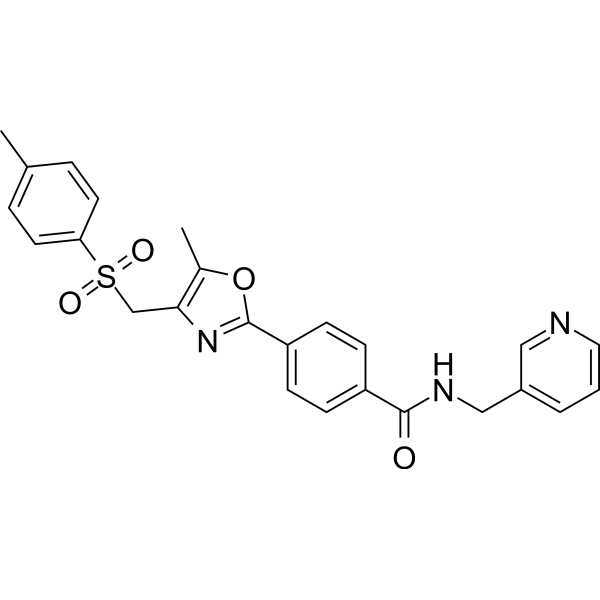
HY-12808
STF-118804Apoptosis Inducer -
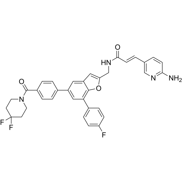
HY-12793
PadnarsertibPAK4/Nampt Inhibitor -

HY-10227
BortezomibProteasome Inhibitor -
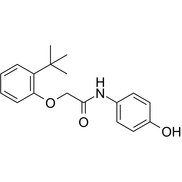
HY-144776
NATNAMPT Activator -
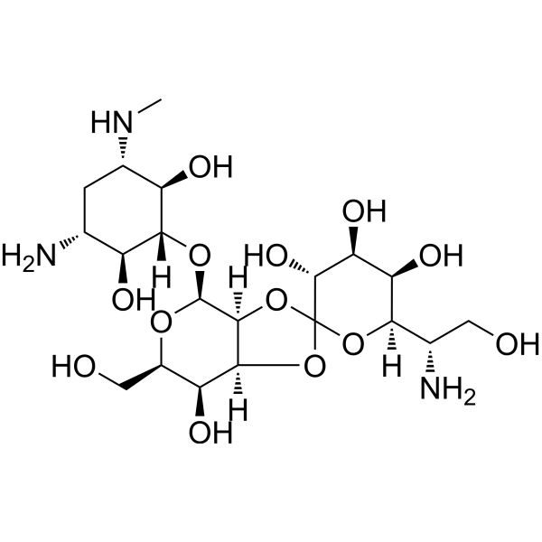
HY-B0490
Hygromycin BAntibiotic
Publications Citing Use of MedChemExpress (MCE) GNE-617
More- Nat Commun. 2023 Dec 13;14(1):8095. [Abstract]
- Cell Death Dis. 2025 Apr 25;16(1):342. [Abstract]
- Genomics. 2019 Dec;111(6):1889-1895. [Abstract]
- Research Square Preprint. 2024 Apr 12.
- Research Square Preprint. 2023 Nov 14.
- Patent. US20180263995A1.
- Oncotarget. 2018 Mar 27;9(23):16451-16461. [Abstract]
 Customer Validation & Images

-
Cell Proliferation/Viability Assay


Biologische Aktivität
Beschreibung
IC50 & Target
IC50: 5 nM (NAMPT)[1]
Cellular Effect
|
Cell Line

|
Type | Value | Description | References |
|---|---|---|---|---|
| A2780 | IC50 |
0.002 μM
Compound: 1; GNE-617
|
Inhibition of NAMPT in human A2780 cells assessed as decrease in cell viability after 72 hrs by SRB assay
Inhibition of NAMPT in human A2780 cells assessed as decrease in cell viability after 72 hrs by SRB assay
|
[PMID: 27541271] |
| A2780 | IC50 |
0.002 μM
Compound: 58
|
Antiproliferative activity against human A2780 cells assessed as growth inhibition after 72 hrs by SRB-based microplate reader analysis
Antiproliferative activity against human A2780 cells assessed as growth inhibition after 72 hrs by SRB-based microplate reader analysis
|
[PMID: 23859118] |
| B16-F10 | IC50 |
71 nM
Compound: 4
|
Antiproliferative activity against mouse B16F10 cells assessed as cell viability after 96 hrs by CyQuant staining-based assay
Antiproliferative activity against mouse B16F10 cells assessed as cell viability after 96 hrs by CyQuant staining-based assay
|
[PMID: 24021463] |
| Calu-6 | IC50 |
5 nM
Compound: 4
|
Antiproliferative activity against human Calu6 cells assessed as cell viability after 96 hrs by CyQuant staining-based assay
Antiproliferative activity against human Calu6 cells assessed as cell viability after 96 hrs by CyQuant staining-based assay
|
[PMID: 24021463] |
| COS-1 | IC50 |
5 nM
Compound: 4
|
Cytotoxicity against African green monkey COS1 cells assessed as cell viability after 96 hrs by CyQuant staining-based assay
Cytotoxicity against African green monkey COS1 cells assessed as cell viability after 96 hrs by CyQuant staining-based assay
|
[PMID: 24021463] |
| COS-7 | IC50 |
9 nM
Compound: 4
|
Cytotoxicity against African green monkey COS7 cells assessed as cell viability after 96 hrs by CyQuant staining-based assay
Cytotoxicity against African green monkey COS7 cells assessed as cell viability after 96 hrs by CyQuant staining-based assay
|
[PMID: 24021463] |
| HCT-116 | IC50 |
2 nM
Compound: 4
|
Antiproliferative activity against human HCT116 cells assessed as cell viability after 96 hrs by CyQuant staining-based assay
Antiproliferative activity against human HCT116 cells assessed as cell viability after 96 hrs by CyQuant staining-based assay
|
[PMID: 24021463] |
| HCT-116 | IC50 |
2 nM
Compound: 58
|
Antiproliferative activity against human HCT116 cells assessed as cell viability after 72 hrs
Antiproliferative activity against human HCT116 cells assessed as cell viability after 72 hrs
|
[PMID: 23859118] |
| HT-1080 | IC50 |
2 nM
Compound: 4
|
Antiproliferative activity against human HT1080 cells assessed as cell viability after 96 hrs by CyQuant staining-based assay
Antiproliferative activity against human HT1080 cells assessed as cell viability after 96 hrs by CyQuant staining-based assay
|
[PMID: 24021463] |
| HT-1080 | IC50 |
2.1 nM
Compound: 58
|
Antiproliferative activity against human HT1080 cells assessed as cell viability after 72 hrs
Antiproliferative activity against human HT1080 cells assessed as cell viability after 72 hrs
|
[PMID: 23859118] |
| MIA PaCa-2 | IC50 |
7 nM
Compound: 4
|
Antiproliferative activity against human MIAPaCa2 cells assessed as cell viability after 96 hrs by CyQuant staining-based assay
Antiproliferative activity against human MIAPaCa2 cells assessed as cell viability after 96 hrs by CyQuant staining-based assay
|
[PMID: 24021463] |
| MIA PaCa-2 | IC50 |
7.4 nM
Compound: 58
|
Antiproliferative activity against human MIAPaCa2 cells assessed as cell viability after 72 hrs
Antiproliferative activity against human MIAPaCa2 cells assessed as cell viability after 72 hrs
|
[PMID: 23859118] |
| PC-3 | EC50 |
1.1 nM
Compound: 4
|
Inhibition of NAMPT in human PC3 cells assessed as reduction in NAD level after 48 hrs by mass spectrometry
Inhibition of NAMPT in human PC3 cells assessed as reduction in NAD level after 48 hrs by mass spectrometry
|
[PMID: 24021463] |
| PC-3 | IC50 |
2.7 nM
Compound: 58
|
Antiproliferative activity against human PC3 cells assessed as cell viability after 72 hrs
Antiproliferative activity against human PC3 cells assessed as cell viability after 72 hrs
|
[PMID: 23859118] |
| PC-3 | IC50 |
3 nM
Compound: 4
|
Antiproliferative activity against human PC3 cells assessed as cell viability after 96 hrs by CyQuant staining-based assay
Antiproliferative activity against human PC3 cells assessed as cell viability after 96 hrs by CyQuant staining-based assay
|
[PMID: 24021463] |
| RF/6A | IC50 |
4 nM
Compound: 4
|
Cytotoxicity against rhesus monkey RF/6A cells assessed as cell viability after 96 hrs by CyQuant staining-based assay
Cytotoxicity against rhesus monkey RF/6A cells assessed as cell viability after 96 hrs by CyQuant staining-based assay
|
[PMID: 24021463] |
| U-251 | IC50 |
1.8 nM
Compound: 58
|
Antiproliferative activity against human U251 cells assessed as cell viability after 72 hrs by SRB assay
Antiproliferative activity against human U251 cells assessed as cell viability after 72 hrs by SRB assay
|
[PMID: 23859118] |
In Vitro
The activity ofGNE-617 hydrochloride is evaluated on a panel 53 non-small cell lung cancer (NSCLC) cell lines in the presence or absence of 10 μM nicotinic acid. GNE-617 inhibits NAMPT IC50 of 18.9 nM in A549 cell.The majority of cell lines exhibit a steep dose response to GNE-617 when evaluated by decrease in ATP or total nucleic acid, and the cytotoxicity is completely rescued by simultaneous addition of nicotinic acid. The majority of the cell lines tested have IC50 values below 100 nM, with approximately half with IC50 values less than 10 nM[1].
MedChemExpress (MCE) has not independently confirmed the accuracy of these methods. They are for reference only.
In Vivo
MedChemExpress (MCE) has not independently confirmed the accuracy of these methods. They are for reference only.


Chemical Information
-
CAS. Nr. 1362154-70-8
-
Appearance Solid
-
Molecular Weight 427.42
-
Formel C21H15F2N3O3S
-
Color White to off-white
-
SMILES
O=C(NCC1=CC=C(S(C2=CC(F)=CC(F)=C2)(=O)=O)C=C1)C3=CN4C(C=C3)=NC=C4
-
Versand
Room temperature in continental US; may vary elsewhere.
-
Speicherung
Powder -20°C 3 years 4°C 2 years In solvent -80°C 2 years -20°C 1 year


Publications (7)
-
Journal Impact Factor
-
Most Recent
-
Nat Commun
Niacin restriction with NAMPT-inhibition is synthetic lethal to neuroendocrine carcinoma. [Abstract]2023 Dec 13;14(1):8095. PMID: 38092728 -
Cell Death Dis
Targeting metabolic vulnerability by combining NAMPT inhibitors and disulfiram for treatment of recurrent ovarian cancer. [Abstract]2025 Apr 25;16(1):342. PMID: 40280967 -
Genomics
Genomic and tumor biological aspects of the anticancer nicotinamide phosphoribosyltransferase inhibitor FK866 in resistant human colorectal cancer cells. [Abstract]2019 Dec;111(6):1889-1895. PMID: 30582964 -
-
-
-
Oncotarget
Cross resistance to diverse anticancer nicotinamide phosphoribosyltransferase inhibitors induced by FK866 treatment. [Abstract]2018 Mar 27;9(23):16451-16461. PMID: 29662658
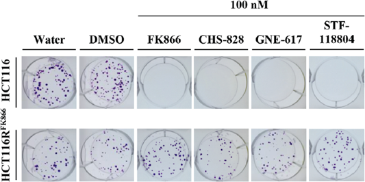
GNE-617 purchased from MedChemExpress. Usage Cited in: Oncotarget. 2018 Mar 27;9(23):16451-16461. [Abstract]
Drug sensitivities of HCT116RFK866 and HCT116 in the colony formation assay. HCT116RFK866 and HCT116 cells are treated with 100 nM each of FK866, CHS-828, GNE-617, STF-118804, and incubated for 10 days.


Lösungsmittel & Löslichkeit
In Vitro:
DMSO : 16.67 mg/mL (39.00 mM; Need ultrasonic; Hygroscopic DMSO has a significant impact on the solubility of product, please use newly opened DMSO)
Please refer to the solubility information to select the appropriate solvent. Once prepared, please aliquot and store the solution to prevent product inactivation from repeated freeze-thaw cycles.
Storage method and period of stock solution: -80°C, 2 years; -20°C, 1 year. When stored at -80°C, please use it within 2 years. When stored at -20°C, please use it within 1 year.
Please refer to the solubility information to select the appropriate solvent. Once prepared, please aliquot and store the solution to prevent product inactivation from repeated freeze-thaw cycles.
Storage method and period of stock solution: -80°C, 2 years; -20°C, 1 year. When stored at -80°C, please use it within 2 years. When stored at -20°C, please use it within 1 year.
Konzentration (Stammlösung) × Volumen (Stammlösung) = Konzentration (Ziellösung) × Volumen (Ziellösung)
In Vivo:
Select the appropriate dissolution method based on your experimental animal and administration route.
- For the following dissolution methods, please ensure to first prepare a clear stock solution using an In Vitro approach and then sequentially add co-solvents:
- To ensure reliable experimental results, the clarified stock solution can be appropriately stored based on storage conditions. As for the working solution for In Vivo experiments, it is recommended to prepare freshly and use it on the same day.
- The percentages shown for the solvents indicate their volumetric ratio in the final prepared solution. If precipitation or phase separation occurs during preparation, heat and/or sonication can be used to aid dissolution.
Add each solvent one by one: 10% DMSO 40% PEG300 5% Tween-80 45% Saline
Solubility: ≥ 1.67 mg/mL (3.91 mM); Clear solution
This protocol yields a clear solution of ≥ 1.67 mg/mL (saturation unknown).
Taking 1 mL working solution as an example, add 100 μL DMSO stock solution (16.7 mg/mL) to 400 μL PEG300, and mix evenly; then add 50 μL Tween-80 and mix evenly; then add 450 μL Saline to adjust the volume to 1 mL.
Preparation of Saline: Dissolve 0.9 g sodium chloride in ddH₂O and dilute to 100 mL to obtain a clear Saline solution.
In Vivo Dissolution Calculator
Please enter the basic information of animal experiments:
-
-
-
-
Recommended: Prepare an additional quantity of animals to account for potential losses during experiments.
Please enter your animal formula composition:
-
%DMSO +
Recommended: Keep the proportion of DMSO in working solution below 2% if your animal is weak.
-
%+
-
+%Tween-80 + +
-
%Saline +
The co-solvents required include: DMSO, . All of co-solvents are available by MedChemExpress (MCE). , Tween 80. All of co-solvents are available by MedChemExpress (MCE).
Working solution concentration: 0.22 mg/mL
Method for preparing stock solution: mg drug dissolved in μL DMSO. Stock solution concentration: mg/mL.
1. Take μL DMSO stock solution;
2. Add μL .
μL , mix evenly;
3. Then add μL Tween 80, mix evenly;
4. Then add μL
Please ensure that the stock solution in the first step is dissolved to a clear state, and add co-solvents in sequence. You can use ultrasonic heating (ultrasonic cleaner, recommended frequency 20-40 kHz), vortexing, etc. to assist dissolution.


Reinheit & Dokumentation
-
Data Sheet (279 KB)
-
SDS (396 KB)
- English - EN (396 KB)
- Français - FR (396 KB)
- Deutsch - DE (396 KB)
- Norwegian - NO (396 KB)
- Español - ES (396 KB)
- Swedish - SV (396 KB)
- Italian - IT (396 KB)
- Korean - KR (396 KB)
- Portuguese - PT (396 KB)
-
Handling Instructions (2659 KB)
Verweise
[1]. Shames DS, et al. Loss of NAPRT1 Expression by Tumor-specific Promoter Methylation Provides a Novel Predictive Biomarker for NAMPT Inhibitors. Clin Cancer Res. 2013 Dec 15;19(24):6912-23. [Content Brief]
[2]. Zabka TS, et al. Retinal toxicity, in vivo and in vitro, associated with inhibition of nicotinamide phosphoribosyltransferase. Toxicol Sci. 2015 Mar;144(1):163-72. [Content Brief]


Complete Stock Solution Preparation Table
Please refer to the solubility information to select the appropriate solvent. Once prepared, please aliquot and store the solution to prevent product inactivation from repeated freeze-thaw cycles.
Storage method and period of stock solution: -80°C, 2 years; -20°C, 1 year. When stored at -80°C, please use it within 2 years. When stored at -20°C, please use it within 1 year.
| Optional Solvent | Concentration Solvent Mass | 1 mg | 5 mg | 10 mg | 25 mg |
|---|---|---|---|---|---|
| DMSO | 1 mM | 2.3396 mL | 11.6981 mL | 23.3962 mL | 58.4905 mL |
| 5 mM | 0.4679 mL | 2.3396 mL | 4.6792 mL | 11.6981 mL | |
| 10 mM | 0.2340 mL | 1.1698 mL | 2.3396 mL | 5.8490 mL | |
| 15 mM | 0.1560 mL | 0.7799 mL | 1.5597 mL | 3.8994 mL | |
| 20 mM | 0.1170 mL | 0.5849 mL | 1.1698 mL | 2.9245 mL | |
| 25 mM | 0.0936 mL | 0.4679 mL | 0.9358 mL | 2.3396 mL | |
| 30 mM | 0.0780 mL | 0.3899 mL | 0.7799 mL | 1.9497 mL |


GNE-617 Related Classifications
HY-15766 Related Classifications
 Powered by Bioz