



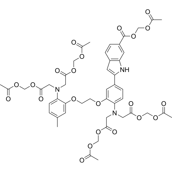
Indo-1 AM
Based on 1 publication(s) in Google Scholar
Calcium is an important part of the human body, usually in the form of calcium, a large number of bones and teeth of the human body, a small amount of blood and tissues. MCE calcium ion detection probe can specifically identify intracellular calcium ions, with high sensitivity, low cytotoxicity, increased AM acetylmethyl ester can enter the cell well, after being sheared by the intracellular esterase stay in the cell to bind to calcium ions, produce strong fluorescence, in addition, Mag-indo-1/AM and Mag-Fluo-4 AM at a certain concentration (usually 5 mM) can effectively identify intracellular magnesium ions.
For research use only. We do not sell to patients.
- Purity : 95.0%
- CAS No.: 112926-02-0
- Formula: C47H51N3O22
- Molecular Weight:1009.91
-
Storage:
-20°C, protect from light
* In solvent : -80°C, 6 months; -20°C, 1 month (protect from light)

To place orders, for customer services and technical support, please contact: MedChemExpress USA
Tel: 609-228-6898 E-mail: [email protected] [email protected]


-
 Biological Activity
-
Chemical Information
- Protocols
- Purity & Documentation
- References
-
 Help & FAQs



Publications Citing Use of MedChemExpress (MCE) Indo-1 AM
More

Biological Activity
Description
In Vitro
Guide (Following is our recommended protocol. This protocol only provides a guideline, and should be modified according to your specific needs).
1. Preparation of Indo-1 AM working solution
1.1 Preparation of the stock solution
Dissolve 1 mg Indo-1 AM in 135 μL DMSO to obtain 10 mM of stock solution.
1.2 Preparation of Indo-1 AM working solution
Dilute the stock solution in HBSS to obtain 1-10 μM of working solution.
Note: Please adjust the concentration of Indo-1 AM working solution according to the actual situation.
2. Cell staining (Suspension cells)
2.1 Centrifuge at 1000 g at 4°C for 3-5 minutes and then discard the supernatant. Wash twice with PBS, 5 minutes each time.The cell density is 1×10⁶/mL
2.2 Add 1 mL of working solution, and then incubate at room temperature for 5-30 minutes.
2.3 Centrifuge at 400 g at 4°C for 3-4 minutes and then discard the supernatant.
2.4 Wash twice with PBS, 5 minutes each time.
2.5 Resuspend cells with serum-free cell culture medium or PBS. Observation by fluorescence microscopy or flow cytometry.
3. Cell staining (Adherent cells)
3.1 Culture adherent cells on sterile coverslips.
3.2 Remove the coverslip from the medium and aspirate excess medium.
3.3 Add 100 μL of working solution, gently shake it to completely cover the cells,and then incubate at room temperature for 5-30 minutes.
3.4 Wash twice with medium, 5 minutes each time. Observation by fluorescence microscopy.
MedChemExpress (MCE) has not independently confirmed the accuracy of these methods. They are for reference only. Further protocols information, click here.
Emission (Em)
410
Excitation (Ex)
364


Chemical Information
-
CAS No. 112926-02-0
-
Appearance Solid
-
Molecular Weight 1009.91
-
Formula C47H51N3O22
-
Color Light yellow to yellow
-
SMILES
O=C(C1=CC2=C(C=C1)C=C(C3=CC=C(N(CC(OCOC(C)=O)=O)CC(OCOC(C)=O)=O)C(OCCOC4=CC(C)=CC=C4N(CC(OCOC(C)=O)=O)CC(OCOC(C)=O)=O)=C3)N2)OCOC(C)=O
-
Synonyms
Indo-1 Acetoxymethyl ester
-
Shipping
Room temperature in continental US; may vary elsewhere.
-
Storage
-20°C, protect from light
* In solvent : -80°C, 6 months; -20°C, 1 month (protect from light)


Publications (1)
-
Journal Impact Factor
-
Most Recent
-
Stem Cell Res Ther
Enhance the therapeutic efficacy of human umbilical cord-derived mesenchymal stem cells in prevention of acute graft-versus-host disease through CRISPLD2 modulation. [Abstract]2025 May 1;16(1):222. PMID: 40312744

Protocols
-
Cell Cytotoxicity Assay
Cytotoxicity assays are usually based on the assessment of cell membrane damage, which can also be indirectly detected by measuring cell viability. Detection methods include MTT assay, CKK-8 assay, LDH assay and ATP assay, etc.
-
Mammalian live/dead viability and cytotoxicity staining
Live/dead viability and cytotoxicity staining assays are based on the simultaneous detection of intracellular esterase activity in metabolically active (viable) cells and membrane integrity loss in non-viable cells. In commonly used dual-staining approaches, membrane-permeant fluorogenic substrates are converted by intracellular esterases into fluorescent products in live cells, while impermeant DNA-binding dyes selectively enter cells with compromised plasma membranes and label nucleic acids in dead or dying cells, enabling discrimination between viable and non-viable populations by fluorescence microscopy or flow cytometry.
-
Calcium Spark Assay
Calcium sparks are localized, transient increases in intracellular calcium concentration ([Ca2+]i) that occur in cardiac myocytes and represent elementary events underlying excitation-contraction coupling. These events are generated by the coordinated opening of clusters of ryanodine receptors (RyRs) on the sarcoplasmic reticulum membrane, leading to a brief release of Ca2+ into the cytosol. The detection and analysis of calcium sparks provide insights into the mechanisms of calcium handling and signaling in cardiac cells. Imaging techniques using fluorescent calcium indicators such as Fluo-3 are employed to visualize these subcellular calcium transients with high spatial and temporal resolution. The protocol is based on established methodologies described in primary literature for both experimental measurement and automated analysis of calcium sparks.
-
Ca2+ Staining Technique
Ca2+ staining is an experimental technique that utilizes specific fluorescent probes (such as Fluo-4 AM, Fura-2, etc.) to qualitatively or quantitatively detect dynamic changes in intracellular Ca2+ concentrations; this is achieved by monitoring the changes in fluorescent signals generated when these probes bind to free intracellular calcium ions. The underlying principle relies primarily on the presence of chelating groups within the probe's molecular structure that possess high affinity for calcium ions.
-
Cell Viability Determination by MTT Colorimetric Assay
The following protocol uses the MTT colorimetric assay as a classic literature-established method for assessing cell viability/metabolic activity in cultured mammalian cells. MTT[3-(4,5-dimethylthiazol-2-yl)-2,5-diphenyltetrazolium bromide] is reduced by metabolically active cells to a colored formazan product; the amount of formazan is quantified spectrophotometrically and provides an indirect measure of metabolically active viable cells. Importantly, MTT reduction reflects cellular oxidoreductase/metabolic activity rather than an absolute direct count of living cells, so changes in cellular metabolism can alter the signal independently of cell number.


Purity & Documentation
-
Data Sheet (278 KB)
-
SDS (252 KB)
- English - EN (252 KB)
- Français - FR (252 KB)
- Deutsch - DE (252 KB)
- Norwegian - NO (252 KB)
- Español - ES (252 KB)
- Swedish - SV (252 KB)
- Italian - IT (252 KB)
- Korean - KR (252 KB)
- Portuguese - PT (252 KB)
-
Handling Instructions (2659 KB)
References
[1]. Andrienko T, et al. Real-Time Fluorescence Measurements of ROS and [Ca2+] in Ischemic / Reperfused Rat Hearts: Detectable Increases Occur only after Mitochondrial Pore Opening and Are Attenuated by Ischemic Preconditioning. PLoS One. 2016 Dec 1;11(12):e01 [Content Brief]
[2]. Andrienko T, et al. Real-Time Fluorescence Measurements of ROS and [Ca2+] in Ischemic / Reperfused Rat Hearts: Detectable Increases Occur only after Mitochondrial Pore Opening and Are Attenuated by Ischemic Preconditioning. PLoS One. 2016 Dec 1;11(12):e0167300. [Content Brief]
[3]. Blatter LA, et al. Intracellular diffusion, binding, and compartmentalization of the fluorescent calcium indicators indo-1 and fura-2. Biophys J. 1990 Dec;58(6):1491-9. [Content Brief]

Calculators
Concentration (start) × Volume (start) = Concentration (final) × Volume (final)


Indo-1 AM Related Classifications
HY-101898 Related Classifications
 Powered by Bioz