



Luciferase (bacterial)

Based on 1 Customer Validation
Luciferase (bacterial) acts as a luminescence inducer and catalyst. Luciferase (bacterial) catalyzes the reduction of methylene blue, quinones, ferricyanide, and FMN using reduced DPN (HY-12452) (or an alternative electron donor for FMN). Luciferase (bacterial) binds to reduced FMN to form a luminescent reaction complex. Luciferase (bacterial) scavenges reactive oxygen species (ROS) and protects bacterial cells from oxidative stress damage. Luciferase (bacterial) is applicable to studies related to oxidative stress.
For research use only. We do not sell to patients.
- CAS No.: 9014-00-0
-
Storage:
Please store the product under the recommended conditions in the Certificate of Analysis.


To place orders, for customer services and technical support, please contact: MedChemExpress USA
Tel: 609-228-6898 E-mail: [email protected] [email protected]


-
 Biological Activity
-
Chemical Information
-
Solvent & Solubility
- Protocols
- Purity & Documentation
- References
-
 Help & FAQs


-
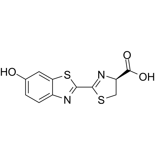
HY-12591A
D-LuciferinSubstrate for Luciferase -
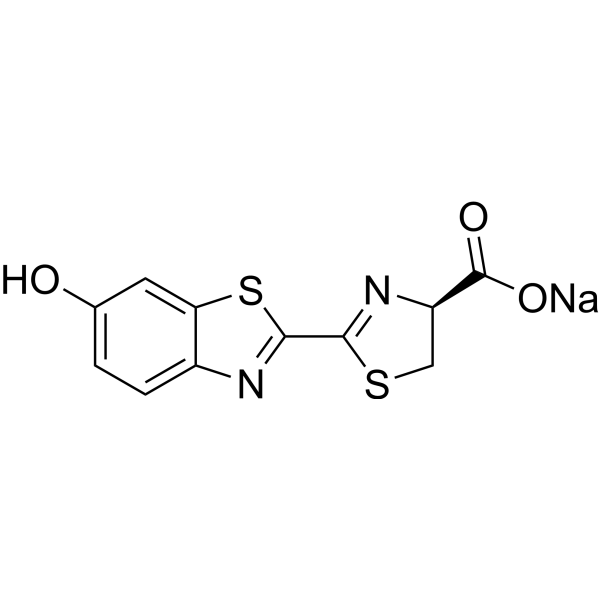
HY-12591
D-Luciferin sodiumSubstrate for Luciferase -
HY-P1004A
Luciferase, fireflyLight-Emitting Enzyme -
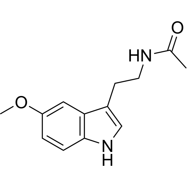
HY-B0075
MelatoninMelatonin Receptor Activator -
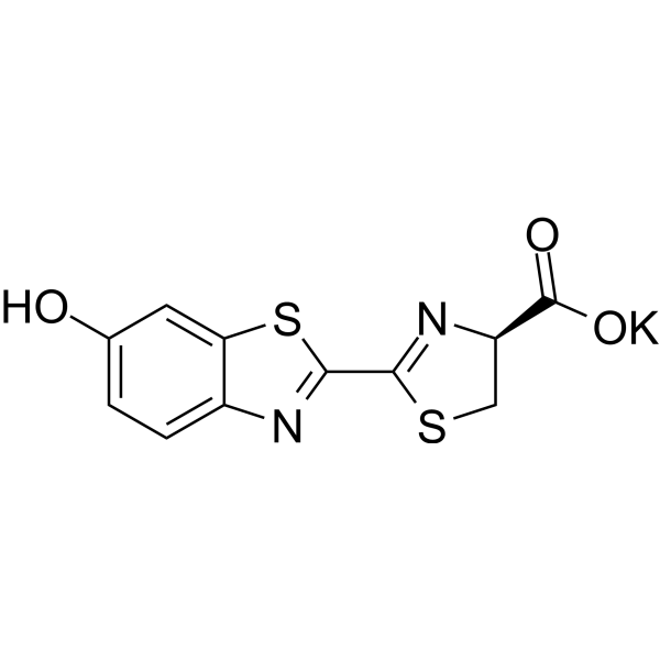
HY-12591B
D-Luciferin potassiumSubstrate for Luciferase -
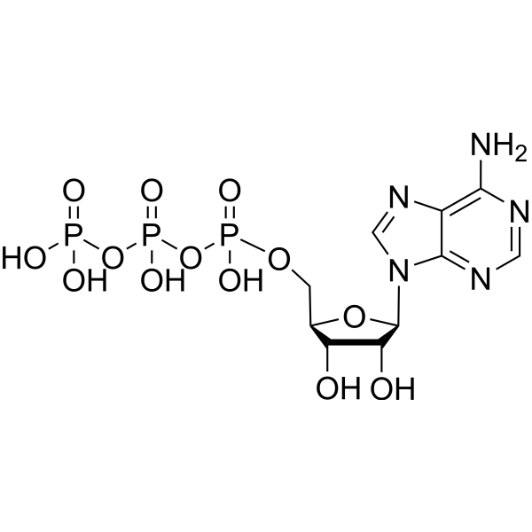
HY-B2176
ATPEndogenous Metabolite -
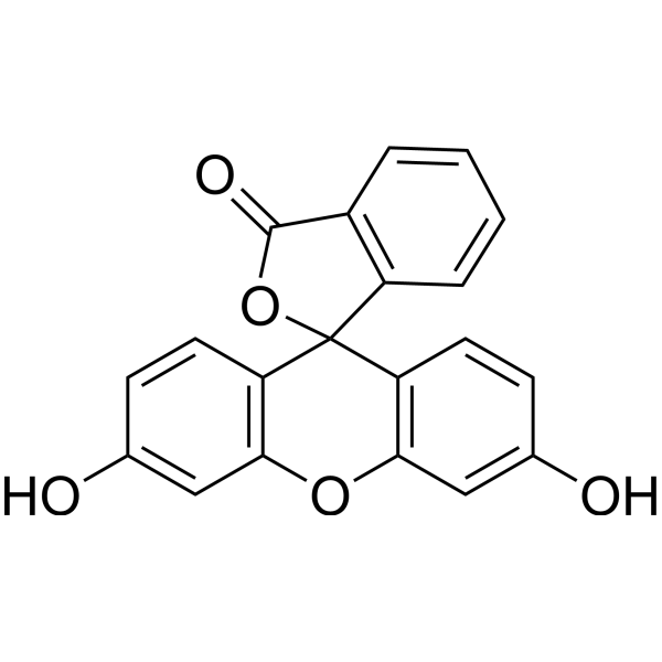
HY-D0251
FluoresceinFluorescent Tracer -
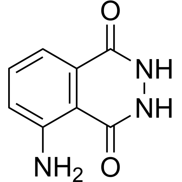
HY-15922
LuminolChemiluminescent Substance -
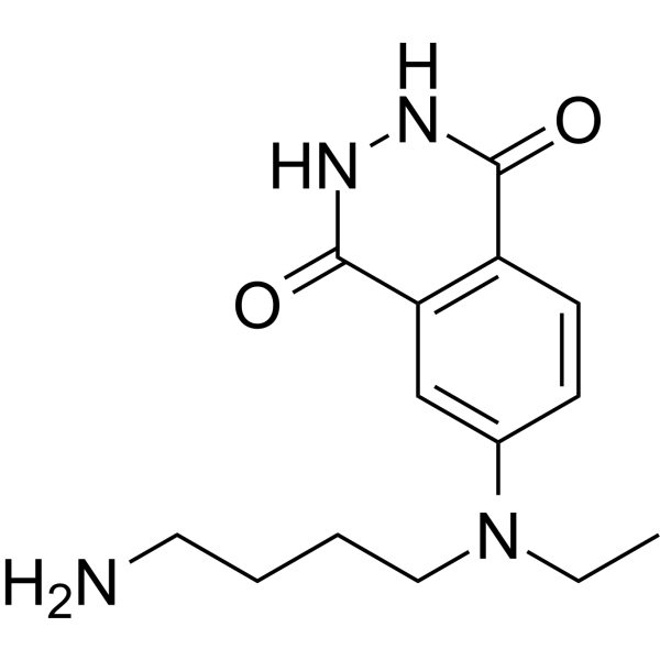
HY-D0890
ABEIBiochemical Assay Reagent


Biological Activity
Description
In Vitro
Luciferase (bacterial) protects Vibrio harveyi luxA and luxB mutant cells against oxidative stress induced by 1.25 mM hydrogen peroxide, 0.14 mM cumene hydroperoxide, 0.3 mM t-butyl hydroperoxide, and 0.2 mM ferrous sulfate[2].
Luciferase (bacterial) catalyzes oxygen-dependent luminescence requiring reduced FMN and long-chain aldehyde, with reduced FMN binding to the enzyme via two sites (dissociation constants K1 = 3.72×10⁻⁶ M and K2 = 2.69×10⁻⁶ M), and supports luminescence with DPNH or TPNH when FMN is present, while aldehyde concentration directly determines total light output[1].
Luciferase (bacterial) catalyzes metal-independent oxidation of DPNH by ferricyanide, Methylene blue (HY-14536), and quinones in the absence of aldehyde and FMN, with ferricyanide competing with FMN for a reduced enzyme intermediate to suppress luminescence until fully reduced[1].
Luciferase (bacterial) exhibits species-specific differences in FMNH2 binding affinity and C4a-(hydro)peroxyflavin formation rates, with V. campbellii Lux displaying the highest FMNH2 binding affinity (Kd = 1.8 μM) and fastest oxygen reaction rate (253 s-1 at 0.26 mM oxygen)[3].
Luciferase (bacterial) (4-27 μM) binds aldehyde at two distinct sites, with high-affinity binding acting as a substrate interaction and lower-affinity binding at higher concentrations inhibiting catalytic activity by blocking FMNH2 binding and reducing C4a-(hydro)peroxyflavin formation[3].
Luciferase (bacterial) has specific active site and flexible loop residues critical for catalytic activity, intermediate stability, and light emission properties; mutations to these residues reduce bioluminescence efficiency, alter intermediate decay rates, or shift emission spectra[3].
Luciferase (bacterial) binds FMNH2 with an apparent Kd of 0.08 μM at room temperature, with initial bioluminescence intensity proportional to FMNH2 concentration and exponential first-order decay kinetics[5].
MedChemExpress (MCE) has not independently confirmed the accuracy of these methods. They are for reference only. Further protocols information, click here.


Chemical Information
-
CAS No. 9014-00-0
-
Appearance Solid
-
Color White to light yellow
-
SMILES
[Luciferase (bacterial)]
-
Structure Classification
-
Initial Source
Neonothopanus nambi fungus
-
Shipping
Room temperature in continental US; may vary elsewhere.
-
Storage
Please store the product under the recommended conditions in the Certificate of Analysis.


Solvent & Solubility
In Vitro:
H2O : 20 mg/mL (Need ultrasonic)

Protocols
-
ROS/oxidative-stress fluorescent staining
ROS/oxidative-stress fluorescent staining uses cell-permeant fluorogenic probes that become fluorescent after oxidation inside cells or tissues; commonly used examples include DCFH-DA/DCFDA for broad cellular oxidant detection, DHE for superoxide-related signal detection, MitoSOX for mitochondrial superoxide-related signal detection, and CellROX probes for oxidative-stress-associated fluorescence readouts. The assay detects probe oxidation rather than a single ROS species unless the probe and analysis method have been chemically validated for that species. DCFH-DA enters cells, is deacetylated by intracellular esterases to DCFH, and produces fluorescent DCF after oxidation, so the readout is used as an operational measure of total cellular oxidative stress rather than a species-specific ROS measurement. DHE and MitoSOX can report superoxide-related oxidation, but red fluorescence alone can include non-specific ethidium-like oxidation products; HPLC or optimized spectral approaches are


Purity & Documentation
-
Data Sheet (276 KB)
-
SDS (251 KB)
- English - EN (251 KB)
- Français - FR (251 KB)
- Deutsch - DE (251 KB)
- Norwegian - NO (251 KB)
- Español - ES (251 KB)
- Swedish - SV (251 KB)
- Italian - IT (251 KB)
- Korean - KR (251 KB)
- Portuguese - PT (251 KB)
-
Handling Instructions (2659 KB)
References
[2]. Szpilewska H, et al. Experimental evidence for the physiological role of bacterial luciferase in the protection of cells against oxidative stress. Current microbiology. 2003 Nov;47(5):379-82. [Content Brief]
[3]. Tinikul R, et al. Bacterial luciferase: Molecular mechanisms and applications. The Enzymes. 2020;47:427-455. [Content Brief]

Calculators
Concentration (start) × Volume (start) = Concentration (final) × Volume (final)


Luciferase (bacterial) Related Classifications
HY-P1004 Related Classifications
 Powered by Bioz