



MK-3328

Based on 1 Customer Validation
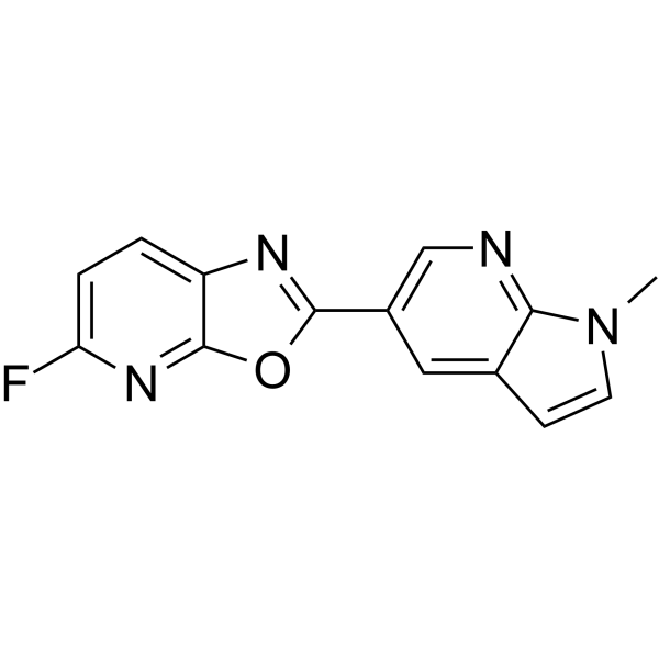
MK-3328 is a β-Amyloid ligand, which exhibits high binding potency with an IC50 of 10.5 nM. MK-3328 is capable of being labelled with 18F for positron emission tomography (PET) imaging.
For research use only. We do not sell to patients.
- Purity : 99.70%
- CAS No.: 1201323-97-8
- Formula: C14H9FN4O
- Molecular Weight:268.25
-
Storage:
4°C, protect from light
* In solvent : -80°C, 6 months; -20°C, 1 month (protect from light)

To place orders, for customer services and technical support, please contact: MedChemExpress USA
Tel: 609-228-6898 E-mail: [email protected] [email protected]


-
 Biological Activity
-
Chemical Information
- Protocols
- Purity & Documentation
- References
-
 Help & FAQs


-
HY-105252A
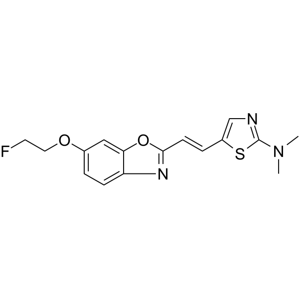
BF 227 -

HY-P1363A
β-Amyloid (1-42), humanAmyloid β Peptide -

HY-P1363B
β-Amyloid (1-42), human, HFIP-treatedβ-Amyloid (1-42) Peptide -
HY-19370
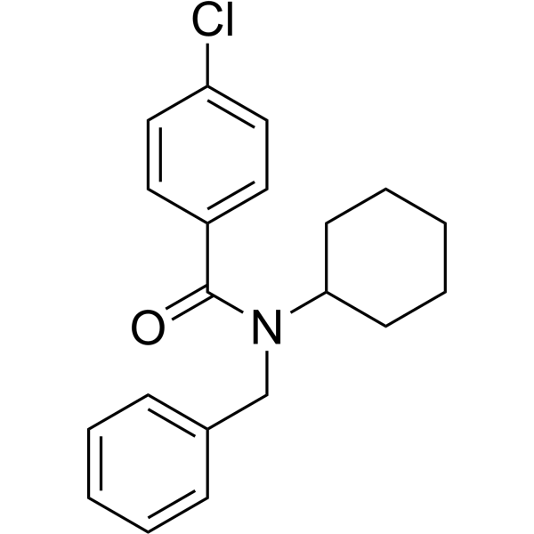
FPS-ZM1Amyloid-β Inhibitor -
HY-18292
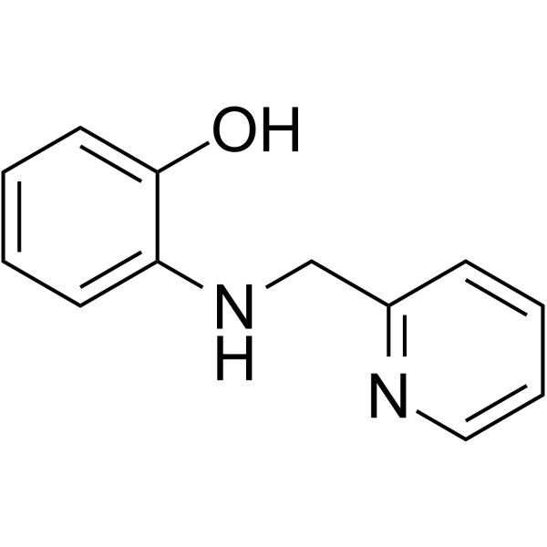
ARN2966APP Translation Modulator -
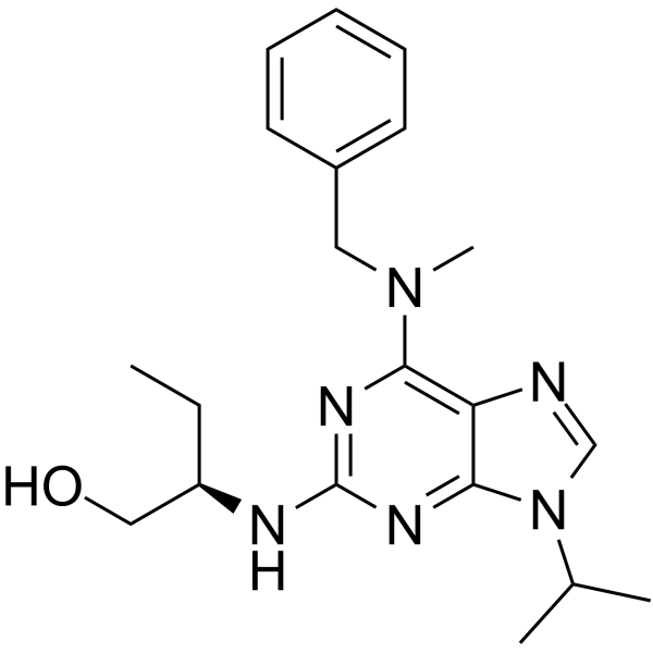
HY-111267
Aftin-4Amyloid-β Agonist -
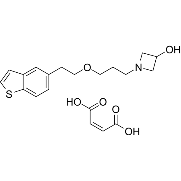
HY-17631A
Edonerpic maleate -
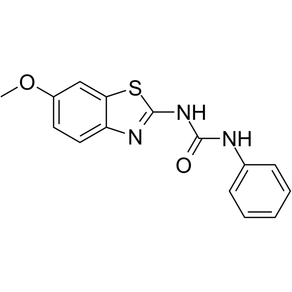
HY-15374
FrentizoleAβ-ABAD Inbihitor -
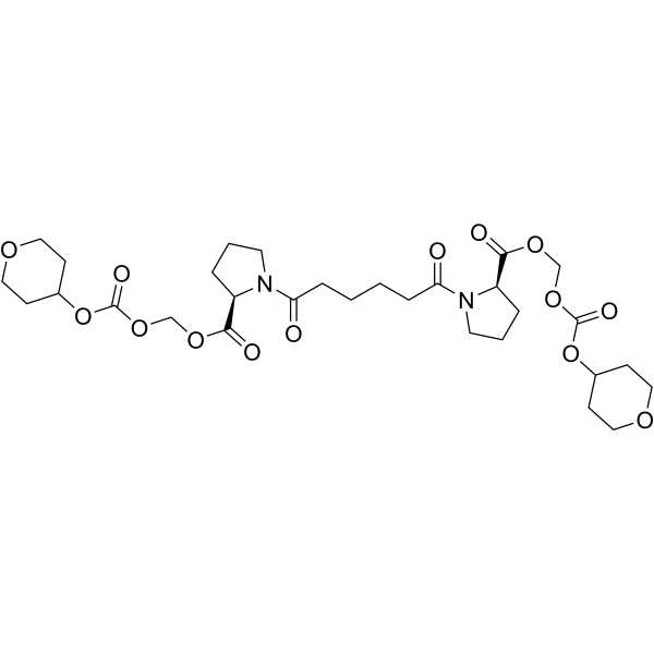
HY-19771
GSK 3039294Miridesap Pro-drug -
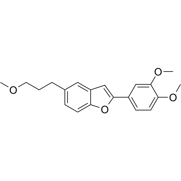
HY-14503
MDR-1339Amyloid-β Inhibitor -
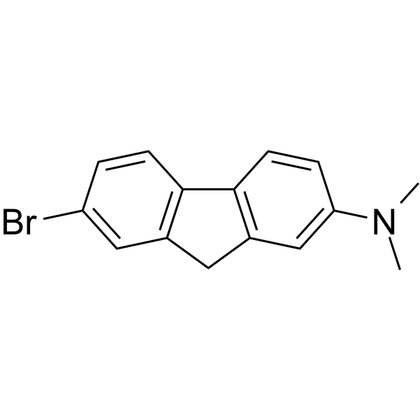
HY-14533
K 01-162Aβ Inhibitor -
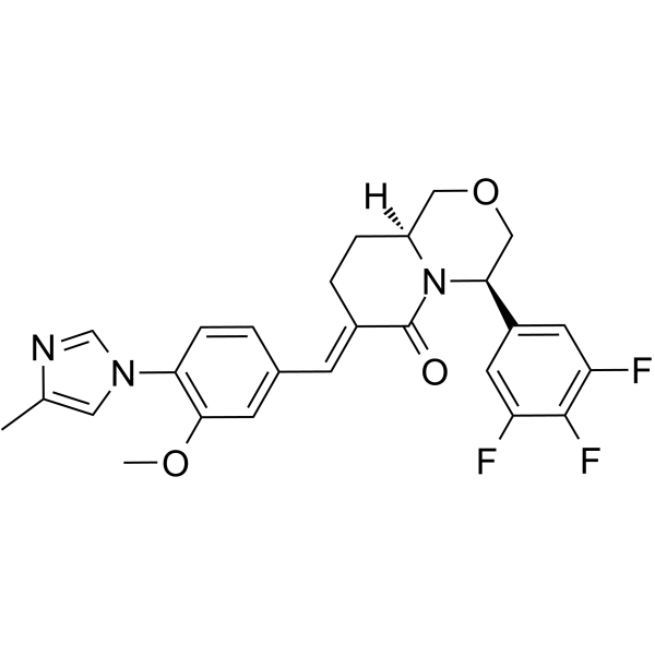
HY-50900
gamma-Secretase ModulatorsAmyloid-β Inhibitor


Biological Activity
Description
IC50 & Target
IC50: 10.5 nM (β-Amyloid)[1]
In Vitro
MK-3328 exhibits amyloid binding potency balanced with low levels of nonspecific binding[1].
MedChemExpress (MCE) has not independently confirmed the accuracy of these methods. They are for reference only. Further protocols information, click here.
In Vivo
MedChemExpress (MCE) has not independently confirmed the accuracy of these methods. They are for reference only.


Chemical Information
-
CAS No. 1201323-97-8
-
Appearance Solid
-
Molecular Weight 268.25
-
Formula C14H9FN4O
-
Color Off-white to light brown
-
SMILES
CN1C=CC2=CC(C3=NC4=CC=C(F)N=C4O3)=CN=C21
-
Shipping
Room temperature in continental US; may vary elsewhere.
-
Storage
4°C, protect from light
* In solvent : -80°C, 6 months; -20°C, 1 month (protect from light)

Protocols
-
Cell Cytotoxicity Assay
Cytotoxicity assays are usually based on the assessment of cell membrane damage, which can also be indirectly detected by measuring cell viability. Detection methods include MTT assay, CKK-8 assay, LDH assay and ATP assay, etc.
-
Amyloid: Congo Red Amyloid Staining
Congo red amyloid staining is a histochemical method used to detect extracellular amyloid deposits in tissue sections based on the affinity of Congo red dye for β-pleated sheet-rich protein aggregates. When bound to amyloid, Congo red produces characteristic apple-green birefringence under polarized light microscopy, which is widely regarded as a diagnostic feature of amyloid deposition in histopathology. The diagnostic principle relies on the combination of dye binding (congophilia) and optical anisotropy under polarized illumination, which distinguishes amyloid from most non-amyloid eosinophilic extracellular deposits in routine histological evaluation. Amyloid identification by Congo red staining remains a cornerstone in diagnostic pathology despite the availability of adjunct methods such as immunohistochemistry and mass spectrometry, particularly because of its ability to localize deposits directly within tissue architecture. The specificity of Congo red-positive deposits is incre


Purity & Documentation
-
Data Sheet (272 KB)
-
SDS (393 KB)
- English - EN (393 KB)
- Français - FR (393 KB)
- Deutsch - DE (393 KB)
- Norwegian - NO (393 KB)
- Español - ES (393 KB)
- Swedish - SV (393 KB)
- Italian - IT (393 KB)
- Korean - KR (393 KB)
- Portuguese - PT (393 KB)
-
Handling Instructions (2659 KB)
References
[1]. Harrison ST, et al. Synthesis and Evaluation of 5-Fluoro-2-aryloxazolo[5,4-b]pyridines as β-Amyloid PET Ligands and Identification of MK-3328. ACS Med Chem Lett. 2011 Apr 18;2(7):498-502. [Content Brief]
[2]. Hostetler ED, et al. [18F]Fluoroazabenzoxazoles as potential amyloid plaque PET tracers: synthesis and in vivo evaluation in rhesus monkey. Nucl Med Biol. 2011 Nov;38(8):1193-203. [Content Brief]

Calculators
Concentration (start) × Volume (start) = Concentration (final) × Volume (final)


MK-3328 Related Classifications
HY-100275 Related Classifications
 Powered by Bioz