



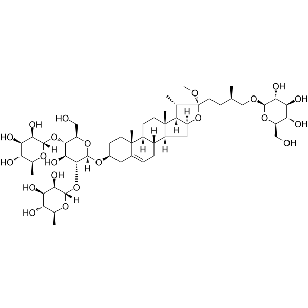
Methyl protodioscin

Based on 1 Customer Validation
Methyl protodioscin (NSC-698790; Smilax saponin B) is a multi-target, selective, steroidal diglycoside inhibitor with antitumor activity that induces cell cycle arrest. The mechanism of action of Methyl protodioscin is complex, involving the induction of G2/M cell cycle arrest, regulation of the Bcl-2/Bax apoptotic pathway, inhibition of the Akt1/c-Myc axis and MAPK/ERK signaling, while simultaneously downregulating ADAM15 and inducing FOXO1 to reduce cholesterol synthesis. It also inhibits the JNK/c-Jun pathway, reducing the production of inflammatory factors (IL-6, TNF-α). Methyl protodioscin exhibits significant antitumor (inhibiting proliferation, migration, invasion, and inducing apoptosis), anti-inflammatory, and anti-restenosis activities. Methyl protodioscin can be used in research on lung cancer, prostate cancer, pancreatic cancer, and other tumors, as well as inflammatory diseases such as airway inflammation and enteritis.
For research use only. We do not sell to patients.
- Purity : 99.67%
- CAS No.: 54522-52-0
- Formula: C52H86O22
- Molecular Weight:1063.23
-
Storage:
4°C, protect from light
* In solvent : -80°C, 6 months; -20°C, 1 month (protect from light)

To place orders, for customer services and technical support, please contact: MedChemExpress USA
Tel: 609-228-6898 E-mail: [email protected] [email protected]


-
 Biological Activity
-
Chemical Information
-
Solvent & Solubility
- Protocols
- Purity & Documentation
- References
-
 Help & FAQs


-
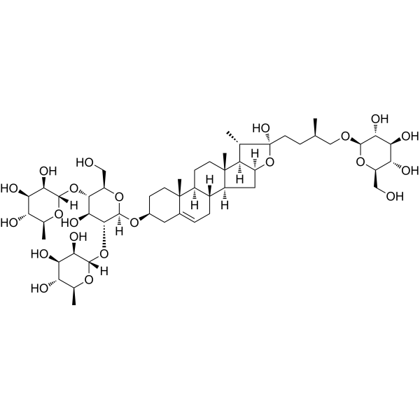
HY-N0799
ProtodioscinEndogenous Metabolite -
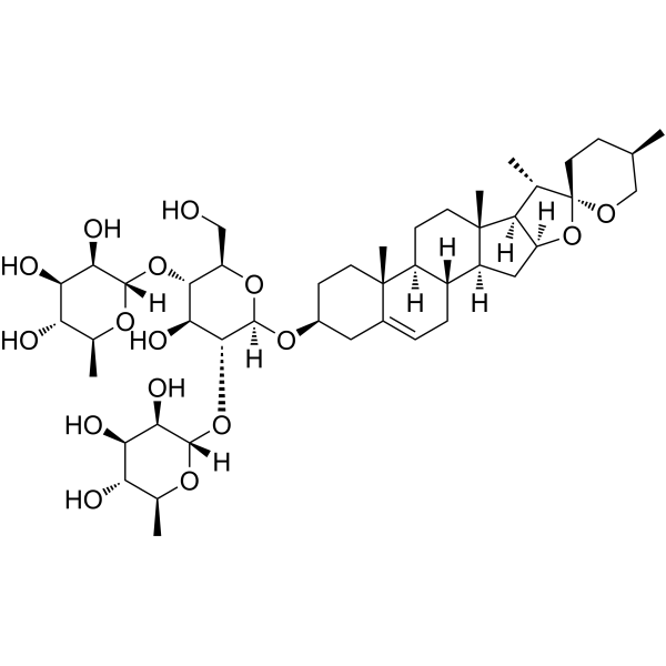
HY-N0124
DioscinAnticancer Agent -
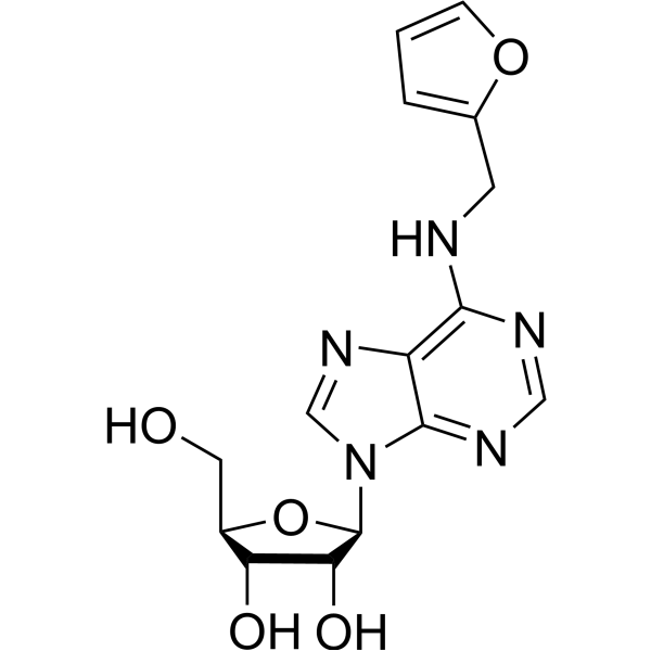
HY-101055
Kinetin ribosideApoptosis Activator -

HY-14648
DexamethasoneGR Agonist -
HY-13519
TRAM-34Potassium Channel Inhibitor -
HY-N4271
ProtogracillinAntithrombotic Agent -

HY-101055R
Kinetin riboside (Standard)Apoptosis Activator


Biological Activity
Description
Cellular Effect
|
Cell Line

|
Type | Value | Description | References |
|---|---|---|---|---|
| A7R5 | IC50 |
9 μM
Compound: 1
|
Antiproliferative activity against rat A7r5 cells assessed as reduction in cell viability after 24 hrs by MTT assay
Antiproliferative activity against rat A7r5 cells assessed as reduction in cell viability after 24 hrs by MTT assay
|
[PMID: 27227546] |
| HL-60 | IC50 |
7.3 μM
Compound: 10
|
Cytotoxicity against human HL60 cells after 72 hrs by MTT assay
Cytotoxicity against human HL60 cells after 72 hrs by MTT assay
|
[PMID: 12828464] |
In Vitro
Methyl protodioscin (10 nM-0.1 μM; 48 h) was screened in vitro against 60 human cancer cell lines, showing strong cytotoxicity against most solid tumor cells (GI50 ≤ 10.0 μM), with the strongest selectivity against colon cancer cells HCT-15 (GI50 = 1.93 μM) and breast cancer cells MDA-MB-435 (GI50 = 1.69 μM), and moderate toxicity against leukemia cells (GI50 10-30 μM); the GI50 values ??of methyl protodioscin against various tumor cells ranged from 1.69 to 50 μM (e.g., MDA-MB-435: 1.69 μM, DU145: approximately 4 μM), and the IC50 against vascular smooth muscle cells A7r5 was approximately 9 μM[1].
Methyl protodioscin (5-20 μM; 48 h) dose-dependently inhibited proliferation in A549 lung cancer cells, inducing G2/M phase cell cycle arrest and apoptosis, downregulating Bcl-2 expression, upregulating Bax expression, activating caspase-3, reducing mitochondrial membrane potential, and promoting cytochrome c release[2].
Methyl protodioscin (1-10 μM; 24-48 h) inhibited proliferation (48-hour IC50 approximately 4 μM), migration, and invasion in DU145 prostate cancer cells, inducing G2/M phase arrest and apoptosis, downregulating SREBP1, SREBP2, and HMGCR mRNA expression, upregulating ABCA1 and FOXO1 protein expression, and inhibiting P-ERK activity[3].
Methyl protodioscin (15-50 μM; 24-48 h) in PANC-1 (IC50 = 34.4 μM) and MIA PaCa-2 (IC50 = 50 μM) In pancreatic cancer cells, methyl protodioscin inhibits proliferation, induces G2/M phase arrest and apoptosis, downregulates the expression of glycolysis-related genes Glut1, HK2, LDHA, and PDK1, and inhibits the Akt1/c-Myc signaling pathway[4].
Methyl protodioscin (1-100 ng/mL; 12-48 h) repairs inflammation-induced barrier function damage in Caco-2 intestinal epithelial cells, promotes crypt formation in mouse intestinal crypt cultures (10 ng/mL being optimal), and upregulates RegIIIc mRNA expression[5].
Methyl protodioscin (3-9 μM; 16-24 h) inhibits proliferation (IC50 approximately 9 μM) and migration in A7r5 vascular smooth muscle cells, induces G0/G1 phase arrest, and downregulates the protein expression and activity of ADAM15, P-FAK, P-ERK, and MMP-2/9[6].
Methyl protodioscin (10-100 μM; 4 h) inhibits IL-1β-induced IL-6, IL-8, and TNF-α production in A549 cells and inhibits JNK/c-Jun activation; 100 μM treatment of MH-S macrophages for 24 hours slightly inhibits NO production, but this is accompanied by slight cytotoxicity[6].
MedChemExpress (MCE) has not independently confirmed the accuracy of these methods. They are for reference only.
In Vivo
Methyl protodioscin (100 mg/kg; intravenous injection; frequency and duration not specified) inhibited tumor growth, reduced 18F-FDG uptake in tumor tissue, and inhibited glycolysis in vivo in a subcutaneous pancreatic cancer MIA PaCa-2 cell xenograft model in BALB/c-nu mice[4].
Methyl protodioscin (25 mg/kg; intraperitoneal injection; once daily; 5 days/10 days) increased the survival rate of 4% DSS-induced mice, promoted mucosal healing in 2.5% DSS-induced mice, reduced colon inflammation scores, decreased NF-κB activation and inflammatory cytokine (TNF-α, IL-17, IL-23) production, and reduced bacterial translocation to mesenteric lymph nodes in a DSS (HY-116282C)-induced colitis model in C57BL/6 mice[5].
Methyl protodioscin (25 mg/kg; intraperitoneal injection; once daily; 7 days) alleviated colon inflammation, reduced bacterial colonization and translocation to mesenteric lymph nodes, and restored body weight and colon length in a Citrobacter rodentium infection-induced colitis model in C57BL/6 mice[5].
Methyl protodioscin (3 μM, 6 μM; topical application; single administration; 2 weeks) significantly reduced neointimal formation, decreased the neointima/media area ratio, and downregulated ADAM15 in vascular tissue in a carotid artery balloon injury model in male rats[6].
Protein expression[6].
Methyl protodioscin (30 mg/kg, 60 mg/kg; oral administration; single dose; 16 h) reduced total cell and neutrophil/macrophage infiltration in bronchoalveolar lavage fluid, decreased mRNA expression of IL-6, IL-1β, and TNF-α in lung tissue, and improved pathological changes such as alveolar wall thickening and alveolar lumen narrowing in a LPS-induced acute lung injury model in male ICR mice[7].
MedChemExpress (MCE) has not independently confirmed the accuracy of these methods. They are for reference only.
- Detection of Apoptosis
- TUNEL staining for apoptotic DNA fragmentation
- Flow cytometric DNA-content cell-cycle staining
- Annexin V plus membrane-impermeant dye apoptosis staining
- BrdU Incorporation Assay
- Cell migration
- Apoptosis Solutions
- Protocol for Cell Cycle
- Cell invasion
- Research Protocol for Inflammation-related Diseases
- Subcutaneous Cell-Line-Derived Xenograft
- Cell Viability Determination by MTT Colorimetric Assay


Chemical Information
-
CAS No. 54522-52-0
-
Appearance Solid
-
Molecular Weight 1063.23
-
Formula C52H86O22
-
Color White to off-white
-
SMILES
C[C@@]1([C@]([C@@H]2C)([H])[C@](O[C@]2(OC)CC[C@@H](C)CO[C@@H]([C@@H]([C@@H](O)[C@@H]3O)O)O[C@@H]3CO)([H])C4)[C@]4([H])[C@@](CC=C5[C@@]6(CC[C@H](O[C@@](O[C@H](CO)[C@@H](O[C@@](O[C@@H](C)[C@H](O)[C@H]7O)([H])[C@@H]7O)[C@@H]8O)([H])[C@@H]8O[C@@](O[C@@H](C)[C@H](O)[C@H]9O)([H])[C@@H]9O)C5)C)([H])[C@]6([H])CC1
-
Synonyms
NSC-698790; Smilax saponin B
-
Structure Classification
-
Initial Source
-
Shipping
Room temperature in continental US; may vary elsewhere.
-
Storage
4°C, protect from light
* In solvent : -80°C, 6 months; -20°C, 1 month (protect from light)


Solvent & Solubility
In Vitro:
DMSO : 100 mg/mL (94.05 mM; Need ultrasonic; Hygroscopic DMSO has a significant impact on the solubility of product, please use newly opened DMSO)
Please refer to the solubility information to select the appropriate solvent. Once prepared, please aliquot and store the solution to prevent product inactivation from repeated freeze-thaw cycles.
Storage method and period of stock solution: -80°C, 6 months; -20°C, 1 month (protect from light). When stored at -80°C, please use it within 6 months. When stored at -20°C, please use it within 1 month.
Please refer to the solubility information to select the appropriate solvent. Once prepared, please aliquot and store the solution to prevent product inactivation from repeated freeze-thaw cycles.
Storage method and period of stock solution: -80°C, 6 months; -20°C, 1 month (protect from light). When stored at -80°C, please use it within 6 months. When stored at -20°C, please use it within 1 month.
Concentration (start) × Volume (start) = Concentration (final) × Volume (final)
In Vivo:
Select the appropriate dissolution method based on your experimental animal and administration route.
- For the following dissolution methods, please ensure to first prepare a clear stock solution using an In Vitro approach and then sequentially add co-solvents:
- To ensure reliable experimental results, the clarified stock solution can be appropriately stored based on storage conditions. As for the working solution for In Vivo experiments, it is recommended to prepare freshly and use it on the same day.
- The percentages shown for the solvents indicate their volumetric ratio in the final prepared solution. If precipitation or phase separation occurs during preparation, heat and/or sonication can be used to aid dissolution.
Add each solvent one by one: 10% DMSO 40% PEG300 5% Tween-80 45% Saline
Solubility: ≥ 3.5 mg/mL (3.29 mM); Clear solution
This protocol yields a clear solution of ≥ 3.5 mg/mL (saturation unknown).
Taking 1 mL working solution as an example, add 100 μL DMSO stock solution (35.0 mg/mL) to 400 μL PEG300, and mix evenly; then add 50 μL Tween-80 and mix evenly; then add 450 μL Saline to adjust the volume to 1 mL.
Preparation of Saline: Dissolve 0.9 g sodium chloride in ddH₂O and dilute to 100 mL to obtain a clear Saline solution.
Add each solvent one by one: 10% DMSO 90% (20% SBE-β-CD in Saline)
Solubility: 3.5 mg/mL (3.29 mM); Suspended solution; Need ultrasonic
This protocol yields a suspended solution of 3.5 mg/mL. Suspended solution can be used for oral and intraperitoneal injection.
Taking 1 mL working solution as an example, add 100 μL DMSO stock solution (35.0 mg/mL) to 900 μL 20% SBE-β-CD in Saline, and mix evenly.
Preparation of 20% SBE-β-CD in Saline (4°C, storage for one week): 2 g SBE-β-CD powder is dissolved in 10 mL Saline, completely dissolve until clear.
In Vivo Dissolution Calculator
Please enter the basic information of animal experiments:
-
-
-
-
Recommended: Prepare an additional quantity of animals to account for potential losses during experiments.
Please enter your animal formula composition:
-
%DMSO +
Recommended: Keep the proportion of DMSO in working solution below 2% if your animal is weak.
-
%+
-
+%Tween-80 + +
-
%Saline +
The co-solvents required include: DMSO, . All of co-solvents are available by MedChemExpress (MCE). , Tween 80. All of co-solvents are available by MedChemExpress (MCE).
Working solution concentration: 0.22 mg/mL
Method for preparing stock solution: mg drug dissolved in μL DMSO. Stock solution concentration: mg/mL. * In solvent : -80°C, 6 months; -20°C, 1 month (protect from light)
1. Take μL DMSO stock solution;
2. Add μL .
μL , mix evenly;
3. Then add μL Tween 80, mix evenly;
4. Then add μL
Please ensure that the stock solution in the first step is dissolved to a clear state, and add co-solvents in sequence. You can use ultrasonic heating (ultrasonic cleaner, recommended frequency 20-40 kHz), vortexing, etc. to assist dissolution.


Purity & Documentation
-
Data Sheet (293 KB)
-
SDS (252 KB)
- English - EN (252 KB)
- Français - FR (252 KB)
- Deutsch - DE (252 KB)
- Norwegian - NO (252 KB)
- Español - ES (252 KB)
- Swedish - SV (252 KB)
- Italian - IT (252 KB)
- Korean - KR (252 KB)
- Portuguese - PT (252 KB)
-
Handling Instructions (2659 KB)
References
[1]. Hu K, et al. The cytotoxicity of methyl protodioscin against human cancer cell lines in vitro. Cancer Invest. 2003 Jun;21(3):389-93. [Content Brief]
[2]. Bai Y, et al. Methyl protodioscin induces G2/M cell cycle arrest and apoptosis in A549 human lung cancer cells. Pharmacogn Mag. 2014 Jul;10(39):318-24. [Content Brief]
[3]. Chen J, et al. Anticancer Activity of Methyl Protodioscin against Prostate Cancer by Modulation of Cholesterol-Associated MAPK Signaling Pathway via FOXO1 Induction. Biol Pharm Bull. 2023;46(4):574-585. [Content Brief]
[4]. Chen L, et al. Natural Compound Methyl Protodioscin Suppresses Proliferation and Inhibits Glycolysis in Pancreatic Cancer. Evid Based Complement Alternat Med. 2018 Mar 15;2018:7343090. [Content Brief]
[5]. Zhang R, et al. Natural compound methyl protodioscin protects against intestinal inflammation through modulation of intestinal immune responses. Pharmacol Res Perspect. 2015 Mar;3(2):e00118. [Content Brief]
[6]. Chung YL, et al. Methyl Protodioscin, a Steroidal Saponin, Inhibits Neointima Formation in Vitro and in Vivo. J Nat Prod. 2016 Jun 24;79(6):1635-44. [Content Brief]
[7]. Lee JH, et al. Methyl Protodioscin from the Roots of Asparagus cochinchinensis Attenuates Airway Inflammation by Inhibiting Cytokine Production. Evid Based Complement Alternat Med. 2015;2015:640846. [Content Brief]


Complete Stock Solution Preparation Table
Please refer to the solubility information to select the appropriate solvent. Once prepared, please aliquot and store the solution to prevent product inactivation from repeated freeze-thaw cycles.
Storage method and period of stock solution: -80°C, 6 months; -20°C, 1 month (protect from light). When stored at -80°C, please use it within 6 months. When stored at -20°C, please use it within 1 month.
| Optional Solvent | Concentration Solvent Mass | 1 mg | 5 mg | 10 mg | 25 mg |
|---|---|---|---|---|---|
| DMSO | 1 mM | 0.9405 mL | 4.7027 mL | 9.4053 mL | 23.5133 mL |
| 5 mM | 0.1881 mL | 0.9405 mL | 1.8811 mL | 4.7027 mL | |
| 10 mM | 0.0941 mL | 0.4703 mL | 0.9405 mL | 2.3513 mL | |
| 15 mM | 0.0627 mL | 0.3135 mL | 0.6270 mL | 1.5676 mL | |
| 20 mM | 0.0470 mL | 0.2351 mL | 0.4703 mL | 1.1757 mL | |
| 25 mM | 0.0376 mL | 0.1881 mL | 0.3762 mL | 0.9405 mL | |
| 30 mM | 0.0314 mL | 0.1568 mL | 0.3135 mL | 0.7838 mL | |
| 40 mM | 0.0235 mL | 0.1176 mL | 0.2351 mL | 0.5878 mL | |
| 50 mM | 0.0188 mL | 0.0941 mL | 0.1881 mL | 0.4703 mL | |
| 60 mM | 0.0157 mL | 0.0784 mL | 0.1568 mL | 0.3919 mL | |
| 80 mM | 0.0118 mL | 0.0588 mL | 0.1176 mL | 0.2939 mL |


Methyl protodioscin Related Classifications
HY-N0863 Related Classifications
 Powered by Bioz