



Mofegiline hydrochloride

Based on 1 Customer Validation
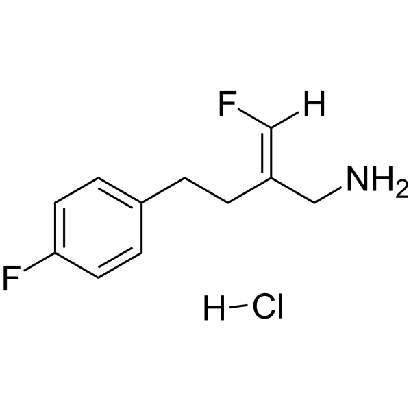
Mofegiline hydrochloride (MDL-72974A) is an orally active and blood-brain barrier-penetrant MAO-B/SSAO selective inhibitor, with an IC50 of 3.6 nM for MAO-B and 10 nM for SSAO. Mofegiline hydrochloride is a cationic amphiphilic compound that can induce indirect sympathomimetic effects, lysosomal accumulation, hERG channel interactions, phospholipidosis, and cell death. Mofegiline hydrochloride protects mice from MPTP-induced depletion of striatal dopamine, DOPAC, and HVA. Mofegiline hydrochloride can be used in research related to Parkinson's disease.
For research use only. We do not sell to patients.
- Purity : 99.57%
- CAS No.: 120635-25-8
- Formula: C11H14ClF2N
- Molecular Weight:233.69
-
Storage:
4°C, sealed storage, away from moisture
* In solvent : -80°C, 6 months; -20°C, 1 month (sealed storage, away from moisture)

To place orders, for customer services and technical support, please contact: MedChemExpress USA
Tel: 609-228-6898 E-mail: [email protected] [email protected]


-
 Biological Activity
-
Chemical Information
-
Solvent & Solubility
- Protocols
- Purity & Documentation
- References
-
 Help & FAQs


-
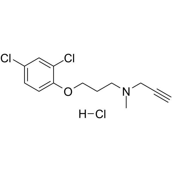
HY-14197A
Clorgyline hydrochlorideMAO-A Inhibitor -
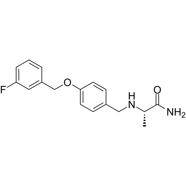
HY-70057
SafinamideMAO-B Inhibitor -
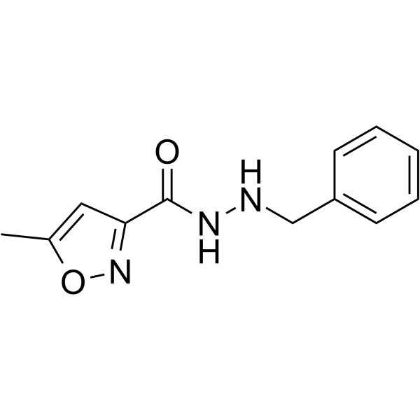
HY-13929
IsocarboxazidMonoamine Oxidase Inhibitor -
HY-B1018A
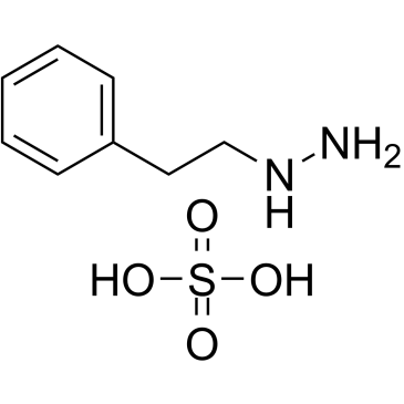
Phenelzine sulfateMAO Inhibitor -
HY-A0091A
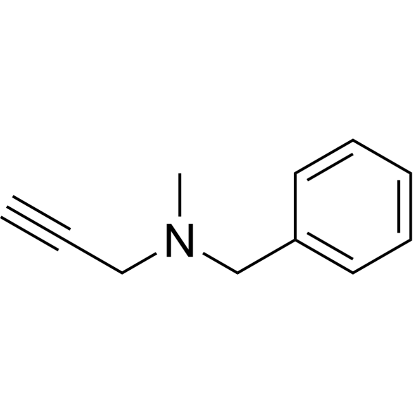
PargylineMonoamine Oxidase Inhibitor -
HY-138625A
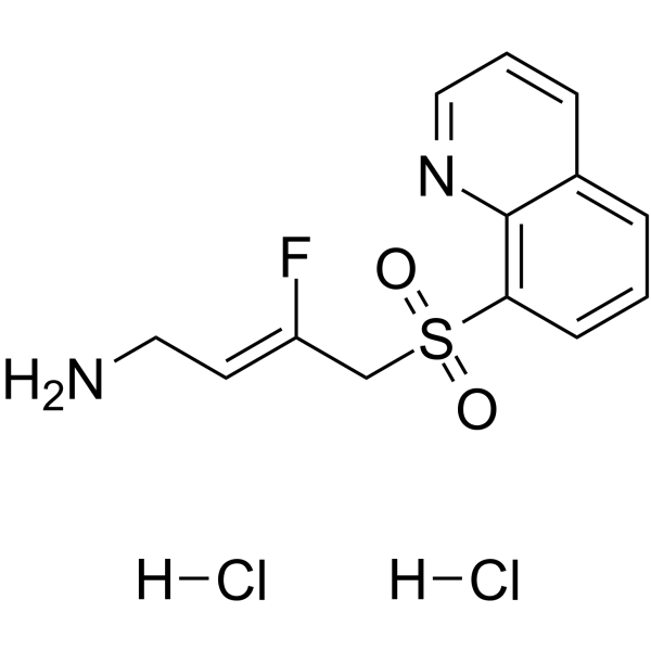
PXS-5505 anhydrousLysyl Oxidase Inhibitor -
HY-14201
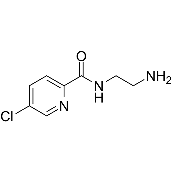
LazabemideMAO-B Inhibitor -
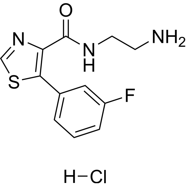
HY-100027A
Ro 41-1049 hydrochlorideMAO-A Inhibitor -
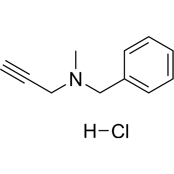
HY-A0091
Pargyline hydrochlorideMonoamine Oxidase Inhibitor -

HY-Y0882
Hydroxylamine hydrochlorideMAO Inhibitor -
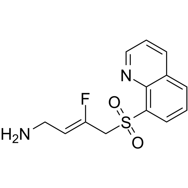
HY-138625
PXS-5505 free baseLysyl Oxidase Inhibitor -
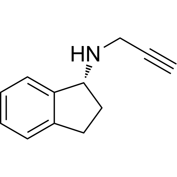
HY-14605A
RasagilineMAO Inhibitor


Biological Activity
Description
IC50 & Target
IC50: 3.6 nM (MAO-B), 680 nM (MAO-A)[1]
In Vitro
Mofegiline hydrochloride (MDL72974A) inhibits rat brain mitochondrial MAO in a concentration and time-dependent fashion[1].
Mofegiline hydrochloride (MDL72974A) inhibits [3H]dopamine (15 nM) uptake with an IC50 value of 31.8 μM, but poorly inhibits [3H]GBR-12935 (1 nM) binding (IC50 >100 μM) in the rat striatum[2].
Mofegiline hydrochloride (MDL72974A) inhibits SSAOs from dog aorta, rat aorta, bovine aorta and human umbilical artery with IC50s of 2 nM, 5 nM, 80 nM and 20 nM, respectively[3].
MedChemExpress (MCE) has not independently confirmed the accuracy of these methods. They are for reference only. Further protocols information, click here.
In Vivo
MedChemExpress (MCE) has not independently confirmed the accuracy of these methods. They are for reference only.
-
Animal Model:Male Sprague-Dawley rats (150-400 g)[1]
-
Dosage:Group 1: 0.1-2.5 mg/kg; Group 2: 0.05-5 mg/kg
-
Administration:Oral gavage; single dose for group 1, as for group 2, once daily for 14 days
-
Result:Showed the inhibition effect on rat brain MAO-A and MAO-B with EC50s of 8 mg/kg and 0.18 mg/kg, respectively, in group 1.
Resulted more potent efficacy on MAO-A inhibition in a daily dosed-manner (group 2) than single dose (group 1) manner, indicating a long half-life of Mofegiline hydrochloride.
-
Animal Model:Mate SwissWebster (CF-W) mice (25-30 g)[1]
-
Dosage:1.25 mg/kg
-
Administration:Intraperitoneal injection; 18 hours prior to administration of MPTP (20 mg/kg; i.p.; 4 times for two-hourly intervals, for 8 days)
-
Result:Rescued MPTP-induced decreases in striatal levels of dopamine (DA), dihydroxyphenylacetic acid (DOPAC) and homovanillic acid (HVA) in mice.
-
Animal Model:
-
Dosage:Group 1: 1.8, 9 mg/kg; Group 2: 0.1, 1 mg/kg
-
Administration:Oral gavage; single dose for group 1, as for group 2, once daily for 14 days
-
Result:Did not significantly potentiate the cardiovascular effects of intraduodenally administered Tyramine (HY-W007606) in anaesthetised rats.


Chemical Information
-
CAS No. 120635-25-8
-
Appearance Solid
-
Molecular Weight 233.69
-
Formula C11H14ClF2N
-
Color White to off-white
-
SMILES
FC1=CC=C(CC/C(CN)=C(F)/[H])C=C1.[H]Cl
-
Synonyms
MDL72974A
-
Shipping
Room temperature in continental US; may vary elsewhere.
-
Storage
4°C, sealed storage, away from moisture
* In solvent : -80°C, 6 months; -20°C, 1 month (sealed storage, away from moisture)


Solvent & Solubility
In Vitro:
DMSO : 110 mg/mL (470.71 mM; Need ultrasonic; Hygroscopic DMSO has a significant impact on the solubility of product, please use newly opened DMSO)
H2O : 25 mg/mL (106.98 mM; Need ultrasonic)
Please refer to the solubility information to select the appropriate solvent. Once prepared, please aliquot and store the solution to prevent product inactivation from repeated freeze-thaw cycles.
Storage method and period of stock solution: -80°C, 6 months; -20°C, 1 month (sealed storage, away from moisture). When stored at -80°C, please use it within 6 months. When stored at -20°C, please use it within 1 month.
* Note: If you choose water as the stock solution, please dilute it to the working solution, then filter and sterilize it with a 0.22 μm filter before use.
Please refer to the solubility information to select the appropriate solvent. Once prepared, please aliquot and store the solution to prevent product inactivation from repeated freeze-thaw cycles.
Storage method and period of stock solution: -80°C, 6 months; -20°C, 1 month (sealed storage, away from moisture). When stored at -80°C, please use it within 6 months. When stored at -20°C, please use it within 1 month.
* Note: If you choose water as the stock solution, please dilute it to the working solution, then filter and sterilize it with a 0.22 μm filter before use.
Concentration (start) × Volume (start) = Concentration (final) × Volume (final)
In Vivo:
Select the appropriate dissolution method based on your experimental animal and administration route.
- For the following dissolution methods, please ensure to first prepare a clear stock solution using an In Vitro approach and then sequentially add co-solvents:
- To ensure reliable experimental results, the clarified stock solution can be appropriately stored based on storage conditions. As for the working solution for In Vivo experiments, it is recommended to prepare freshly and use it on the same day.
- The percentages shown for the solvents indicate their volumetric ratio in the final prepared solution. If precipitation or phase separation occurs during preparation, heat and/or sonication can be used to aid dissolution.
Add each solvent one by one: 10% DMSO 40% PEG300 5% Tween-80 45% Saline
Solubility: ≥ 2.75 mg/mL (11.77 mM); Clear solution
This protocol yields a clear solution of ≥ 2.75 mg/mL (saturation unknown).
Taking 1 mL working solution as an example, add 100 μL DMSO stock solution (27.5 mg/mL) to 400 μL PEG300, and mix evenly; then add 50 μL Tween-80 and mix evenly; then add 450 μL Saline to adjust the volume to 1 mL.
Preparation of Saline: Dissolve 0.9 g sodium chloride in ddH₂O and dilute to 100 mL to obtain a clear Saline solution.
Add each solvent one by one: 10% DMSO 90% (20% SBE-β-CD in Saline)
Solubility: ≥ 2.75 mg/mL (11.77 mM); Clear solution
This protocol yields a clear solution of ≥ 2.75 mg/mL (saturation unknown).
Taking 1 mL working solution as an example, add 100 μL DMSO stock solution (27.5 mg/mL) to 900 μL 20% SBE-β-CD in Saline, and mix evenly.
Preparation of 20% SBE-β-CD in Saline (4°C, storage for one week): 2 g SBE-β-CD powder is dissolved in 10 mL Saline, completely dissolve until clear.
For the following dissolution methods, please prepare the working solution directly:
It is recommended to prepare fresh solutions and use them promptly within a short period of time.
The percentages shown for the solvents indicate their volumetric ratio in the final prepared solution. If precipitation or phase separation occurs during preparation, heat and/or sonication can be used to aid dissolution.
Add each solvent one by one: PBS
Solubility: 10 mg/mL (42.79 mM); Clear solution; Need ultrasonic
In Vivo Dissolution Calculator
Please enter the basic information of animal experiments:
-
-
-
-
Recommended: Prepare an additional quantity of animals to account for potential losses during experiments.
Working solution concentration: 0.22 mg/mL
This product has good water solubility, please refer to the measured solubility data in water/PBS/Saline for details.

Protocols
-
Cell Cytotoxicity Assay
Cytotoxicity assays are usually based on the assessment of cell membrane damage, which can also be indirectly detected by measuring cell viability. Detection methods include MTT assay, CKK-8 assay, LDH assay and ATP assay, etc.
-
Mammalian live/dead viability and cytotoxicity staining
Live/dead viability and cytotoxicity staining assays are based on the simultaneous detection of intracellular esterase activity in metabolically active (viable) cells and membrane integrity loss in non-viable cells. In commonly used dual-staining approaches, membrane-permeant fluorogenic substrates are converted by intracellular esterases into fluorescent products in live cells, while impermeant DNA-binding dyes selectively enter cells with compromised plasma membranes and label nucleic acids in dead or dying cells, enabling discrimination between viable and non-viable populations by fluorescence microscopy or flow cytometry.
-
Human pluripotent stem cell midbrain dopaminergic neuron differentiation
Human pluripotent stem cells are directed toward midbrain dopaminergic neurons by first inducing a neural floor-plate-like progenitor state, then patterning cells with ventralizing SHH signaling and midbrain/WNT-FGF cues, and finally maturing progenitors into neurons expressing dopaminergic markers such as TH, NURR1/NR4A2, PITX3, DAT/SLC6A3, VMAT2/SLC18A2, GIRK2/KCNJ6, FOXA2, LMX1A, and EN1. The main readouts are loss of pluripotency, acquisition of FOXA2+/LMX1A+ midbrain floor-plate progenitors, emergence of βIII-tubulin+/MAP2+ neurons, and production of TH+ dopaminergic neurons with molecular, dopamine-release, and electrophysiological features of midbrain dopaminergic identity.
-
Apoptosis Solutions
Apoptosis is a regulated, generally non-lytic cell-death pathway that removes unwanted, damaged, infected, or abnormal cells through coordinated morphological changes, caspase activation, DNA fragmentation, and membrane remodeling. The intrinsic apoptosis pathway is controlled mainly by mitochondrial outer membrane permeabilization, BCL-2 family proteins, cytochrome c release, apoptosome formation, caspase-9 activation, and downstream executioner caspase-3/7 activation. The extrinsic apoptosis pathway is initiated by death receptors such as Fas, TNFR, and TRAIL receptors, which recruit adaptor proteins and activate caspase-8 before engaging executioner caspases or mitochondrial amplification through BID cleavage. Apoptosis is linked to many phenotypes, including cancer cell killing, tissue homeostasis, immune regulation, neurodegeneration, infection response, and treatment-induced cytotoxicity; unresolved questions include how apoptosis interacts with necroptosis, pyroptosis, ferroptos
-
Lysosome and acidic-vesicle live-cell staining
Lysosome and acidic-vesicle live-cell staining detects acidic intracellular compartments by using membrane-permeant acidotropic probes that accumulate in low-pH vesicles, including lysosomes, late endosomes, autolysosomes, and acidic phagosomes. LysoTracker staining is commonly used as an intensity-based readout of acidic lysosomal compartment abundance or enlargement, while acridine orange produces green fluorescence in less concentrated compartments and red fluorescence after concentration-dependent accumulation in acidic vesicular organelles. Loss or reduction of acridine-orange red signal can be used as a readout of lysosomal membrane permeabilization or reduced acidic-vesicle integrity. This protocol is designed for live cultured cells and can be adapted for fluorescence microscopy, high-content imaging, plate-reader readout, or flow cytometry when the selected literature supports the readout. Because these dyes report acidotropic accumulation rather than lysosome identity alone,


Purity & Documentation
-
Data Sheet (305 KB)
-
SDS (393 KB)
- English - EN (393 KB)
- Français - FR (393 KB)
- Deutsch - DE (393 KB)
- Norwegian - NO (393 KB)
- Español - ES (393 KB)
- Swedish - SV (393 KB)
- Italian - IT (393 KB)
- Korean - KR (393 KB)
- Portuguese - PT (393 KB)
-
Handling Instructions (2659 KB)
References
[1]. Zreika M, et al. MDL 72,974: a potent and selective enzyme-activated irreversible inhibitor of monoamine oxidase type B with potential for use in Parkinson's disease. J Neural Transm Park Dis Dement Sect. 1989;1(4):243-54. [Content Brief]
[2]. Fang J, et al. Effect of L-deprenyl, its structural analogues and some monoamine oxidase inhibitors on dopamine uptake. Neuropharmacology. 1994 Jun;33(6):763-8. [Content Brief]
[3]. Dow J, et al. Novel carbamate metabolites of mofegiline, a primary amine monoamine oxidase B inhibitor, in dogs and humans. Drug Metab Dispos. 1994 Sep-Oct;22(5):738-49. [Content Brief]
[4]. Yu PH, et al. Inhibition of a type B monoamine oxidase inhibitor, (E)-2-(4-fluorophenethyl)-3-fluoroallylamine (MDL-72974A), on semicarbazide-sensitive amine oxidases isolated from vascular tissues and sera of different species. Biochem Pharmacol. 1992 Jan 22;43(2):307-12. [Content Brief]


Complete Stock Solution Preparation Table
Please refer to the solubility information to select the appropriate solvent. Once prepared, please aliquot and store the solution to prevent product inactivation from repeated freeze-thaw cycles.
Storage method and period of stock solution: -80°C, 6 months; -20°C, 1 month (sealed storage, away from moisture). When stored at -80°C, please use it within 6 months. When stored at -20°C, please use it within 1 month.
| Optional Solvent | Concentration Solvent Mass | 1 mg | 5 mg | 10 mg | 25 mg |
|---|---|---|---|---|---|
| H2O / DMSO | 1 mM | 4.2792 mL | 21.3959 mL | 42.7917 mL | 106.9793 mL |
| 5 mM | 0.8558 mL | 4.2792 mL | 8.5583 mL | 21.3959 mL | |
| 10 mM | 0.4279 mL | 2.1396 mL | 4.2792 mL | 10.6979 mL | |
| 15 mM | 0.2853 mL | 1.4264 mL | 2.8528 mL | 7.1320 mL | |
| 20 mM | 0.2140 mL | 1.0698 mL | 2.1396 mL | 5.3490 mL | |
| 25 mM | 0.1712 mL | 0.8558 mL | 1.7117 mL | 4.2792 mL | |
| 30 mM | 0.1426 mL | 0.7132 mL | 1.4264 mL | 3.5660 mL | |
| 40 mM | 0.1070 mL | 0.5349 mL | 1.0698 mL | 2.6745 mL | |
| 50 mM | 0.0856 mL | 0.4279 mL | 0.8558 mL | 2.1396 mL | |
| 60 mM | 0.0713 mL | 0.3566 mL | 0.7132 mL | 1.7830 mL | |
| 80 mM | 0.0535 mL | 0.2674 mL | 0.5349 mL | 1.3372 mL | |
| 100 mM | 0.0428 mL | 0.2140 mL | 0.4279 mL | 1.0698 mL |
* Note: If you choose water as the stock solution, please dilute it to the working solution, then filter and sterilize it with a 0.22 μm filter before use.


Mofegiline hydrochloride Related Classifications
HY-16677A Related Classifications
 Powered by Bioz