



YHO-13177
Based on 2 publication(s) in Google Scholar
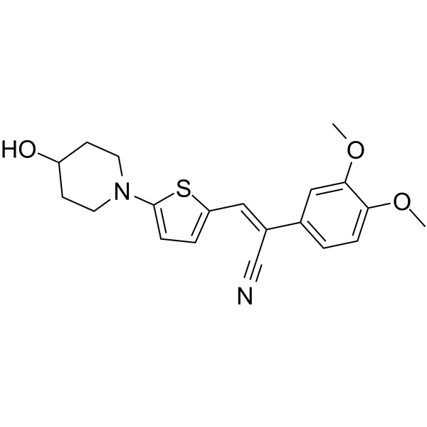
YHO-13177, a acrylonitrile derivative, is an orally active, potent and specific inhibitor of breast cancer resistance protein (BCRP) and ABCG2 with an IC50 value of 10 nM. YHO-13177 potentiates the cytotoxicity of SN-38 in HCT116 and A549 cells that express BCRP. YHO-13177 combined with Irinotecan (HY-16562) significantly suppresses the tumor growth in an HCT116/BCRP xenograft model.
For research use only. We do not sell to patients.
- Purity : 99.65%
- CAS No.: 912287-56-0
- Formula: C20H22N2O3S
- Molecular Weight:370.47
-
Storage:Powder -20°C, 3 years , 4°C, 2 years ; In solvent -80°C, 2 years , -20°C, 1 year

To place orders, for customer services and technical support, please contact: MedChemExpress USA
Tel: 609-228-6898 E-mail: [email protected] [email protected]


-
 Biological Activity
-
Chemical Information
-
Solvent & Solubility
- Protocols
- Purity & Documentation
- References
-
 Help & FAQs


-
HY-10010
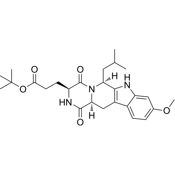
Ko 143BCRP/ABCG2 Inhibitor -
HY-10550
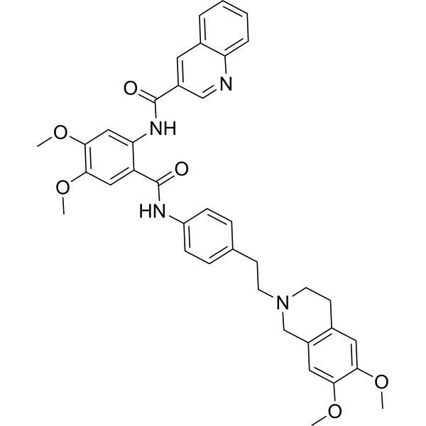
TariquidarP-gp Inhibitor -
HY-50879
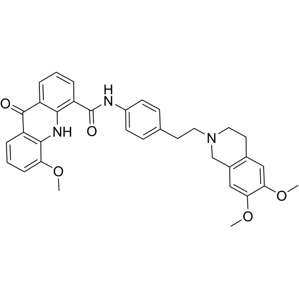
ElacridarP-gp And BCRP Dual-Inhibitor -
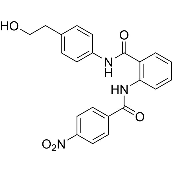
HY-19753
KS176BCRP Inhibitor -
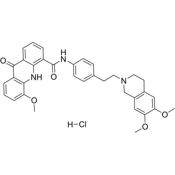
HY-50880
Elacridar hydrochlorideP-gp And BCRP Dual-Inhibitor -
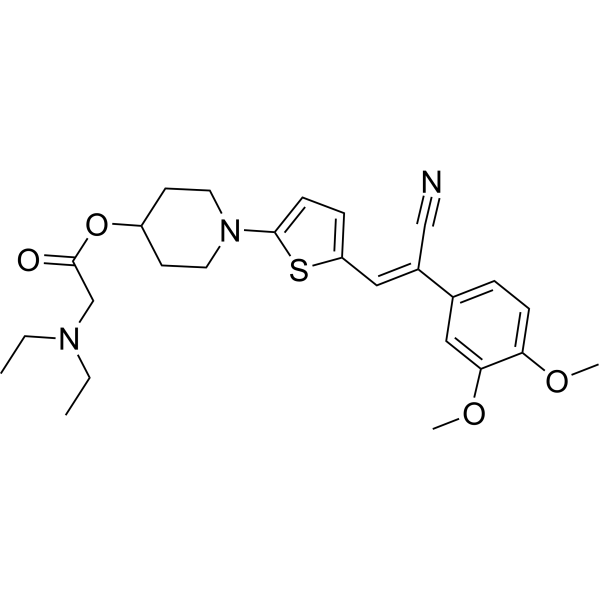
HY-12758A
YHO-13351 free base -
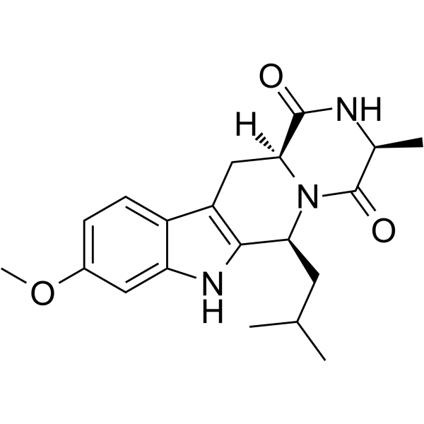
HY-100390
(S)-ML753286BCRP Inhibitor -
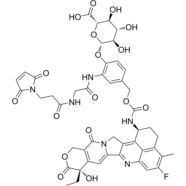
HY-153179
Mal-Gly-PAB-Exatecan-D-glucuronic acidADC Intermediate Compound -
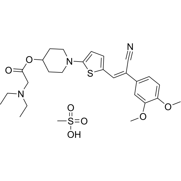
HY-12758
YHO-13351ABCG2 Inhibitor -
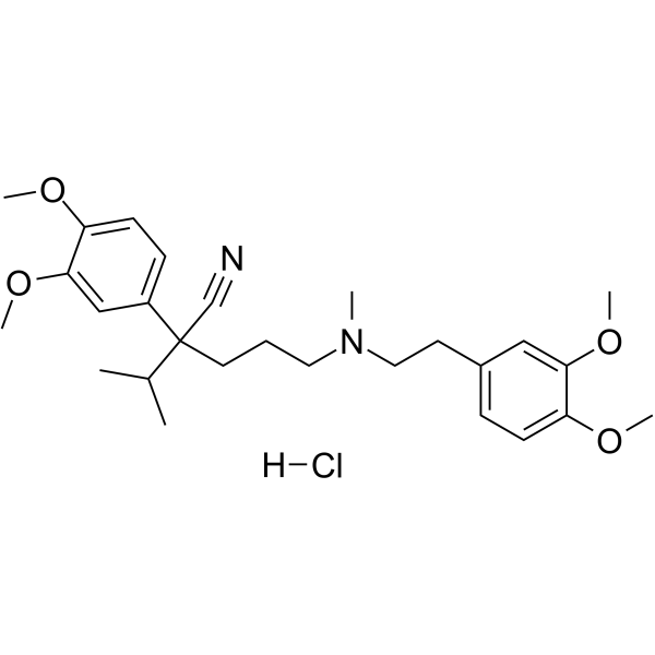
HY-A0064
Verapamil hydrochlorideCalcium Channel Blocker -
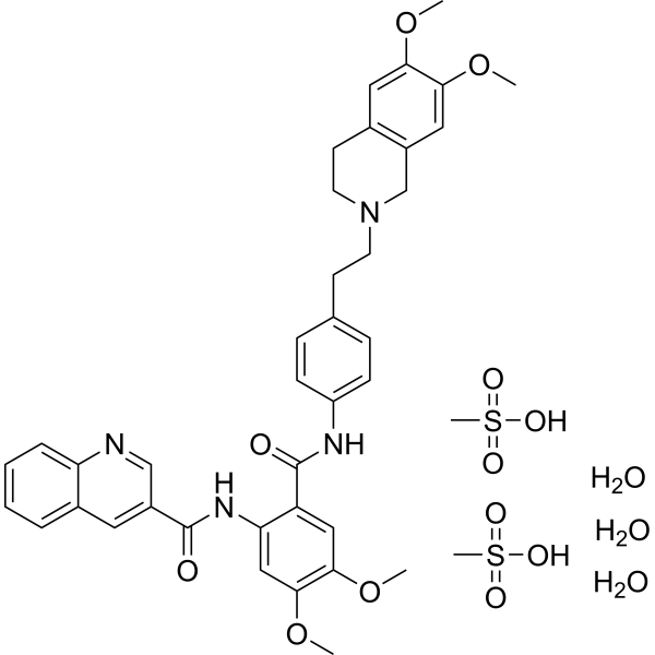
HY-10550A
Tariquidar methanesulfonate, hydrateP-gp Inhibitor -
HY-N2143
Fumitremorgin CABCG2/BRCP Inhibitor
Publications Citing Use of MedChemExpress (MCE) YHO-13177
More Customer Validation & Images

-
IF


Biological Activity
Description
Cellular Effect
|
Cell Line

|
Type | Value | Description | References |
|---|---|---|---|---|
| HCT-116 | IC50 |
10 nM
Compound: 14; YHO-13177
|
Cytotoxicity against human HCT-116 cells assessed as cell viability measured as mitochondrial NADH-dependent dehydrogenase activity incubated for 72 to 120 hrs
Cytotoxicity against human HCT-116 cells assessed as cell viability measured as mitochondrial NADH-dependent dehydrogenase activity incubated for 72 to 120 hrs
|
[PMID: 37847948] |
In Vitro
YHO-13177 (0.01 to 1 μM, 96 h) reverses BCRP-mediated drug resistance and sensitivity in a concentration-dependent manner in various BCRP-transduced cancer cells[1].
YHO-13177 (0.01 to 1 μM, 96 h) decreases the expression of BCRP protein in HCT116 and HCT116/BCRP cells, or A549 and A549/SN4 cells[1].
YHO-13177 (0-0.1 μM, 96 h) reduces the side population cell ratio in HeLa cells in a concentration-dependent manner[2].
MedChemExpress (MCE) has not independently confirmed the accuracy of these methods. They are for reference only. Further protocols information, click here.
-
Cell Line:HCT116 and A549 cells
-
Concentration:0.01 to 1 μM
-
Incubation Time:96 h
-
Result:Apparently decreased the level of BCRP protein at more than 24 hours.
In Vivo
MedChemExpress (MCE) has not independently confirmed the accuracy of these methods. They are for reference only.
-
Animal Model:HCT116/BCRP xenograft mouse model[1]
-
Dosage:19.7 μM or 27.3 μM
-
Administration:19.7 μM, i.v., or 27.3 μM, p.o., a single dose for 25 days
-
Result:Was maintained for at least 8 hours in plasma.


Chemical Information
-
CAS No. 912287-56-0
-
Appearance Solid
-
Molecular Weight 370.47
-
Formula C20H22N2O3S
-
Color Light yellow to yellow
-
SMILES
N#C/C(C1=CC=C(OC)C(OC)=C1)=C\C2=CC=C(N3CCC(O)CC3)S2
-
Shipping
Room temperature in continental US; may vary elsewhere.
-
Storage
Powder -20°C 3 years 4°C 2 years In solvent -80°C 2 years -20°C 1 year


Publications (2)
-
Journal Impact Factor
-
Most Recent
-
Drug Deliv
2017 Nov;24(1):1453-1459. PMID: 28949254
YHO-13177 purchased from MedChemExpress. Usage Cited in: Drug Deliv. 2017 Nov;24(1):1453-1459. [Abstract]
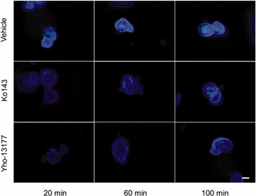
Fluorescent substrates accumulate in the organoids. The organoids are incubated in Hoechst 33342 with or without YHO-13177 or Ko143 for 20, 60 and 100 minutes, respectively. Ko143 and YHO-13177 notably decrease the fluorescence intensity of Hoechst 33342 in the organoids.
-
Crit Rev Anal Chem
A Critical Review on Advancement in Analytical Strategies for the Quantification of Clinically Relevant Biological Transporters. [Abstract]2022;52(7):1557-1571. PMID: 33691566


Solvent & Solubility
In Vitro:
DMSO : 33.33 mg/mL (89.97 mM; Need ultrasonic; Hygroscopic DMSO has a significant impact on the solubility of product, please use newly opened DMSO)
Please refer to the solubility information to select the appropriate solvent. Once prepared, please aliquot and store the solution to prevent product inactivation from repeated freeze-thaw cycles.
Storage method and period of stock solution: -80°C, 2 years; -20°C, 1 year. When stored at -80°C, please use it within 2 years. When stored at -20°C, please use it within 1 year.
Please refer to the solubility information to select the appropriate solvent. Once prepared, please aliquot and store the solution to prevent product inactivation from repeated freeze-thaw cycles.
Storage method and period of stock solution: -80°C, 2 years; -20°C, 1 year. When stored at -80°C, please use it within 2 years. When stored at -20°C, please use it within 1 year.
Concentration (start) × Volume (start) = Concentration (final) × Volume (final)
In Vivo:
Select the appropriate dissolution method based on your experimental animal and administration route.
- For the following dissolution methods, please ensure to first prepare a clear stock solution using an In Vitro approach and then sequentially add co-solvents:
- To ensure reliable experimental results, the clarified stock solution can be appropriately stored based on storage conditions. As for the working solution for In Vivo experiments, it is recommended to prepare freshly and use it on the same day.
- The percentages shown for the solvents indicate their volumetric ratio in the final prepared solution. If precipitation or phase separation occurs during preparation, heat and/or sonication can be used to aid dissolution.
Add each solvent one by one: 10% DMSO 40% PEG300 5% Tween-80 45% Saline
Solubility: ≥ 2.5 mg/mL (6.75 mM); Clear solution
This protocol yields a clear solution of ≥ 2.5 mg/mL (saturation unknown).
Taking 1 mL working solution as an example, add 100 μL DMSO stock solution (25.0 mg/mL) to 400 μL PEG300, and mix evenly; then add 50 μL Tween-80 and mix evenly; then add 450 μL Saline to adjust the volume to 1 mL.
Preparation of Saline: Dissolve 0.9 g sodium chloride in ddH₂O and dilute to 100 mL to obtain a clear Saline solution.
In Vivo Dissolution Calculator
Please enter the basic information of animal experiments:
-
-
-
-
Recommended: Prepare an additional quantity of animals to account for potential losses during experiments.
Please enter your animal formula composition:
-
%DMSO +
Recommended: Keep the proportion of DMSO in working solution below 2% if your animal is weak.
-
%+
-
+%Tween-80 + +
-
%Saline +
The co-solvents required include: DMSO, . All of co-solvents are available by MedChemExpress (MCE). , Tween 80. All of co-solvents are available by MedChemExpress (MCE).
Working solution concentration: 0.22 mg/mL
Method for preparing stock solution: mg drug dissolved in μL DMSO. Stock solution concentration: mg/mL.
1. Take μL DMSO stock solution;
2. Add μL .
μL , mix evenly;
3. Then add μL Tween 80, mix evenly;
4. Then add μL
Please ensure that the stock solution in the first step is dissolved to a clear state, and add co-solvents in sequence. You can use ultrasonic heating (ultrasonic cleaner, recommended frequency 20-40 kHz), vortexing, etc. to assist dissolution.

Protocols
-
Cell Cytotoxicity Assay
Cytotoxicity assays are usually based on the assessment of cell membrane damage, which can also be indirectly detected by measuring cell viability. Detection methods include MTT assay, CKK-8 assay, LDH assay and ATP assay, etc.
-
Mammalian live/dead viability and cytotoxicity staining
Live/dead viability and cytotoxicity staining assays are based on the simultaneous detection of intracellular esterase activity in metabolically active (viable) cells and membrane integrity loss in non-viable cells. In commonly used dual-staining approaches, membrane-permeant fluorogenic substrates are converted by intracellular esterases into fluorescent products in live cells, while impermeant DNA-binding dyes selectively enter cells with compromised plasma membranes and label nucleic acids in dead or dying cells, enabling discrimination between viable and non-viable populations by fluorescence microscopy or flow cytometry.
-
Patient-Derived Xenograft (PDX)
Patient-derived xenograft (PDX) models are generated by engrafting primary human tumor tissue directly into immunodeficient mice, allowing in vivo propagation of patient tumor biology without initial in vitro adaptation. These models are used to preserve key histopathological and molecular characteristics of the original tumor and enable assessment of tumor growth dynamics and therapeutic response in a living organism. The biological readout is tumor engraftment and subsequent growth in the murine host, which reflects the ability of human tumor cells to survive, vascularize, and expand in an immunocompromised microenvironment.
-
Patient-Derived Orthotopic Xenograft (PDOX)
Patient-derived orthotopic xenograft (PDOX) modeling implants fresh patient tumor tissue or patient-derived tumor cells into the anatomically corresponding organ or tissue site of immunodeficient mice, usually by surgical orthotopic implantation, to preserve patient tumor histology, local microenvironmental context, invasion, metastatic behavior, and treatment-response features better than subcutaneous implantation. PDOX readouts include tumor engraftment, orthotopic tumor growth, local invasion, metastasis, recurrence after resection, histologic similarity to the donor tumor, biomarker retention, molecular concordance, survival, and response or resistance to therapy. PDOX models are used for preclinical drug testing and individualized therapy evaluation, but engraftment success varies by tumor type and specimen quality.
-
Splenic/Portal-Vein Liver Metastasis Xenograft
Splenic and portal-vein liver metastasis xenograft models deliver tumor cells into the portal circulation so that cells reach the liver first and form hepatic metastatic lesions; splenic injection uses the spleen as an access route to the portal system, while direct portal-vein injection introduces cells into the portal vein without requiring splenectomy. The assay detects liver colonization, intrahepatic tumor growth, tumor distribution, treatment response, survival, and liver-metastasis microenvironment changes; readouts include bioluminescence or fluorescence imaging, gross liver nodule counts, liver weight or tumor burden, histology, and survival.
-
Subcutaneous Cell-Line-Derived Xenograft
Subcutaneous cell-line-derived xenograft (CDX) models are established by implanting cultured human cancer cell lines into immunodeficient mice, where the injected cells form localized tumors that can be monitored in vivo as a measure of tumorigenic potential, growth kinetics, and treatment response. These models are widely used in oncology research because they allow reproducible tumor formation and enable comparative assessment of tumor growth between different cell lines or genetic manipulations in a controlled in vivo microenvironment. Subcutaneous implantation of cancer cells in immunodeficient mice is a standard approach for evaluating tumor growth behavior and therapeutic response across multiple cancer types, including prostate, esophageal, pancreatic, and colon cancer models.
-
Breast Cancer Modeling
Breast cancer is a heterogeneous cancer, and it has been distinguished into four subtypes: luminal A, luminal B, HER2-positive and basal-like. Molecular mutations, epigenetic alterations, hormone exposure and immune microenvironment are related to the progression of breast cancer.
-
Orthotopic Cell-Line Xenograft
Orthotopic cell-line xenograft models involve implantation of human cancer cell lines into the anatomically corresponding organ of immunodeficient mice to reproduce tumor growth within a native microenvironment, enabling more clinically relevant tumor behavior compared with subcutaneous models. These models are widely used because orthotopic placement better recapitulates tumor progression, including invasion and metastatic spread, which are often underrepresented in heterotopic implantation systems. Compared with conventional xenografts, orthotopic implantation is described as more technically complex but provides improved simulation of tumor-microenvironment interactions and metastatic behavior, making it particularly valuable for translational oncology research. Surgical orthotopic implantation approaches have been emphasized as enabling faithful reproduction of clinical cancer features, including metastasis and disease progression patterns that align with the tumor’s organ of origi
-
Cell Viability Determination by MTT Colorimetric Assay
The following protocol uses the MTT colorimetric assay as a classic literature-established method for assessing cell viability/metabolic activity in cultured mammalian cells. MTT[3-(4,5-dimethylthiazol-2-yl)-2,5-diphenyltetrazolium bromide] is reduced by metabolically active cells to a colored formazan product; the amount of formazan is quantified spectrophotometrically and provides an indirect measure of metabolically active viable cells. Importantly, MTT reduction reflects cellular oxidoreductase/metabolic activity rather than an absolute direct count of living cells, so changes in cellular metabolism can alter the signal independently of cell number.
-
Intraperitoneal/Peritoneal Dissemination Xenograft
Intraperitoneal (IP) or peritoneal dissemination xenograft models are based on the introduction of human cancer cells into the peritoneal cavity of immunodeficient mice, where they attach to peritoneal surfaces, form multicellular aggregates or spheroids, and progressively generate disseminated tumor nodules that mimic advanced peritoneal metastatic disease. These models are widely used to study ovarian cancer progression, tumor-microenvironment interactions, and intraperitoneal therapeutic responses, often incorporating bioluminescence or fluorescence imaging to longitudinally monitor tumor burden in vivo. The biological principle relies on the capacity of tumor cells such as SKOV3 or related ovarian carcinoma lines to survive in suspension, aggregate within ascites-like fluid, adhere to mesothelial surfaces, and invade peritoneal organs, thereby recapitulating human peritoneal carcinomatosis patterns observed in advanced disease.


Purity & Documentation
-
Data Sheet (275 KB)
-
SDS (393 KB)
- English - EN (393 KB)
- Français - FR (393 KB)
- Deutsch - DE (393 KB)
- Norwegian - NO (393 KB)
- Español - ES (393 KB)
- Swedish - SV (393 KB)
- Italian - IT (393 KB)
- Korean - KR (393 KB)
- Portuguese - PT (393 KB)
-
Handling Instructions (2659 KB)
References
[1]. Yamazaki R, et al. Novel acrylonitrile derivatives, YHO-13177 and YHO-13351, reverse BCRP/ABCG2-mediated drug resistance in vitro and in vivo. Mol Cancer Ther. 2011 Jul;10(7):1252-63. [Content Brief]
[2]. Shishido Y, et al. ABCG2 inhibitor YHO-13351 sensitizes cancer stem/initiating-like side population cells to irinotecan. Anticancer Res. 2013 Apr;33(4):1379-86. [Content Brief]
[3]. Yu S, et al. The mechanisms of multidrug resistance of breast cancer and research progress on related reversal agents. Bioorg Med Chem. 2023 Nov 15;95:117486. [Content Brief]


Complete Stock Solution Preparation Table
Please refer to the solubility information to select the appropriate solvent. Once prepared, please aliquot and store the solution to prevent product inactivation from repeated freeze-thaw cycles.
Storage method and period of stock solution: -80°C, 2 years; -20°C, 1 year. When stored at -80°C, please use it within 2 years. When stored at -20°C, please use it within 1 year.
| Optional Solvent | Concentration Solvent Mass | 1 mg | 5 mg | 10 mg | 25 mg |
|---|---|---|---|---|---|
| DMSO | 1 mM | 2.6993 mL | 13.4964 mL | 26.9927 mL | 67.4818 mL |
| 5 mM | 0.5399 mL | 2.6993 mL | 5.3985 mL | 13.4964 mL | |
| 10 mM | 0.2699 mL | 1.3496 mL | 2.6993 mL | 6.7482 mL | |
| 15 mM | 0.1800 mL | 0.8998 mL | 1.7995 mL | 4.4988 mL | |
| 20 mM | 0.1350 mL | 0.6748 mL | 1.3496 mL | 3.3741 mL | |
| 25 mM | 0.1080 mL | 0.5399 mL | 1.0797 mL | 2.6993 mL | |
| 30 mM | 0.0900 mL | 0.4499 mL | 0.8998 mL | 2.2494 mL | |
| 40 mM | 0.0675 mL | 0.3374 mL | 0.6748 mL | 1.6870 mL | |
| 50 mM | 0.0540 mL | 0.2699 mL | 0.5399 mL | 1.3496 mL | |
| 60 mM | 0.0450 mL | 0.2249 mL | 0.4499 mL | 1.1247 mL | |
| 80 mM | 0.0337 mL | 0.1687 mL | 0.3374 mL | 0.8435 mL |


YHO-13177 Related Classifications
HY-12757 Related Classifications
 Powered by Bioz