



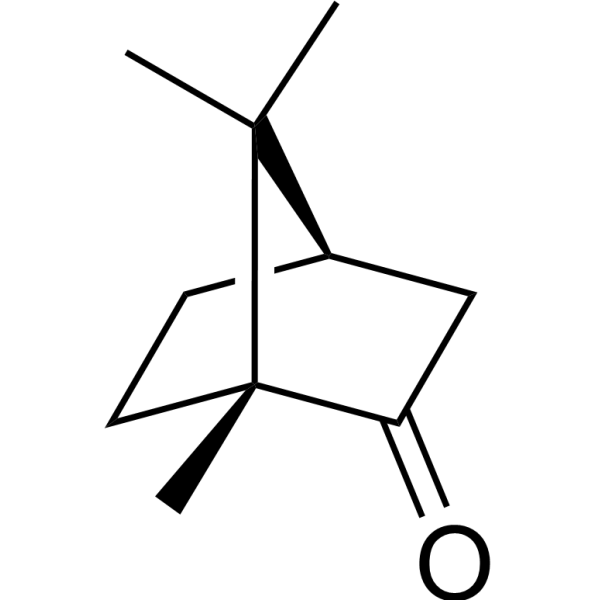
(+)-Camphor
Based on 1 publication(s) in Google Scholar
(+)-Camphor (D-(+)-Camphor; (1R)-(+)-Camphor) is an isomer of Camphor. Camphor is an agonist of monoterpenoid transient receptor potential (TRP) channels (such as TRPV1, TRPV3, TRPM8) and an inhibitor of TRPA1 channels. Camphor's derivatives have multiple biological activities, including antibacterial, antiviral, antioxidant, analgesic and anticancer. Camphor can selectively activate cold-sensitive TRP channels and inhibit TRPA1-mediated nociceptive signals. Camphor stimulates the cold-sensing nerve endings in the skin and regulates the activity of ion channels to exert analgesic, anti-inflammatory and anti-itching effects. It also has anti-proliferative and anti-mutagenic activities on tumor cells, which may be related to inhibiting ribosome function or inducing cell apoptosis. Camphor can be absorbed through the skin and (+)-Camphor can be used in the study of muscle and joint pain and inflammation.
For research use only. We do not sell to patients.
- Purity : 99.51%
- CAS No.: 464-49-3
- Formula: C10H16O
- Molecular Weight:152.24
-
Storage:Powder -20°C, 3 years , 4°C, 2 years ; In solvent -80°C, 2 years , -20°C, 1 year

To place orders, for customer services and technical support, please contact: MedChemExpress USA
Tel: 609-228-6898 E-mail: [email protected] [email protected]


-
 Biological Activity
-
Chemical Information
-
Solvent & Solubility
- Protocols
- Purity & Documentation
- References
-
 Help & FAQs


-

HY-B1173R
(+)-Camphor (Standard)Bacterial Inhibitor -
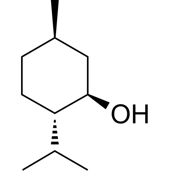
HY-75161
(-)-MentholEndogenous Metabolite -
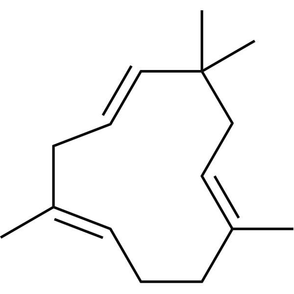
HY-N6968
α-HumuleneCOX Inhibitor -
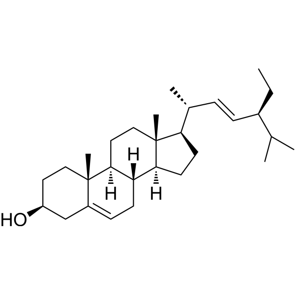
HY-N0131
StigmasterolPlant Sterol -

HY-75161R
(-)-Menthol (Standard)Endogenous Metabolite -
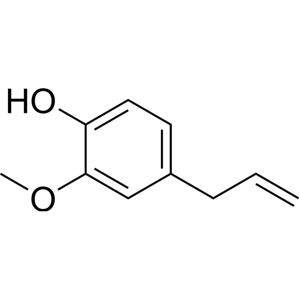
HY-N0337
Eugenol,98% (stabilized with TBC)Antioxidant -
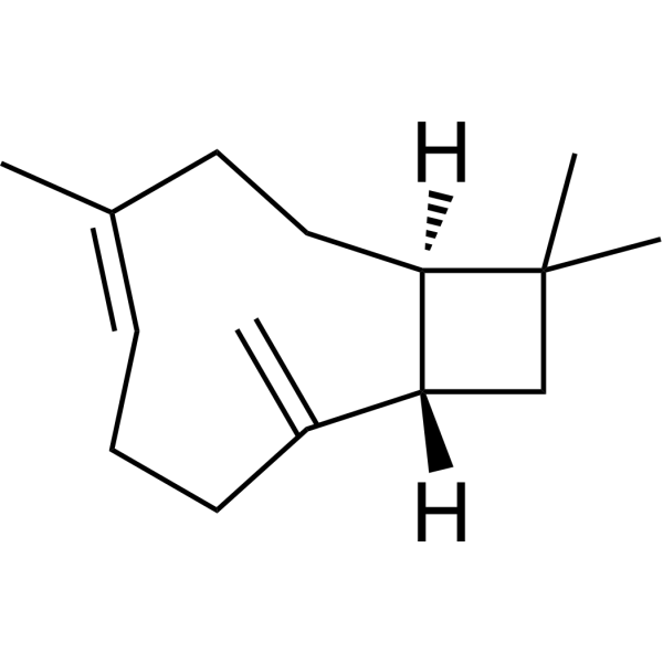
HY-N1415
β-CaryophylleneCannabinoid Receptor Agonist -
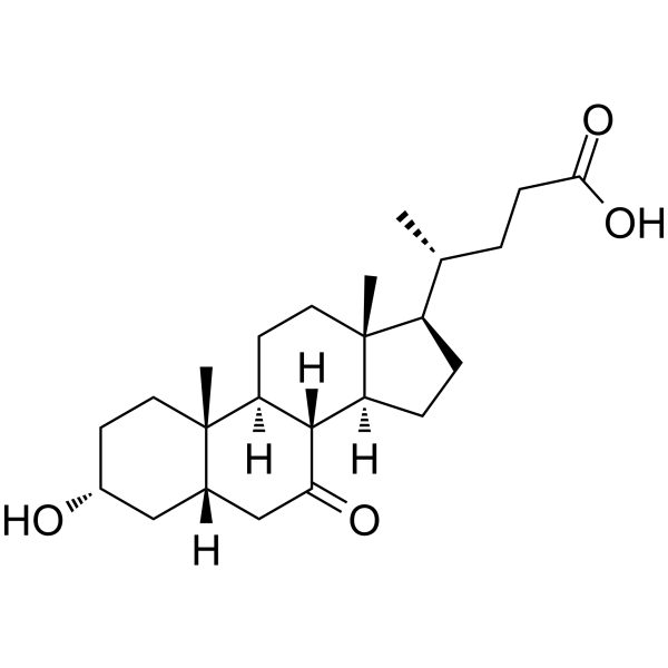
HY-W018512
7-Ketolithocholic acid -
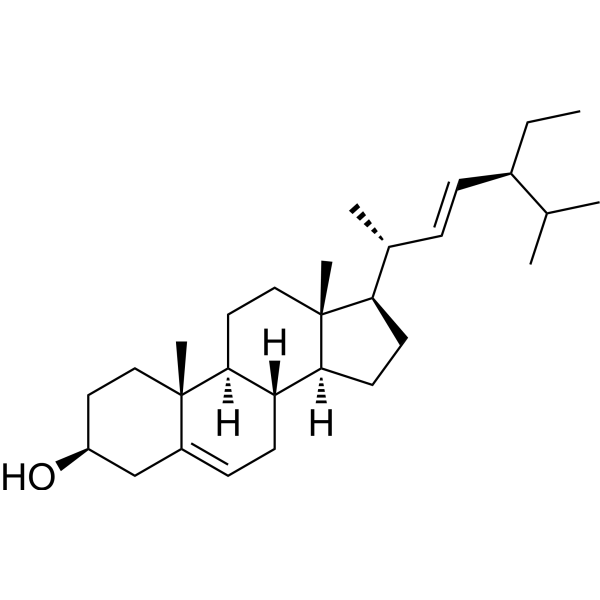
HY-N0131R
Stigmasterol (Standard)Standard of Stigmasterol -
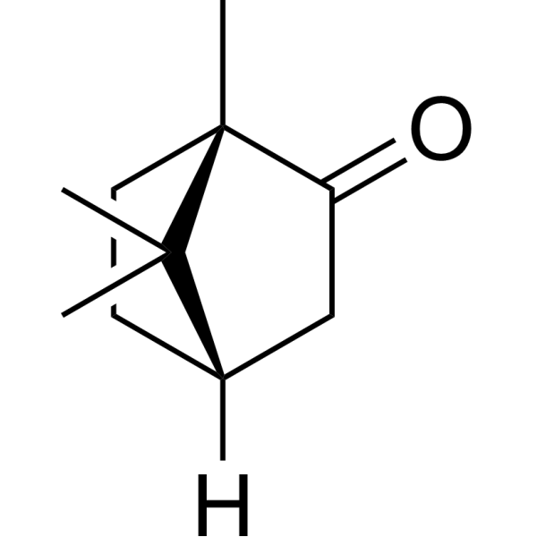
HY-W015579
(-)-CamphorEnantiome -
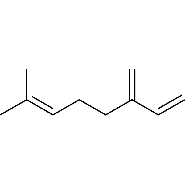
HY-N0803
Myrcene,75% (stabilized with BHT)NF-κB Inhibitor
Publications Citing Use of MedChemExpress (MCE) (+)-Camphor
More

Biological Activity
Description
Cellular Effect
|
Cell Line

|
Type | Value | Description | References |
|---|---|---|---|---|
| MDCK | EC50 |
1644.7 μM
Compound: 1
|
Antiviral activity against Influenza virus A/California/07/09 (H1N1)pdm09 infected in MDCK cells assessed as inhibition of viral replication incubated for 1 hr prior to viral infection measured after 48 hrs by hemagglutination reaction
Antiviral activity against Influenza virus A/California/07/09 (H1N1)pdm09 infected in MDCK cells assessed as inhibition of viral replication incubated for 1 hr prior to viral infection measured after 48 hrs by hemagglutination reaction
|
[PMID: 23993669] |
| MDCK | ED50 |
1644.7 μM
Compound: 1
|
Antiviral activity against rimantidine, amantadine-resistant Influenza A virus (A/California/07/09(H1N1)) pdm09 infected in MDCK cells assessed as inhibition of viral replication by hemagglutinin titer assay
Antiviral activity against rimantidine, amantadine-resistant Influenza A virus (A/California/07/09(H1N1)) pdm09 infected in MDCK cells assessed as inhibition of viral replication by hemagglutinin titer assay
|
[PMID: 24631360] |
In Vitro
(+)-Camphor and its derivatives exhibit antiproliferative and antimutagenic activities in human cancer cell culture experiments (e.g., YC8 lymphoma, melanoma, colon cancer, gastric cancer, liver cancer, breast cancer, and leukemia cells) by inhibiting ribosome function or inducing apoptosis.
(+)-Camphor leaf extract has a protective effect against DNA damage and chromosomal abnormalities induced by the herbicide Atrazine (HY-N7091) in vitro, reducing DNA damage in a time-dependent manner[1].
MedChemExpress (MCE) has not independently confirmed the accuracy of these methods. They are for reference only. Further protocols information, click here.


Chemical Information
-
CAS No. 464-49-3
-
Appearance Solid
-
Molecular Weight 152.24
-
Formula C10H16O
-
Color White to off-white
-
SMILES
CC1(C)[C@@]2(C)CC[C@@H]1CC2=O
-
Synonyms
D-(+)-Camphor; (1R)-(+)-Camphor
-
Structure Classification
-
Initial Source
-
Shipping
Room temperature in continental US; may vary elsewhere.
-
Storage
Powder -20°C 3 years 4°C 2 years In solvent -80°C 2 years -20°C 1 year


Publications (1)
-
Journal Impact Factor
-
Most Recent
-
Vet Microbiol
The Chinese medicine monomer Schisandrin C inhibits PRRSV infection by regulating the OGT-PI3K/AKT/mTOR signaling pathway. [Abstract]2026 May:316:110992. PMID: 41865607


Solvent & Solubility
In Vitro:
DMSO : ≥ 50 mg/mL (328.43 mM; Hygroscopic DMSO has a significant impact on the solubility of product, please use newly opened DMSO)
* "≥" means soluble, but saturation unknown.
Please refer to the solubility information to select the appropriate solvent. Once prepared, please aliquot and store the solution to prevent product inactivation from repeated freeze-thaw cycles.
Storage method and period of stock solution: -80°C, 2 years; -20°C, 1 year. When stored at -80°C, please use it within 2 years. When stored at -20°C, please use it within 1 year.
Please refer to the solubility information to select the appropriate solvent. Once prepared, please aliquot and store the solution to prevent product inactivation from repeated freeze-thaw cycles.
Storage method and period of stock solution: -80°C, 2 years; -20°C, 1 year. When stored at -80°C, please use it within 2 years. When stored at -20°C, please use it within 1 year.
Concentration (start) × Volume (start) = Concentration (final) × Volume (final)
In Vivo:
Select the appropriate dissolution method based on your experimental animal and administration route.
- For the following dissolution methods, please ensure to first prepare a clear stock solution using an In Vitro approach and then sequentially add co-solvents:
- To ensure reliable experimental results, the clarified stock solution can be appropriately stored based on storage conditions. As for the working solution for In Vivo experiments, it is recommended to prepare freshly and use it on the same day.
- The percentages shown for the solvents indicate their volumetric ratio in the final prepared solution. If precipitation or phase separation occurs during preparation, heat and/or sonication can be used to aid dissolution.
Add each solvent one by one: 10% DMSO 40% PEG300 5% Tween-80 45% Saline
Solubility: ≥ 2.5 mg/mL (16.42 mM); Clear solution
This protocol yields a clear solution of ≥ 2.5 mg/mL (saturation unknown).
Taking 1 mL working solution as an example, add 100 μL DMSO stock solution (25.0 mg/mL) to 400 μL PEG300, and mix evenly; then add 50 μL Tween-80 and mix evenly; then add 450 μL Saline to adjust the volume to 1 mL.
Preparation of Saline: Dissolve 0.9 g sodium chloride in ddH₂O and dilute to 100 mL to obtain a clear Saline solution.
Add each solvent one by one: 10% DMSO 90% (20% SBE-β-CD in Saline)
Solubility: ≥ 2.5 mg/mL (16.42 mM); Clear solution
This protocol yields a clear solution of ≥ 2.5 mg/mL (saturation unknown).
Taking 1 mL working solution as an example, add 100 μL DMSO stock solution (25.0 mg/mL) to 900 μL 20% SBE-β-CD in Saline, and mix evenly.
Preparation of 20% SBE-β-CD in Saline (4°C, storage for one week): 2 g SBE-β-CD powder is dissolved in 10 mL Saline, completely dissolve until clear.
In Vivo Dissolution Calculator
Please enter the basic information of animal experiments:
-
-
-
-
Recommended: Prepare an additional quantity of animals to account for potential losses during experiments.
Please enter your animal formula composition:
-
%DMSO +
Recommended: Keep the proportion of DMSO in working solution below 2% if your animal is weak.
-
%+
-
+%Tween-80 + +
-
%Saline +
The co-solvents required include: DMSO, . All of co-solvents are available by MedChemExpress (MCE). , Tween 80. All of co-solvents are available by MedChemExpress (MCE).
Working solution concentration: 0.22 mg/mL
Method for preparing stock solution: mg drug dissolved in μL DMSO. Stock solution concentration: mg/mL.
1. Take μL DMSO stock solution;
2. Add μL .
μL , mix evenly;
3. Then add μL Tween 80, mix evenly;
4. Then add μL
Please ensure that the stock solution in the first step is dissolved to a clear state, and add co-solvents in sequence. You can use ultrasonic heating (ultrasonic cleaner, recommended frequency 20-40 kHz), vortexing, etc. to assist dissolution.

Protocols
-
Research Protocol for Infectious Diseases
Infectious-disease experiments test how pathogens interact with host barriers, innate immune receptors, inflammatory signaling, pathogen replication, and tissue injury; pattern-recognition receptors such as TLRs, RIG-I-like receptors, NOD-like receptors, and inflammasomes detect microbial molecules and activate NF-κB, interferon, and cytokine responses. The central hypothesis is that infection severity reflects the balance between pathogen burden and host response: protective inflammation restricts pathogen growth, whereas excessive or mislocalized inflammation contributes to tissue damage and disease phenotype. Unresolved questions include which host pathways are protective versus pathogenic, why some infection models fail to translate to human disease, and which combined readouts best predict clinically relevant infection outcomes.
-
Apoptosis
Apoptosis, also called programmed cell death, is generally characterized by distinct morphological characteristics.
-
TUNEL staining for apoptotic DNA fragmentation
TUNEL staining detects DNA strand breaks by using terminal deoxynucleotidyl transferase to add labeled nucleotides to exposed 3′-OH DNA termini, generating either microscopic staining in fixed cells or tissue sections, or fluorescence/cytometric signal in cell suspensions. TUNEL positivity reflects DNA fragmentation but should not be interpreted alone as definitive apoptosis, because TUNEL can also label necrotic, autolytic, mechanically damaged, or DNA-repair-associated DNA breaks.
-
Annexin V plus membrane-impermeant dye apoptosis staining
Annexin V-based apoptosis assays rely on the detection of phosphatidylserine (PS) externalization from the inner leaflet of the plasma membrane to the outer leaflet, an early biochemical hallmark of apoptosis. Fluorescently labeled Annexin V binds PS in a calcium-dependent manner, enabling identification of early apoptotic cells by flow cytometry or fluorescence microscopy. When combined with a membrane-impermeant DNA-binding dye (e. g. , propidium iodide), this approach allows discrimination between viable (Annexin V−/dye−), early apoptotic (Annexin V+/dye−), and late apoptotic or necrotic (Annexin V+/dye+) cell populations by assessing membrane integrity and PS exposure.
-
Apoptosis Solutions
Apoptosis is a regulated, generally non-lytic cell-death pathway that removes unwanted, damaged, infected, or abnormal cells through coordinated morphological changes, caspase activation, DNA fragmentation, and membrane remodeling. The intrinsic apoptosis pathway is controlled mainly by mitochondrial outer membrane permeabilization, BCL-2 family proteins, cytochrome c release, apoptosome formation, caspase-9 activation, and downstream executioner caspase-3/7 activation. The extrinsic apoptosis pathway is initiated by death receptors such as Fas, TNFR, and TRAIL receptors, which recruit adaptor proteins and activate caspase-8 before engaging executioner caspases or mitochondrial amplification through BID cleavage. Apoptosis is linked to many phenotypes, including cancer cell killing, tissue homeostasis, immune regulation, neurodegeneration, infection response, and treatment-induced cytotoxicity; unresolved questions include how apoptosis interacts with necroptosis, pyroptosis, ferroptos
-
Research Protocol for Inflammation-related Diseases
The NLRP3 inflammasome is a cytosolic innate immune signaling platform that integrates priming signals and danger-signal activation to promote caspase-1 activation, maturation of IL-1β and IL-18, and gasdermin D-mediated pyroptotic cell death. The core experimental logic is to determine whether inflammatory disease phenotypes are driven by increased NLRP3 expression, ASC-containing inflammasome assembly, caspase-1 cleavage, GSDMD cleavage, and extracellular release of IL-1β/IL-18 rather than by nonspecific cell injury alone. The pathway is strongly linked to inflammation-related disease phenotypes because monosodium urate crystals activate NALP3/NLRP3 inflammasome signaling in gout-like crystal inflammation, cholesterol crystals activate NLRP3 inflammasomes in atherogenesis models, and DSS-induced intestinal inflammation has been reported to involve NLRP3 inflammasome activity. However, experimental colitis studies also show context-dependent protective effects of NLRP3 inflammasome co
-
Genotoxicity/Mutagenicity Study
The bacterial reverse mutation assay detects point mutations that restore amino-acid prototrophy in auxotrophic Salmonella typhimurium or Escherichia coli tester strains; after exposure to a test article, mutagenic activity is read out as an increased number of revertant colonies on minimal agar compared with the vehicle control. The assay uses tester strains with different mutation targets so that base-substitution and frameshift mutagens can be detected, and testing is performed with and without exogenous mammalian metabolic activation because some chemicals require biotransformation to become mutagenic.
-
Cell Viability Determination by MTT Colorimetric Assay
The following protocol uses the MTT colorimetric assay as a classic literature-established method for assessing cell viability/metabolic activity in cultured mammalian cells. MTT[3-(4,5-dimethylthiazol-2-yl)-2,5-diphenyltetrazolium bromide] is reduced by metabolically active cells to a colored formazan product; the amount of formazan is quantified spectrophotometrically and provides an indirect measure of metabolically active viable cells. Importantly, MTT reduction reflects cellular oxidoreductase/metabolic activity rather than an absolute direct count of living cells, so changes in cellular metabolism can alter the signal independently of cell number.


Purity & Documentation
-
Data Sheet (278 KB)
-
SDS (544 KB)
- English - EN (544 KB)
- Français - FR (544 KB)
- Deutsch - DE (544 KB)
- Norwegian - NO (544 KB)
- Español - ES (544 KB)
- Swedish - SV (544 KB)
- Italian - IT (544 KB)
- Korean - KR (544 KB)
- Portuguese - PT (544 KB)
-
Handling Instructions (2659 KB)
References


Complete Stock Solution Preparation Table
Please refer to the solubility information to select the appropriate solvent. Once prepared, please aliquot and store the solution to prevent product inactivation from repeated freeze-thaw cycles.
Storage method and period of stock solution: -80°C, 2 years; -20°C, 1 year. When stored at -80°C, please use it within 2 years. When stored at -20°C, please use it within 1 year.
| Optional Solvent | Concentration Solvent Mass | 1 mg | 5 mg | 10 mg | 25 mg |
|---|---|---|---|---|---|
| DMSO | 1 mM | 6.5686 mL | 32.8429 mL | 65.6858 mL | 164.2144 mL |
| 5 mM | 1.3137 mL | 6.5686 mL | 13.1372 mL | 32.8429 mL | |
| 10 mM | 0.6569 mL | 3.2843 mL | 6.5686 mL | 16.4214 mL | |
| 15 mM | 0.4379 mL | 2.1895 mL | 4.3791 mL | 10.9476 mL | |
| 20 mM | 0.3284 mL | 1.6421 mL | 3.2843 mL | 8.2107 mL | |
| 25 mM | 0.2627 mL | 1.3137 mL | 2.6274 mL | 6.5686 mL | |
| 30 mM | 0.2190 mL | 1.0948 mL | 2.1895 mL | 5.4738 mL | |
| 40 mM | 0.1642 mL | 0.8211 mL | 1.6421 mL | 4.1054 mL | |
| 50 mM | 0.1314 mL | 0.6569 mL | 1.3137 mL | 3.2843 mL | |
| 60 mM | 0.1095 mL | 0.5474 mL | 1.0948 mL | 2.7369 mL | |
| 80 mM | 0.0821 mL | 0.4105 mL | 0.8211 mL | 2.0527 mL | |
| 100 mM | 0.0657 mL | 0.3284 mL | 0.6569 mL | 1.6421 mL |


(+)-Camphor Related Classifications
HY-B1173 Related Classifications
 Powered by Bioz