



β-Amyloid (1-16)

Based on 1 Customer Validation
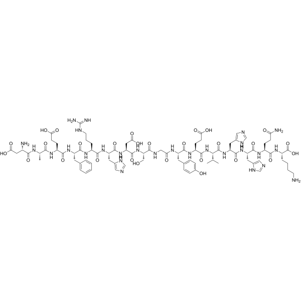
β-Amyloid (1-16) is a β-Amyloid protein fragment involved in metal binding. Beta-amyloid is a peptide that forms amyloid plaques in the brains of Alzheimer's disease (AD) patients.
For research use only. We do not sell to patients.
- Purity : 99.98%
- CAS No.: 131580-10-4
- Formula: C84H119N27O28
- Molecular Weight:1955.01
-
Storage:
Sealed storage, away from moisture.
Powder -80°C, 2 years , -20°C, 1 year* The compound is unstable in solutions, freshly prepared is recommended.

To place orders, for customer services and technical support, please contact: MedChemExpress USA
Tel: 609-228-6898 E-mail: [email protected] [email protected]


-
 Biological Activity
-
Chemical Information
- Protocols
- Purity & Documentation
- References
-
 Help & FAQs


-
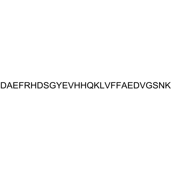
HY-P1468
β-Amyloid (1-28)Amyloid-β Peptide -
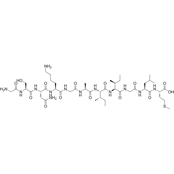
HY-P0128
β-Amyloid (25-35)Amyloid β Peptide -
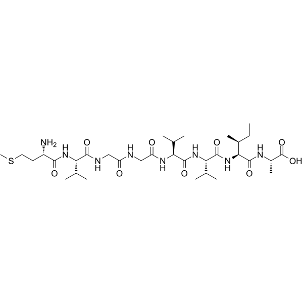
HY-P1903
β-Amyloid (35-42)Peptide Fragment -

HY-P1363A
β-Amyloid (1-42), humanAmyloid β Peptide -

HY-P1363B
β-Amyloid (1-42), human, HFIP-treatedβ-Amyloid (1-42) Peptide -
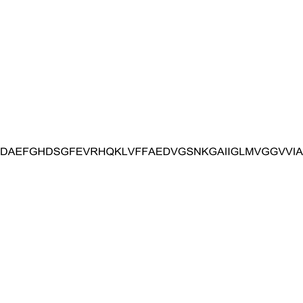
HY-P1388
β-Amyloid (1-42), (rat/mouse)β-Amyloid (1-42) Peptide -
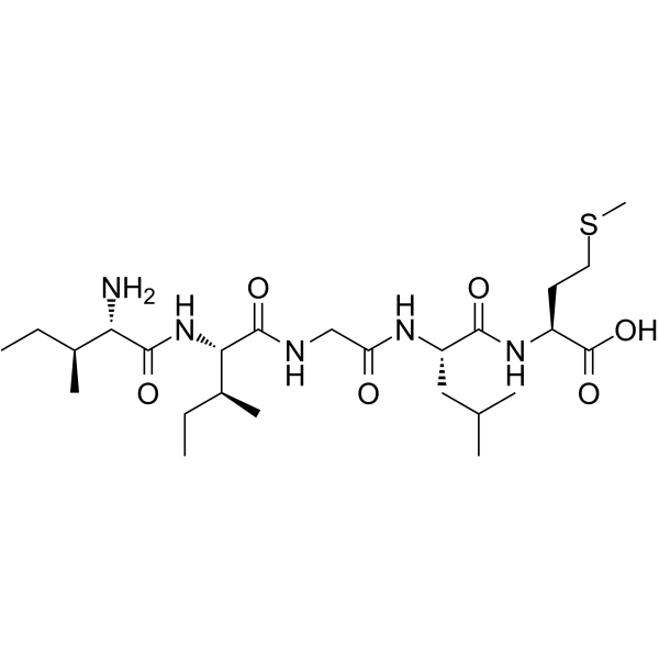
HY-P1517
β-Amyloid (31-35)β-Amyloid Peptide Fragment -
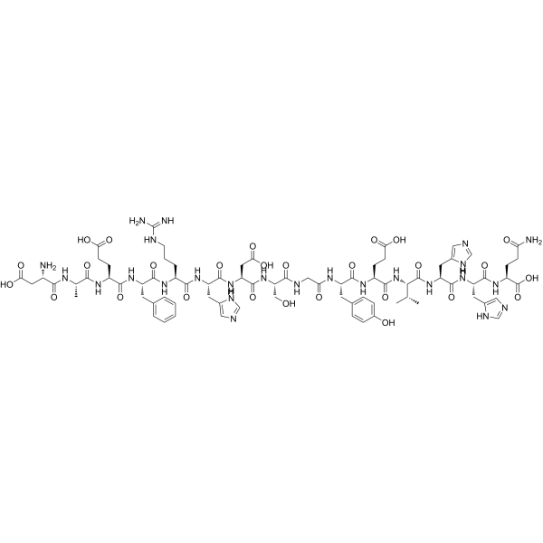
HY-P1046
β-Amyloid (1-15)Peptide Fragment -
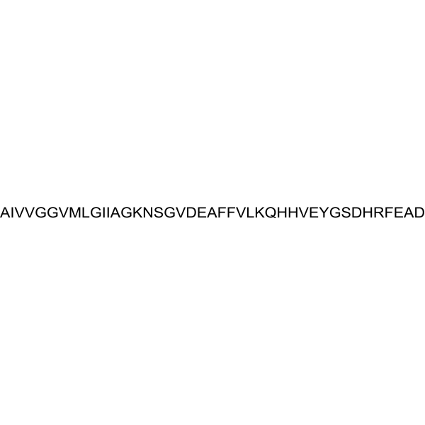
HY-P1362
β-Amyloid (42-1), humanInactive Form of Aβ(1-42) -
HY-N0615
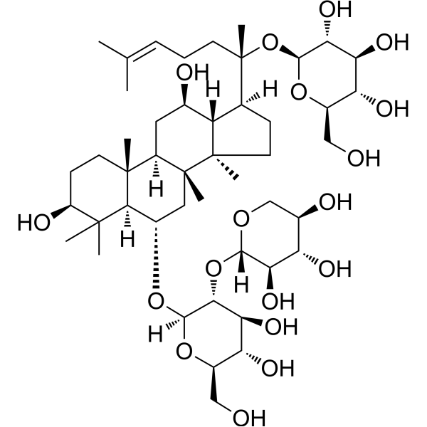
Notoginsenoside R1Apoptosis Inhibitor -
HY-114174
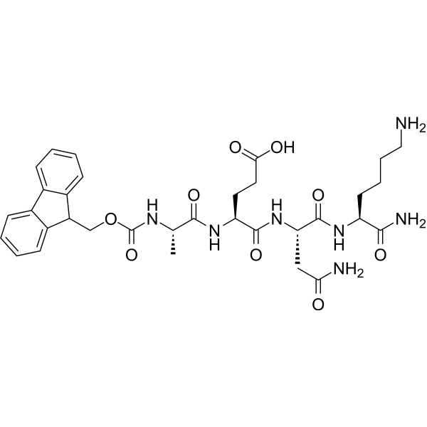
Fmoc-Ala-Glu-Asn-Lys-NH2AEP Inhibitor -

HY-P1474
β-Amyloid (22-35)β-Amyloid


Biological Activity
Description
IC50 & Target
Amyloid-β[1]
In Vivo
MedChemExpress (MCE) has not independently confirmed the accuracy of these methods. They are for reference only.


Chemical Information
-
CAS No. 131580-10-4
-
Appearance Solid
-
Molecular Weight 1955.01
-
Formula C84H119N27O28
-
Color White to off-white
-
Synonyms
Amyloid β-Protein (1-16)
-
Sequence
Asp-Ala-Glu-Phe-Arg-His-Asp-Ser-Gly-Tyr-Glu-Val-His-His-Gln-Lys
-
Sequence Shortening
DAEFRHDSGYEVHHQK
-
Shipping
Room temperature in continental US; may vary elsewhere.
-
Storage
Sealed storage, away from moisture
Powder -80°C 2 years -20°C 1 year * The compound is unstable in solutions, freshly prepared is recommended.

Protocols
-
Amyloid: Congo Red Amyloid Staining
Congo red amyloid staining is a histochemical method used to detect extracellular amyloid deposits in tissue sections based on the affinity of Congo red dye for β-pleated sheet-rich protein aggregates. When bound to amyloid, Congo red produces characteristic apple-green birefringence under polarized light microscopy, which is widely regarded as a diagnostic feature of amyloid deposition in histopathology. The diagnostic principle relies on the combination of dye binding (congophilia) and optical anisotropy under polarized illumination, which distinguishes amyloid from most non-amyloid eosinophilic extracellular deposits in routine histological evaluation. Amyloid identification by Congo red staining remains a cornerstone in diagnostic pathology despite the availability of adjunct methods such as immunohistochemistry and mass spectrometry, particularly because of its ability to localize deposits directly within tissue architecture. The specificity of Congo red-positive deposits is incre
-
Alzheimer’s Disease Modeling
Alzheimer’s Disease (AD) is a neurodegenerative disorder characterized by a progressive decline in cognitive functions and loss of specific types of neurons and synapses. Alzheimer's symptoms can be simulated in mice by injecting drugs (such as Aβ) or genetically modified.


Purity & Documentation
-
Data Sheet (263 KB)
-
SDS (252 KB)
- English - EN (252 KB)
- Français - FR (252 KB)
- Deutsch - DE (252 KB)
- Norwegian - NO (252 KB)
- Español - ES (252 KB)
- Swedish - SV (252 KB)
- Italian - IT (252 KB)
- Korean - KR (252 KB)
- Portuguese - PT (252 KB)
-
Handling Instructions (2659 KB)
References
[1]. Kowalik-Jankowska T, et al. Coordination abilities of the 1-16 and 1-28 fragments of beta-amyloid peptide towards copper(II) ions: a combined potentiometric and spectroscopic study. J Inorg Biochem. 2003 Jul 1;95(4):270-82. [Content Brief]
[2]. Minicozzi V, et al. Identifying the minimal copper- and zinc-binding site sequence in amyloid-beta peptides. J Biol Chem. 2008 Apr 18;283(16):10784-92. [Content Brief]
[3]. Istrate AN, et al. NMR solution structure of rat aβ(1-16): toward understanding the mechanism of rats' resistance to Alzheimer's disease. Biophys J. 2012 Jan 4;102(1):136-43. [Content Brief]

Calculators
Concentration (start) × Volume (start) = Concentration (final) × Volume (final)


β-Amyloid (1-16) Related Classifications
HY-P1466 Related Classifications
 Powered by Bioz