



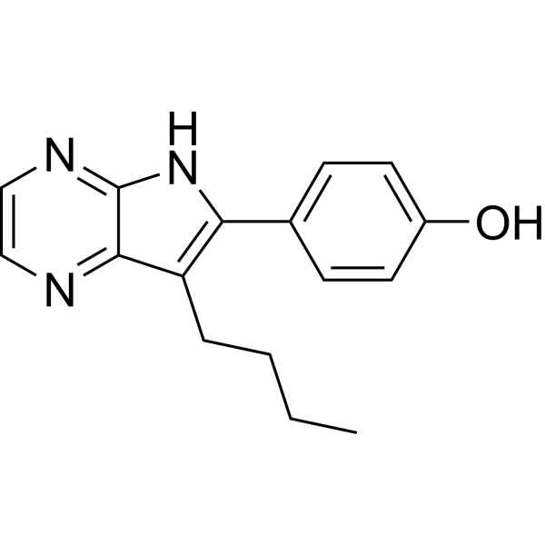
Aloisine A
Aloisine A (RP107) is a a potent cyclin-dependent kinase (CDK) inhibitor with IC50s of 0.15 μM, 0.12 μM, 0.4 μM, 0.16 μM for CDK1/cyclin B, CDK2/cyclin A, CDK2/cyclin E, CDK5/p35, respectively. Aloisine A ininhibits GSK-3α (IC50=0.5 μM) and GSK-3β (IC50=1.5 μM). Aloisine A stimulates wild-type CFTR and mutated CFTR, with submicromolar affinity by a cAMP-independent mechanism. Aloisine A has the potential for CFTR-related diseases, including cystic fibrosis research.
For research use only. We do not sell to patients.
- CAS No.: 496864-16-5
- Formula: C16H17N3O
- Molecular Weight:267.33
-
Storage:
Please store the product under the recommended conditions in the Certificate of Analysis.

To place orders, for customer services and technical support, please contact: MedChemExpress USA
Tel: 609-228-6898 E-mail: [email protected] [email protected]


-
 Biological Activity
-
Chemical Information
- Protocols
- Purity & Documentation
- References
-
 Help & FAQs




Biological Activity
Description
IC50 & Target
[1]|
CDK1/cyclinB 0.15 μM (IC50) |
CDK2/cyclinA 0.12 μM (IC50) |
CDK2/cyclinE 0.4 μM (IC50) |
CDK5/p35 0.16 μM (IC50) |
GSK-3β 0.5 μM (IC50) |
GSK-3α 1.5 μM (IC50) |
JNK 3-10 μM (IC50) |
ERK1 18 μM (IC50) |
ERK2 22 μM (IC50) |
Cellular Effect
|
Cell Line

|
Type | Value | Description | References |
|---|---|---|---|---|
| Calu-3 | EC50 |
140 nM
Compound: K00059, Aloisine
|
Activity at human wild type CFTR expressed in Calu3 cells assessed as forskolin stimulated iodide flux
Activity at human wild type CFTR expressed in Calu3 cells assessed as forskolin stimulated iodide flux
|
[PMID: 18077363] |
| CHO | EC50 |
150 nM
Compound: K00059, Aloisine
|
Activity at human wild type CFTR expressed in CHO cells assessed as forskolin stimulated iodide flux
Activity at human wild type CFTR expressed in CHO cells assessed as forskolin stimulated iodide flux
|
[PMID: 18077363] |
| NT2 | IC50 |
10500 nM
Compound: K00059, Aloisine
|
Cell cycle arrest in human NT2 cells assessed as accumulation at G2 phase
Cell cycle arrest in human NT2 cells assessed as accumulation at G2 phase
|
[PMID: 18077363] |
| NT2 | IC50 |
7 μM
Compound: K00059, Aloisine
|
Cell cycle arrest in human NT2 cells assessed as accumulation at G1 phase
Cell cycle arrest in human NT2 cells assessed as accumulation at G1 phase
|
[PMID: 18077363] |
| Sf9 | IC50 |
0.65 μM
Compound: 39
|
Inhibition of glycogen synthase kinase-3 beta, purified from porcine brain or expressed in and purified from insect Sf9 cells
Inhibition of glycogen synthase kinase-3 beta, purified from porcine brain or expressed in and purified from insect Sf9 cells
|
[PMID: 12519061] |
In Vitro
Aloisine A ininhibits erk1 (IC50=18 µM), erk2 (IC50=22 µM), c-Jun N-terminal kinase (JNK; IC50~3-10 µM). Aloisine A has no effect on protein kinase C α, β1, β2, γ, δ, ε, η, ξ (all IC50>100 µM)[1].
Aloisine A (0.1, 1, 10, 100 µM) completely blocks the proliferation of dividing NT2 cells (IC50=7 μM) and differentiated postmitotic neurons (hNT; IC50=10.5 μM), and very few cells actually die[1].
Aloisine A acts as a submicromolar activator of wild-type (WT)-CFTR [human airway epithelial Calu-3 and WT-CFTR-Chinese hamster ovary (CHO) cells], G551D-CFTR (G551D-CFTR-CHO cells), and F508del-CFTR (in temperature-corrected human airway epithelial F508del/F508del CF15 cells)[2].
MedChemExpress (MCE) has not independently confirmed the accuracy of these methods. They are for reference only.


Chemical Information
-
CAS No. 496864-16-5
-
Molecular Weight 267.33
-
Formula C16H17N3O
-
SMILES
OC1=CC=C(C2=C(CCCC)C3=NC=CN=C3N2)C=C1
-
Synonyms
RP107
-
Shipping
Room temperature in continental US; may vary elsewhere.
-
Storage
Please store the product under the recommended conditions in the Certificate of Analysis.


Purity & Documentation
References
[1]. Yvette Mettey, et al. Aloisines, a new family of CDK/GSK-3 inhibitors. SAR study, crystal structure in complex with CDK2, enzyme selectivity, and cellular effects. J Med Chem. 2003 Jan 16;46(2):222-36. [Content Brief]
[2]. Sabrina Noel, et al. Discovery of pyrrolo[2,3-b]pyrazines derivatives as submicromolar affinity activators of wild type, G551D, and F508del cystic fibrosis transmembrane conductance regulator chloride channels. J Pharmacol Exp Ther. 2006 Oct;319(1):349-59. [Content Brief]

Calculators
Concentration (start) × Volume (start) = Concentration (final) × Volume (final)


Aloisine A Related Classifications
HY-112363 Related Classifications
 Powered by Bioz