



Anti-Mouse Pan-endothelial Cell Antigen/MECA-32 Antibody (MECA-32)
Anti-Mouse Pan-endothelial Cell Antigen/MECA-32 Antibody (MECA-32) is an anti-mouse Pan-endothelial Cell Antigen/MECA-32 IgG2a monoclonal antibody. Anti-Mouse Pan-endothelial Cell Antigen/MECA-32 Antibody (MECA-32) can label endothelial cells. Anti-Mouse Pan-endothelial Cell Antigen/MECA-32 Antibody (MECA-32) is often used for immunohistochemistry, delivery of siRNA and immunofluorescence.
For research use only. We do not sell to patients.
- Molecular Weight:150 kDa
-
Storage:
Please store the product under the recommended conditions in the Certificate of Analysis.

To place orders, for customer services and technical support, please contact: MedChemExpress USA
Tel: 609-228-6898 E-mail: [email protected] [email protected]


-
 Biological Activity
-
Chemical Information
- Protocols
- Purity & Documentation
- References
-
 Help & FAQs




Biological Activity
Description
Isotype
Rat IgG2a kappa
Recommend Isotype Controls
Species Reactivity
Mouse
Conjugated
Unconjugated
Reconsititution
The product can be reconstituted/diluted with sterile PBS or saline.
Format
-
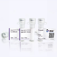
Product Image
Application
Immunohistochemistry (frozen); Flow cytometry; Western blot; Immunofluorescence


Chemical Information
-
Molecular Weight 150 kDa
-
SMILES
[Anti-Mouse Pan-endothelial Cell Antigen/MECA-32 Antibody (MECA-32)]
-
Formulation
Please refer to the lot-specific COA for specific buffer information.
-
Storage
Please store the product under the recommended conditions in the Certificate of Analysis.

Protocols
-
Polymer-Based Two-Step IHC Detection
Polymer-based two-step IHC detects tissue antigens by first binding an unlabeled primary antibody to the antigen and then applying an HRP-polymer secondary reagent that carries multiple secondary antibodies and HRP molecules on a polymer backbone; the localized HRP converts chromogens such as DAB or AEC into visible deposits for light-microscopic interpretation. The method is \"two-step\" because the primary antibody step is followed directly by the polymer-enzyme secondary reagent, rather than by separate secondary-antibody and avidin-biotin complex steps; published comparisons reported similar or higher sensitivity than several multistep systems and avoidance of endogenous-biotin interference.
-
Avidin-Biotin/Streptavidin-Biotin IHC
Avidin-biotin or streptavidin-biotin immunohistochemistry detects tissue antigens by binding a primary antibody to the target antigen, then detecting that antibody with a biotinylated antibody and an avidin-biotin-enzyme or streptavidin-enzyme detection complex; the enzyme reaction produces a visible chromogenic deposit at the antigen site for light-microscopic localization. The classic ABC method uses the high-affinity avidin-biotin interaction to bridge biotinylated secondary antibody and biotinylated peroxidase, and early comparative studies reported stronger immunoperoxidase staining than PAP-based methods in formalin-fixed tissue sections.
-
Antibody-based immunofluorescence/immunocytochemistry staining
Antibody-based immunofluorescence/immunocytochemistry detects the cellular or subcellular localization of a target antigen by binding a primary antibody to the target and detecting that antibody directly with a fluorophore-conjugated primary antibody or indirectly with a fluorophore-conjugated secondary antibody. Indirect immunofluorescence can amplify signal because multiple secondary antibodies can bind one primary antibody. The assay readout is fluorescence intensity and localization measured by fluorescence or confocal microscopy, and the result reflects antigen distribution only when the antibody has been validated for the target, sample type, fixation condition, and imaging workflow. Antibody specificity must not be assumed from catalog information alone, and appropriate validation or control experiments are required for serious interpretation.
- Immunocytochemistry/Immunofluorescence
-
Endothelial Tube Formation Assay
Endothelial tube formation assay evaluates the ability of endothelial cells to attach, migrate, align, and organize into capillary-like networks when cultured on gelled basement membrane extract or Matrigel; the readout is the morphology and quantity of tube-like networks, which reflects an in vitro endothelial morphogenesis step related to angiogenesis. Basement membrane extract/Matrigel provides laminin-rich extracellular matrix cues that support endothelial differentiation into capillary-like structures, but it can contain biologically active growth factors, so growth-factor-reduced matrix is preferred when testing defined angiogenic stimulators or inhibitors.
-
RNA interference technology
RNA interference (RNAi) is a cellular mechanism that inhibits gene expression by suppressing gene transcription or activating RNA degradation. This mechanism was discovered in plants in 1998 by Andrew Fire and Craig Mello. Today, this phenomenon can be observed in almost all eukaryotes, including protozoa, flies, nematodes, insects, parasites, and mammals.


Purity & Documentation
References
[1]. Volk-Draper L, et al. Paclitaxel therapy promotes breast cancer metastasis in a TLR4-dependent manner. Cancer Res. 2014 Oct 1;74(19):5421-34. [Content Brief]
[2]. Florence JM, et al. Disrupting the Btk Pathway Suppresses COPD-Like Lung Alterations in Atherosclerosis Prone ApoE-/- Mice Following Regular Exposure to Cigarette Smoke. Int J Mol Sci. 2018 Jan 24;19(2):343. [Content Brief]
[3]. Meegan JE, et al. Blocking P2X7 receptor with AZ 10606120 exacerbates vascular hyperpermeability and inflammation in murine polymicrobial sepsis. Physiol Rep. 2022 Jun;10(11):e15290. [Content Brief]

Calculators
Concentration (start) × Volume (start) = Concentration (final) × Volume (final)


Anti-Mouse Pan-endothelial Cell Antigen/MECA-32 Antibody (MECA-32) Related Classifications
HY-P990174 Related Classifications
 Powered by Bioz