



Antibacterial agent 154
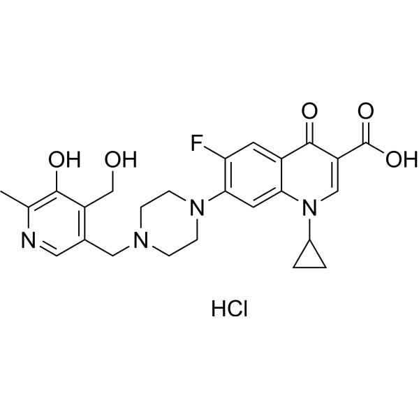
Antibacterial agent 154 (compound 7) is a derivative of Fluoroqinolones and is an orally effective antibacterial agent. Antibacterial agent 154 inhibits Gram-positive and Gram-negative bacteria. Antibacterial agent 154 demonstrated in vivo efficacy in a mouse model of staphylococcal sepsis.
For research use only. We do not sell to patients.
- CAS No.: 2163048-45-9
- Formula: C25H28ClFN4O5
- Molecular Weight:518.97
-
Storage:
Please store the product under the recommended conditions in the Certificate of Analysis.

To place orders, for customer services and technical support, please contact: MedChemExpress USA
Tel: 609-228-6898 E-mail: [email protected] [email protected]


-
 Biological Activity
-
Chemical Information
- Protocols
- Purity & Documentation
- References
-
 Help & FAQs




Biological Activity
Description
In Vitro
Antibacterial agent 154 (compound 7) inhibits Gram-positive bacteria M. luteus, B. subtilis, S. aureus, and S. epidermidis. The MICs are 4 μg/mL, 2 μg/mL, 2 μg/mL, and 2 μg/ mL; the MICs for inhibiting Gram-negative bacteria P. aerugenosa, E. coli, and S. typhimurium are 2 μg/mL and 2 μg/mL respectively[1].
MedChemExpress (MCE) has not independently confirmed the accuracy of these methods. They are for reference only. Further protocols information, click here.


Chemical Information
-
CAS No. 2163048-45-9
-
Molecular Weight 518.97
-
Formula C25H28ClFN4O5
-
SMILES
OC1=C(C)N=CC(CN2CCN(C3=C(F)C=C4C(N(C5CC5)C=C(C(O)=O)C4=O)=C3)CC2)=C1CO.Cl
-
Shipping
Room temperature in continental US; may vary elsewhere.
-
Storage
Please store the product under the recommended conditions in the Certificate of Analysis.

Protocols
-
Research Protocol for Infectious Diseases
Infectious-disease experiments test how pathogens interact with host barriers, innate immune receptors, inflammatory signaling, pathogen replication, and tissue injury; pattern-recognition receptors such as TLRs, RIG-I-like receptors, NOD-like receptors, and inflammasomes detect microbial molecules and activate NF-κB, interferon, and cytokine responses. The central hypothesis is that infection severity reflects the balance between pathogen burden and host response: protective inflammation restricts pathogen growth, whereas excessive or mislocalized inflammation contributes to tissue damage and disease phenotype. Unresolved questions include which host pathways are protective versus pathogenic, why some infection models fail to translate to human disease, and which combined readouts best predict clinically relevant infection outcomes.
-
LPS-Induced Endotoxemia/Systemic Inflammation
Lipopolysaccharide (LPS)-induced endotoxemia is a widely used in vivo model of acute systemic inflammation in which LPS, a Gram-negative bacterial endotoxin, activates innate immune signaling primarily through TLR4, leading to rapid and transient induction of pro-inflammatory cytokines such as TNF-α, IL-6, and IL-1β in circulation and tissues. This cytokine surge is commonly used as a measurable readout of systemic inflammatory activation and immune dysregulation, and is typically assessed within hours after intraperitoneal LPS administration in mouse models of endotoxemia. The model captures key features of systemic inflammatory response syndrome, including cytokine release, immune cell activation, and downstream tissue responses, and has been used to evaluate anti-inflammatory interventions such as cytokine modulation, lipid mediators, and immune cell-targeting therapies.
-
Gram Staining of Tissue Sections
Gram staining of tissue sections is a histochemical technique used to differentiate Gram-positive and Gram-negative bacteria within histological specimens based on differences in bacterial cell wall structure and dye retention, adapted from classical bacteriological Gram staining into tissue-compatible “histological Gram stain” variants. In tissue applications, modifications of the Brown-Hopps and Brown-Brenn methods are commonly used to improve differentiation of microorganisms embedded within host connective tissue and to reduce overstaining or loss of Gram-negative signal, which are known limitations of earlier approaches. The principle relies on crystal violet-iodine complex retention in Gram-positive organisms and subsequent decolorization and counterstaining steps that allow contrast visualization of Gram-negative organisms against tissue background.
-
How to Choose the Right Model Animal
Choosing the right model animal is a validity-driven decision in which the species, strain, sex, age, genetic background, disease-induction method, outcome measures, and welfare burden must match the scientific question rather than laboratory tradition or convenience. A model should be selected by judging face validity, construct validity, and predictive validity: whether it resembles the human phenotype, whether it reproduces relevant mechanisms, and whether results are likely to predict human biology or treatment response. Animal studies often fail to translate because of species differences, weak disease resemblance, poor experimental design, inadequate reporting, publication bias, and underuse of randomization, blinding, and sample-size justification. Unresolved questions include how to rank competing models objectively, how much human-disease complexity must be reproduced for a given objective, and when non-animal systems such as organoids, ex vivo tissue, or computational models


Purity & Documentation
References

Calculators
Concentration (start) × Volume (start) = Concentration (final) × Volume (final)


Antibacterial agent 154 Related Classifications
HY-155250 Related Classifications
 Powered by Bioz