



FOXP3 Antibody (YA858)
(Synonyms: FOXP3; IPEX; JM2; Forkhead box protein P3; Scurfin)
Based on 1 Customer Validation
FOXP3 Antibody (YA858) is a Rabbit-derived and non-conjugated IgG monoclonal antibody, targeting to FOXP3.
-
Host:
Rabbit
-
Isotype:
IgG
-
Application:
WB, IHC-P
-
Reactivity :
Human
-
Formulation:
Supplied in Rabbit IgG in 10mM phosphate buffered saline , pH 7.4, 150mM sodium chloride, 0.05% BSA, 0.02% sodium azide and 50% glycerol.
-
Conjugation:
Non-conjugated


To place orders, for customer services and technical support, please contact: MedChemExpress USA
Tel: 609-228-6898 E-mail: [email protected] [email protected]





Applications
| Application |
WB

WB: Western Blot
|
IHC-P
IHC-P: Immunohistochemistry-Paraffin
|
|---|---|---|
| Dilution Ratio | 1:500-1:1000 | 1:50-1:100 |


Product Details
FOXP3 Antibody (YA858) is a Rabbit-derived and non-conjugated IgG monoclonal antibody, targeting to FOXP3.
-
Host Rabbit

-
Clonality Recombinant,Monoclonal

-
Species ReactivityHuman
-
Observed Molecular WeightObserved band size: 47 kDa
Note: Due to possible protein modifications or aggregation, the molecular weight should be confirmed by actual measurement, and the predicted value is for reference only.

-
Calculated Molecular Weight Predicted band size: 47 kDa

A synthesized peptide derived from human FOXP3 aa250-431.
Endogenous
Non-conjugated
Unmodified
IgG


Product Properties
-
Appearance
Solution
-
Formulation
Supplied in Rabbit IgG in 10mM phosphate buffered saline , pH 7.4, 150mM sodium chloride, 0.05% BSA, 0.02% sodium azide and 50% glycerol.
-
Concentration
Batch-dependent, Please check the COA for the concentration of each lot. Check Lot Concentration
-
Storage & Stability
Stored at -20°C for 1 year. Avoid repeated freeze / thaw cycles.
-
Shipping
Shipping with blue ice.


Verification Images
-
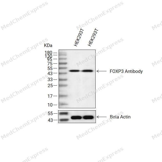 Western blot analysis of extracts from HEK293T(lane 2(20ug) and HEK293T(lane 3(40ug) using FOXP3 Antibody (HY-P80965) Rabbit mAb. Proteins were transferred to a PVDF membrane and blocked with 5% non-fat milk in TBST for 2 hour at room temperature. The primary antibody (1/1000) and Loading control antibody (Beta Actin, HY-P83730, 1/10000) was used in 5% non-fat milk in TBST at 4°C overnight. Goat Anti-Mouse/Rabbit IgG-HRP Secondary Antibody (1/10000) was used for 1 hour at room temperature.
 -
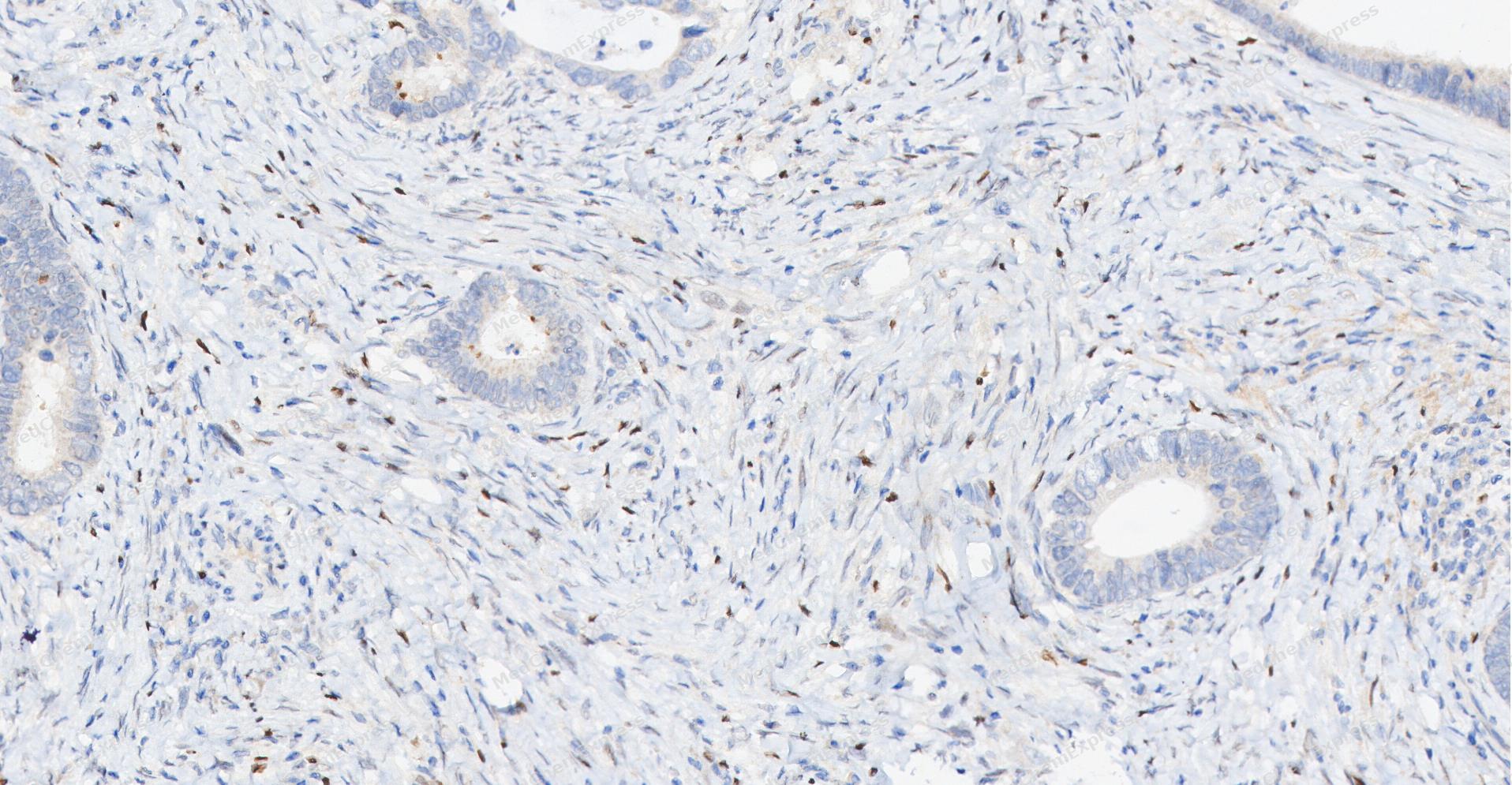 Immunohistochemical analysis of paraffin-embedded human Colon cancer tissue using FOXP3 antibody was performed. The section was pretreated using high-temperature and high-pressure mediated EDTA antigen retrieval buffer (pH 9.0), for 5 minutes. The tissues were incubated with primary antibody (HY-P80965, 1:100 dilution) at room temperature for 60 minutes. Detection was performed using an HRP conjugated compact polymer system. DAB was used as the chromogen. The tissues were counterstained with hematoxylin and mounted with neutral balsam mounting medium.
 -
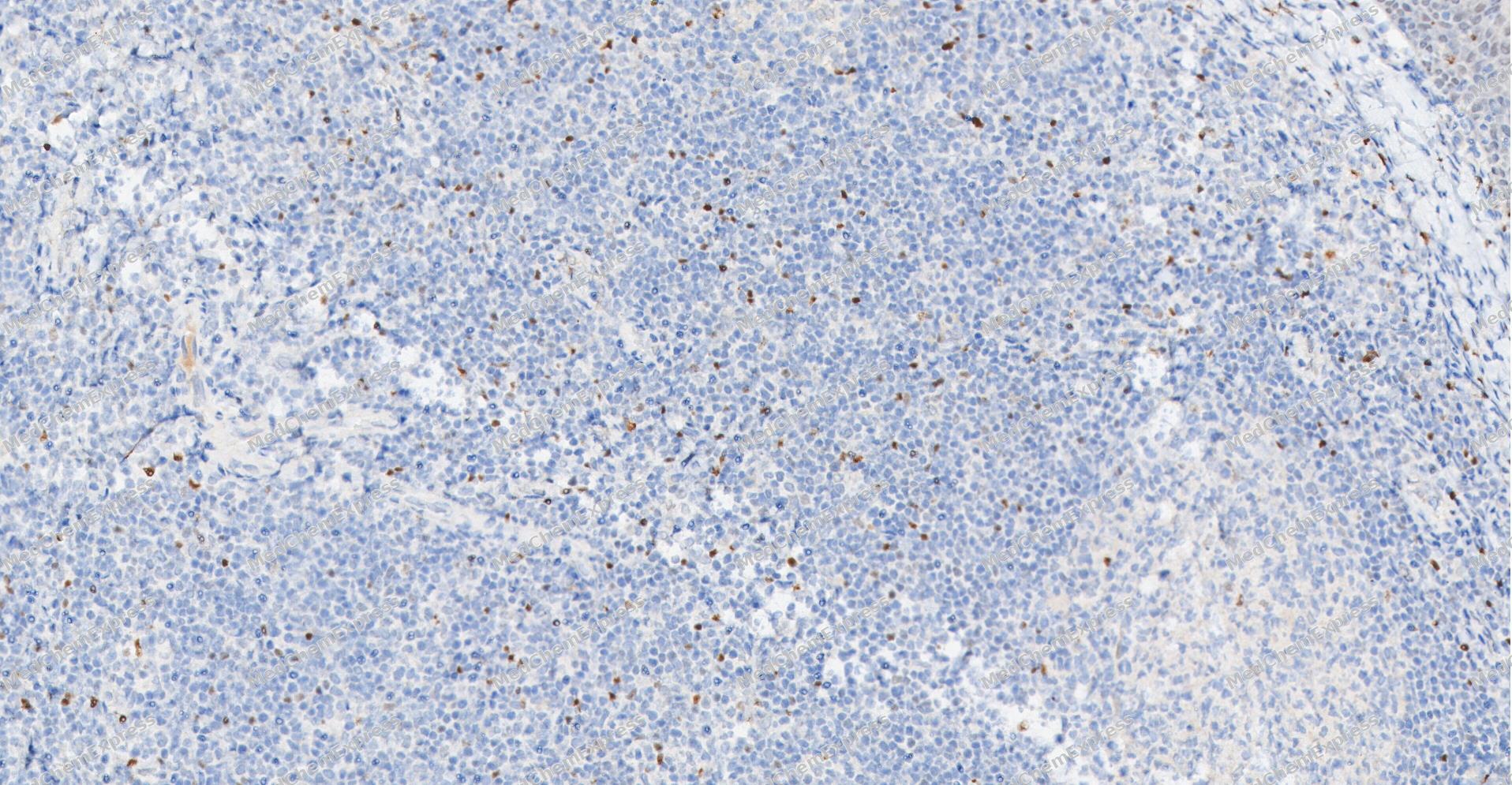 Immunohistochemical analysis of paraffin-embedded human Tonsil tissue using FOXP3 antibody was performed. The section was pretreated using high-temperature and high-pressure mediated EDTA antigen retrieval buffer (pH 9.0), for 5 minutes. The tissues were incubated with primary antibody (HY-P80965, 1:100 dilution) at room temperature for 60 minutes. Detection was performed using an HRP conjugated compact polymer system. DAB was used as the chromogen. The tissues were counterstained with hematoxylin and mounted with neutral balsam mounting medium.
 -
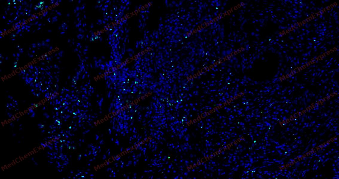 Tyramide signaling amplification based immunofluorescence was performed on paraffin-embedded human Lung Adenocarcinoma tissue using FOXP3 antibody. Antigen retrieval was performed in EDTA buffer pH 9.0 (95 °C, 20 min) followed by cooling to RT. Then incubated with primary antibody (HY-P80965, 1:100 dilution) at room temperature for 60 minutes and HRP conjugated secondary antibody for 10 minutes. Fluorescence was then developed with TSA520. The tissues were counterstained with DAPI and mounted with Anti-fade mounting medium.
 -
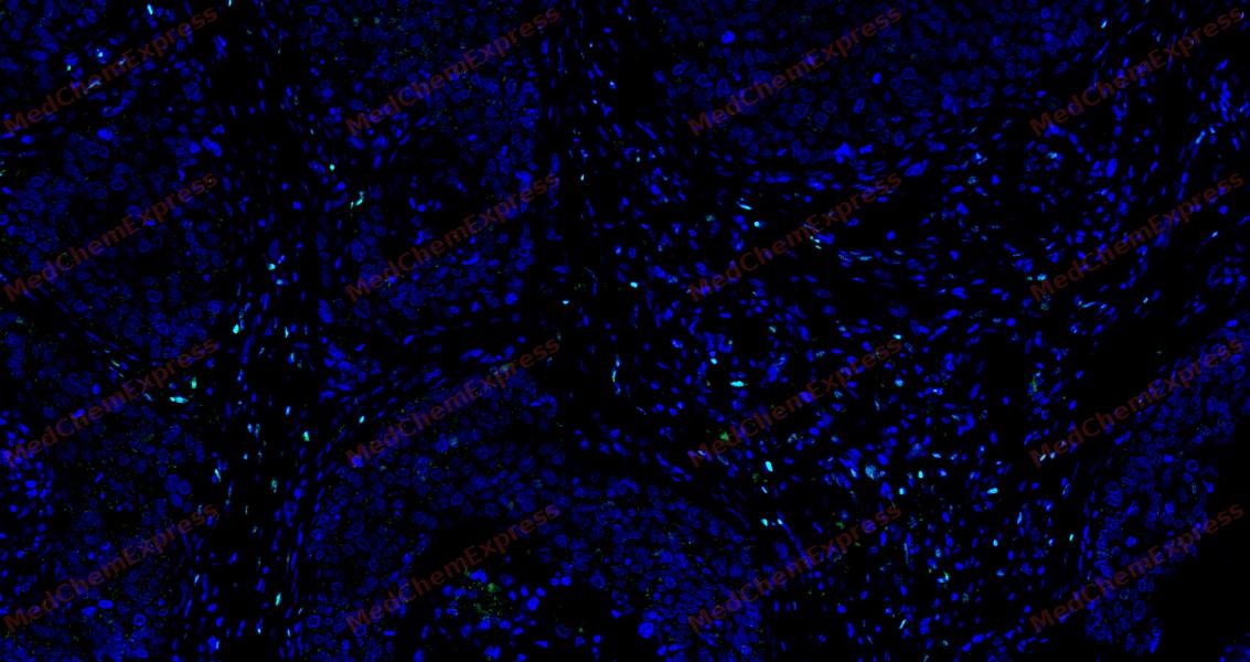 Tyramide signaling amplification based immunofluorescence was performed on paraffin-embedded human Esophageal Carcinoma tissue using FOXP3 antibody. Antigen retrieval was performed in EDTA buffer pH 9.0 (95 °C, 20 min) followed by cooling to RT. Then incubated with primary antibody (HY-P80965, 1:100 dilution) at room temperature for 60 minutes and HRP conjugated secondary antibody for 10 minutes. Fluorescence was then developed with TSA520. The tissues were counterstained with DAPI and mounted with Anti-fade mounting medium.


Background
-
Function
FOXP3, so called scurfin, belongs to the forkhead box protein P (FOXP) family. FOXP3 is critically involved in differentiation and function of regulatory T cells or CD4+/CD25+ regulatory T (Treg) cells for cancer immunotherapy. FOXP3 modulates Treg development and functions by immune evasion of tumor cells through imbalance of immunoediting and immunosurveillance in some cancers. FOXP3 is associated with IL-17, RUNX1, STAT3, NF-κB, FOXO3 and other cofactors such as EOS (Ikzf4), interferon regulatory factor 4 (IRF4), special AT-rich sequence-binding protein-1 (SATB1) and GATA1.
-
Subcellular Localization
Nucleus; Cytoplasm
-
Expression
Induction: Down-regulated in regulatory T-cells (Treg) during inflammation. Up-regulated by FOXO3
-
Isoforms & Post-Translational Modification
FOXP3 has four isomers: Q9BZS1-1: 47244 Da (predicted); Q9BZS1-2: 43410 Da (predicted); Q9BZS1-3: 49843 Da (predicted); Q9BZS1-4: 44407 Da (predicted).
Polyubiquitinated, leading to its proteasomal degradation in regulatory T-cells (Treg) which is mediated by STUB1 in a HSPA1A/B-dependent manner. -
Subunit
Homodimer
-
SwissProt ID
-
Synonyms
FOXP3; IPEX; JM2; Forkhead box protein P3; Scurfin


Documentation
-
Data Sheet (261 KB)
-
SDS (251 KB)
- English - EN (251 KB)
- Français - FR (251 KB)
- Deutsch - DE (251 KB)
- Norwegian - NO (251 KB)
- Español - ES (251 KB)
- Swedish - SV (251 KB)
- Italian - IT (251 KB)
- Korean - KR (251 KB)
- Portuguese - PT (251 KB)
-
User Guide for Antibodies (1077 KB)
References
[1]. Kim JH, et al. Molecular networks of FOXP family: dual biologic functions, interplay with other molecules and clinical implications in cancer progression. Mol Cancer. 2019 Dec 9;18(1):180. [Content Brief]
[2]. Co M, et al. FOXP transcription factors in vertebrate brain development, function, and disorders. Wiley Interdiscip Rev Dev Biol. 2020 Sep;9(5):e375. [Content Brief]

 Powered by Bioz
