



Bcl2 A1 Antibody (YA2743)
(Synonyms: BCL2A1; ACC-1; ACC-2; Bcl-2-like protein 5; Bcl-2-related protein A1; BCL2-related protein A1; BFL1; Bcl2-L-5; BCL2L5; GRS; HBPA1; Protein BFL-1; Protein GRS)
Based on 1 Customer Validation
Bcl2 A1 Antibody (YA2743) is a Rabbit-derived and non-conjugated IgG monoclonal antibody, targeting to Bcl2 A1.
-
Host:
Rabbit
-
Isotype:
IgG
-
Application:
WB, IHC-P, FC
-
Reactivity :
Human, Mouse, Rat
-
Formulation:
Supplied in rabbit IgG in 10 mM PBS, pH 7.4, 150 mM sodium chloride, 0.05% BSA, 0.02% sodium azide and 50% glycerol.
-
Conjugation:
Non-conjugated


To place orders, for customer services and technical support, please contact: MedChemExpress USA
Tel: 609-228-6898 E-mail: [email protected] [email protected]





Applications
| Application |
WB

WB: Western Blot
|
IHC-P
IHC-P: Immunohistochemistry-Paraffin
|
FC
FC: Flow Cytometry
|
|---|---|---|---|
| Dilution Ratio | 1:1000-1:2000 | 1:100-1:200 | 1:50-1:100 |


Product Details
Bcl2 A1 Antibody (YA2743) is a Rabbit-derived and non-conjugated IgG monoclonal antibody, targeting to Bcl2 A1.
-
Host Rabbit

-
Clonality Recombinant,Monoclonal

-
Species ReactivityHuman, Mouse, Rat
-
Observed Molecular WeightObserved band size: 27 kDa
Note: Due to possible protein modifications or aggregation, the molecular weight should be confirmed by actual measurement, and the predicted value is for reference only.

-
Calculated Molecular Weight Predicted band size: 20 kDa

A synthesized peptide derived from human BCL2A1 aa1-60.
Endogenous
Affinity Chromatography
Non-conjugated
Unmodified
IgG


Product Properties
-
Appearance
Liquid
-
Formulation
Supplied in rabbit IgG in 10 mM PBS, pH 7.4, 150 mM sodium chloride, 0.05% BSA, 0.02% sodium azide and 50% glycerol.
-
Concentration
Batch-dependent, Please check the COA for the concentration of each lot. Check Lot Concentration
-
Storage & Stability
Stored at -20°C for 1 year. Avoid repeated freeze / thaw cycles.
-
Shipping
Shipping with blue ice.


Verification Images
-
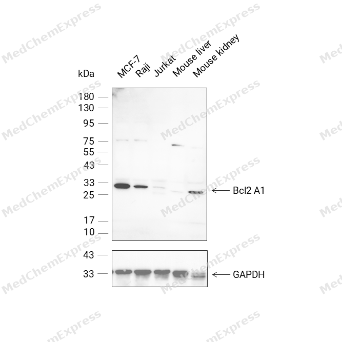 Western blot analysis was performed on protein extracts (25 μg) from MCF-7 (lane 1), Raji (lane 2), Jurkat (lane 3), Mouse liver (lane 4), and Mouse kidney (lane 5) using Bcl2 A1 antibody. Proteins were transferred onto a 0.45 μm PVDF membrane using the Trans-Blot® Turbo™ system for 13 min. The membrane was then blocked with 5% nonfat milk in TBST (HY-K1025) for 1 h at room temperature. Thhe primary antibody (1:1000) and loading control antibody GAPDH Antibody (HRP) (HY-P80954A) (1:2500) were diluted in 5% nonfat milk in TBST and incubated with the membrane overnight at 4°C. After washing, the membrane of primary antibody was incubated with HRP-conjugated goat anti-rabbit/mouse IgG secondary antibody (HY-P8001/HY-P8004) (1:5000) diluted in 5% nonfat milk in TBST for 1 h at room temperature. Protein bands were visualized using an Ultra High Sensitivity ECL detection kit (HY-K1005).
 -
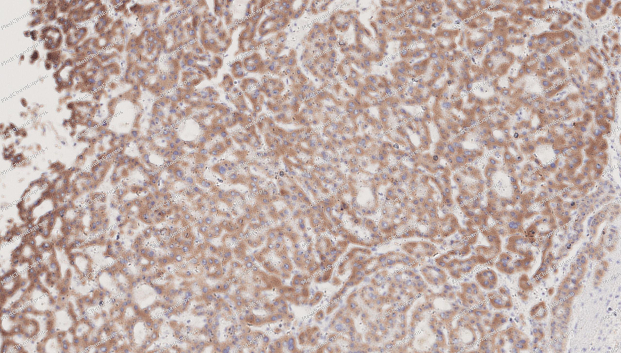 Immunohistochemical analysis of paraffin-embedded human Hepatocellular Carcinoma tissue using Bcl2 A1 antibody was performed. The section was pretreated using high-temperature mediated EDTA antigen retrieval buffer (pH 9.0), for 20 minutes. The tissues were incubated with primary antibody (HY-P82998, 1:100 dilution) at room temperature for 20 minutes. Detection was performed using an HRP conjugated compact polymer system. DAB was used as the chromogen. The tissues were counterstained with hematoxylin and mounted with neutral balsam mounting medium.
 -
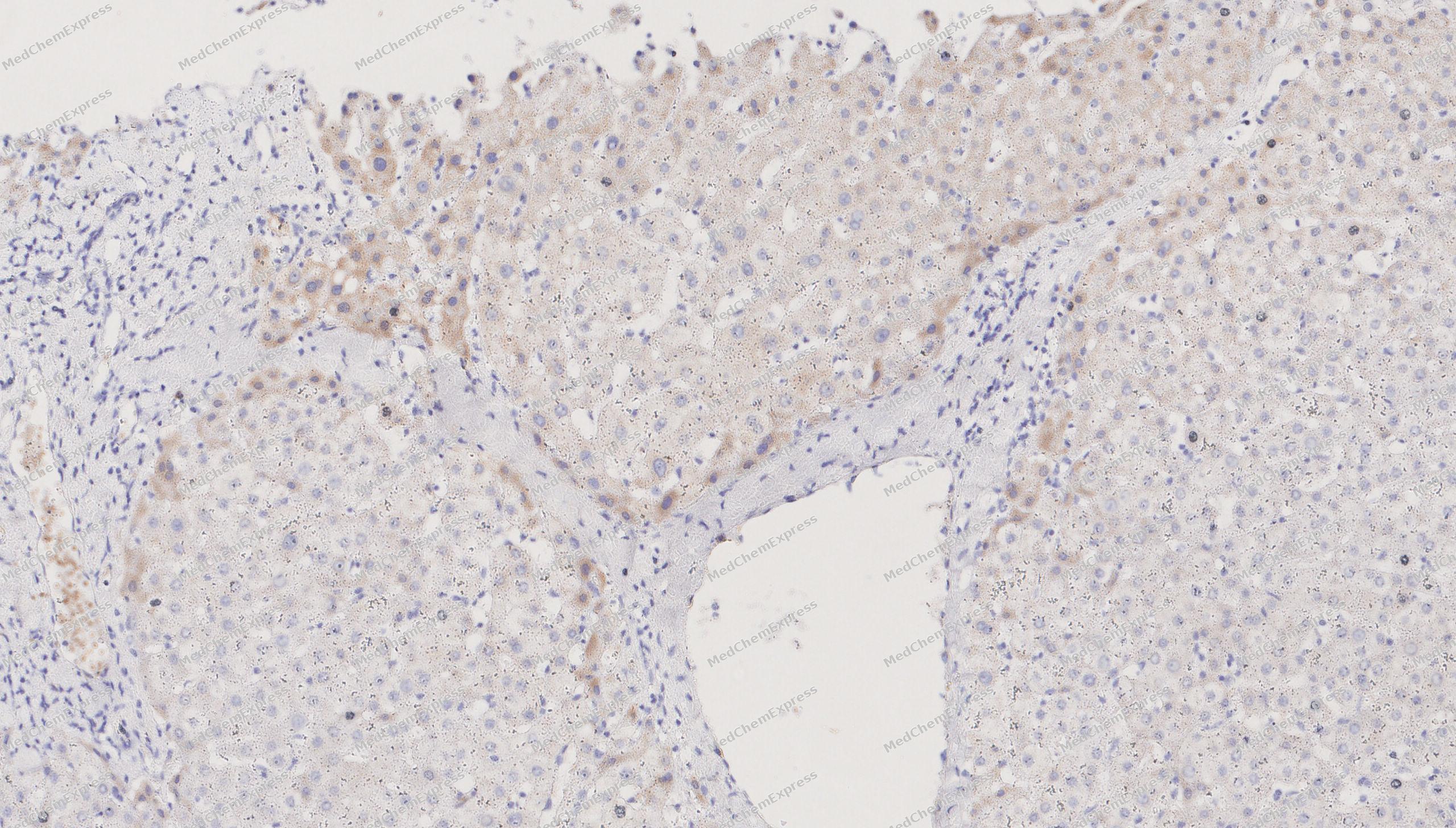 Immunohistochemical analysis of paraffin-embedded human Liver tissue using Bcl2 A1 antibody was performed. The section was pretreated using high-temperature mediated EDTA antigen retrieval buffer (pH 9.0), for 20 minutes. The tissues were incubated with primary antibody (HY-P82998, 1:100 dilution) at room temperature for 20 minutes. Detection was performed using an HRP conjugated compact polymer system. DAB was used as the chromogen. The tissues were counterstained with hematoxylin and mounted with neutral balsam mounting medium.
 -
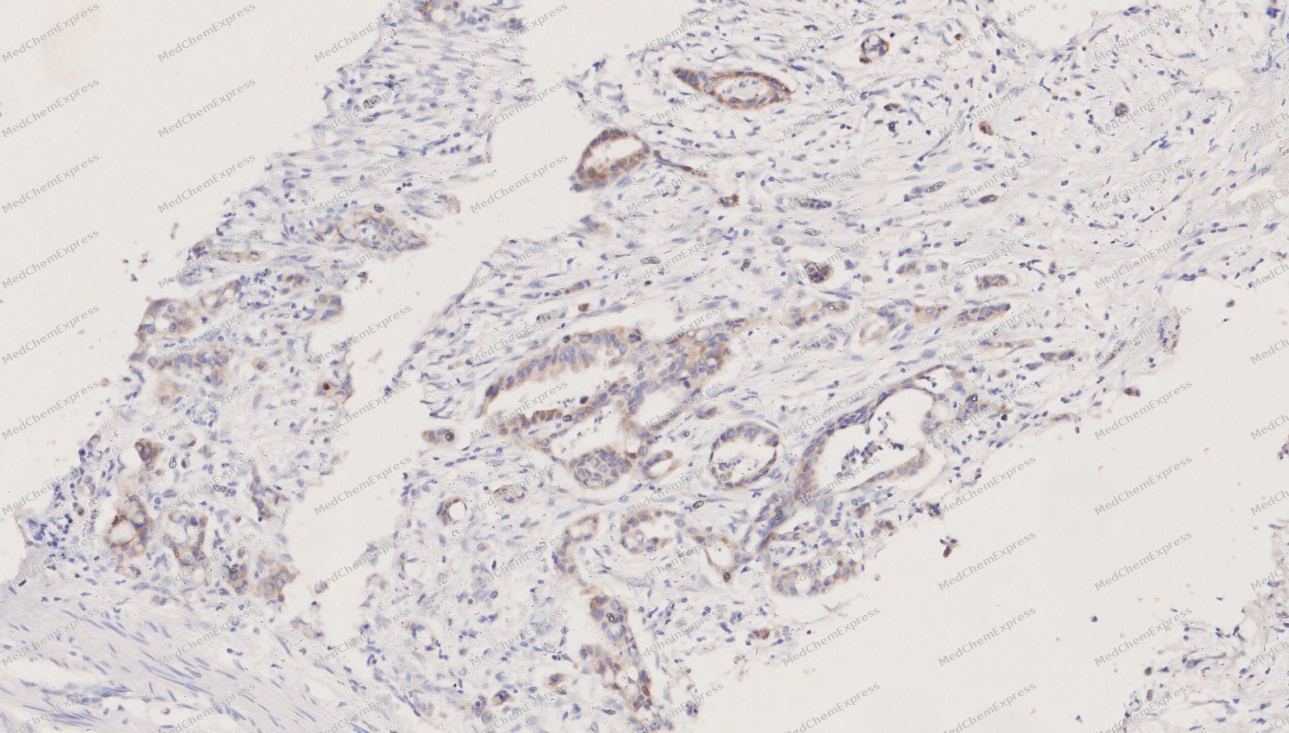 Immunohistochemical analysis of paraffin-embedded human Colorectal cancer tissue using Bcl2 A1 antibody was performed. The section was pretreated using high-temperature mediated EDTA antigen retrieval buffer (pH 9.0), for 20 minutes. The tissues were incubated with primary antibody (HY-P82998, 1:100 dilution) at room temperature for 20 minutes. Detection was performed using an HRP conjugated compact polymer system. DAB was used as the chromogen. The tissues were counterstained with hematoxylin and mounted with neutral balsam mounting medium.


Background
-
Function
Bcl2 A1 is a Retards apoptosis induced by IL-3 deprivation. May function in the response of hemopoietic cells to external signals and in maintaining endothelial survival during infection. Can inhibit apoptosis induced by serum starvation in the mammary epithelial cell line HC11
-
Subcellular Localization
Cytoplasm
-
Expression
Tissue_specificity:Seems to be restricted to the hematopoietic compartment. Expressed in peripheral blood, spleen, and bone marrow, at moderate levels in lung, small intestine and testis, at a minimal levels in other tissues. Also found in vascular smooth muscle cells and hematopoietic malignancies
Induction:By phorbol ester and inflammatory cytokines, such as TNF or IL1B/interleukin-1 beta, but not by growth factors -
Isoforms & Post-Translational Modification
Q16548 has 2 isomers: Q16548-1: 20132 Da (predicted); Q16548-2: 18746 Da (predicted).
-
Subunit
Interacts directly with BAK1, BID, BMF and BBC3 (By similarity). Interacts directly with BCL2L11/BIM. Interacts with BAX isoform Sigma. Interacts directly with PMAIP1. Interacts with RTL10/BOP. Interacts with ING4 (By similarity). Interacts with UBQLN4 (PubMed:34245648)
-
SwissProt ID
-
Synonyms
BCL2A1; ACC-1; ACC-2; Bcl-2-like protein 5; Bcl-2-related protein A1; BCL2-related protein A1; BFL1; Bcl2-L-5; BCL2L5; GRS; HBPA1; Protein BFL-1; Protein GRS
-
Research Field
Cell Biology


Documentation
-
Data Sheet (232 KB)
-
SDS (252 KB)
- English - EN (252 KB)
- Français - FR (252 KB)
- Deutsch - DE (252 KB)
- Norwegian - NO (252 KB)
- Español - ES (252 KB)
- Swedish - SV (252 KB)
- Italian - IT (252 KB)
- Korean - KR (252 KB)
- Portuguese - PT (252 KB)
-
User Guide for Antibodies (1077 KB)

 Powered by Bioz
