



c-Kit Antibody (YA502)
(Synonyms: CD117, Sl, Kit, Mast/stem cell growth factor receptor Kit, SCFR, Proto-oncogene c-Kit, Tyrosine-protein kinase Kit)
Based on 1 Customer Validation
c-Kit Antibody (YA502) is a Rabbit-derived and non-conjugated IgG monoclonal antibody, targeting to c-Kit.
-
Host:
Rabbit
-
Isotype:
IgG
-
Application:
WB, IHC-P
-
Reactivity :
Human
-
Formulation:
Supplied in 1*TBS (pH7.4), 0.05% BSA and 40% Glycerol. Preservative: 0.05% Sodium Azide.
-
Conjugation:
Non-conjugated


To place orders, for customer services and technical support, please contact: MedChemExpress USA
Tel: 609-228-6898 E-mail: [email protected] [email protected]





Applications
| Application |
WB

WB: Western Blot
|
IHC-P
IHC-P: Immunohistochemistry-Paraffin
|
|---|---|---|
| Dilution Ratio | 1:500 | 1:50-1:200 |


Product Details
c-Kit Antibody (YA502) is a Rabbit-derived and non-conjugated IgG monoclonal antibody, targeting to c-Kit.
-
Host Rabbit

-
Clonality Recombinant,Monoclonal

-
Species ReactivityHuman
-
Observed Molecular WeightObserved band size: 145 kDa
Note: Due to possible protein modifications or aggregation, the molecular weight should be confirmed by actual measurement, and the predicted value is for reference only.

-
Calculated Molecular Weight Predicted band size: 110 kDa

Synthetic peptide within C-terminal human KIT.
Endogenous
Protein A affinity purified.
Non-conjugated
Unmodified
IgG


Product Properties
-
Appearance
Solution
-
Formulation
Supplied in 1*TBS (pH7.4), 0.05% BSA and 40% Glycerol. Preservative: 0.05% Sodium Azide.
-
Concentration
Batch-dependent, Please check the COA for the concentration of each lot. Check Lot Concentration
-
Storage & Stability
Stored at -20°C for 1 year. Avoid repeated freeze / thaw cycles.
-
Shipping
Shipping with blue ice.


Verification Images
-
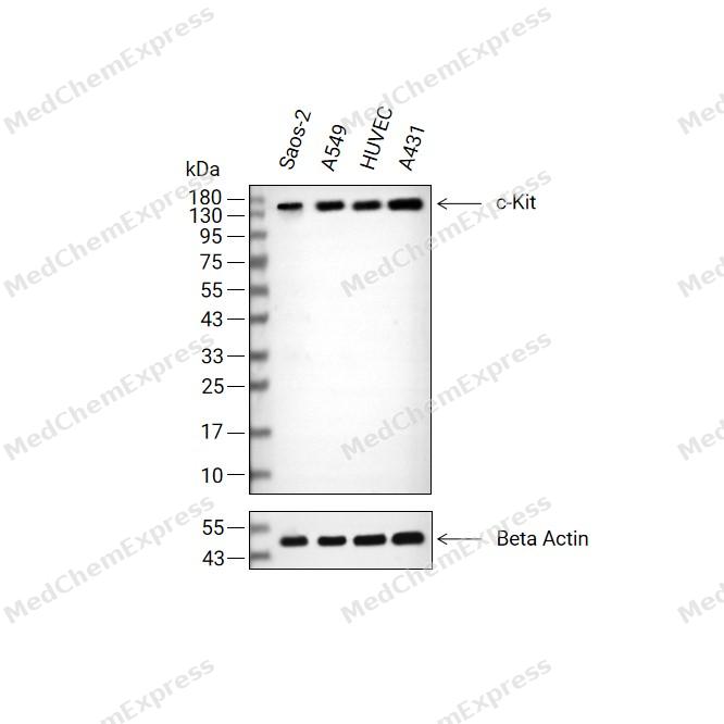 Western blot analysis of extracts from Saos-2(lane 2(20μg) , A549(lane 3(20μg) ,HUVEC(lane 4(20μg)and A431( lane 5(20μg) using c-Kit Antibody (HY-P80085). Proteins were transferred to a PVDF membrane and blocked with 5% non-fat milk in TBST for 2 hour at room temperature. The primary antibody ( 1/500) and Loading control antibody (Beta Actin, HY-P80993,1/10000) was used in 5% non-fat milk in TBST at 4°C overnight. Goat Anti-Rabbit IgG-HRP Secondary Antibody (HY-P8001,1/10,000) was used for 1 hour at room temperature.
 -
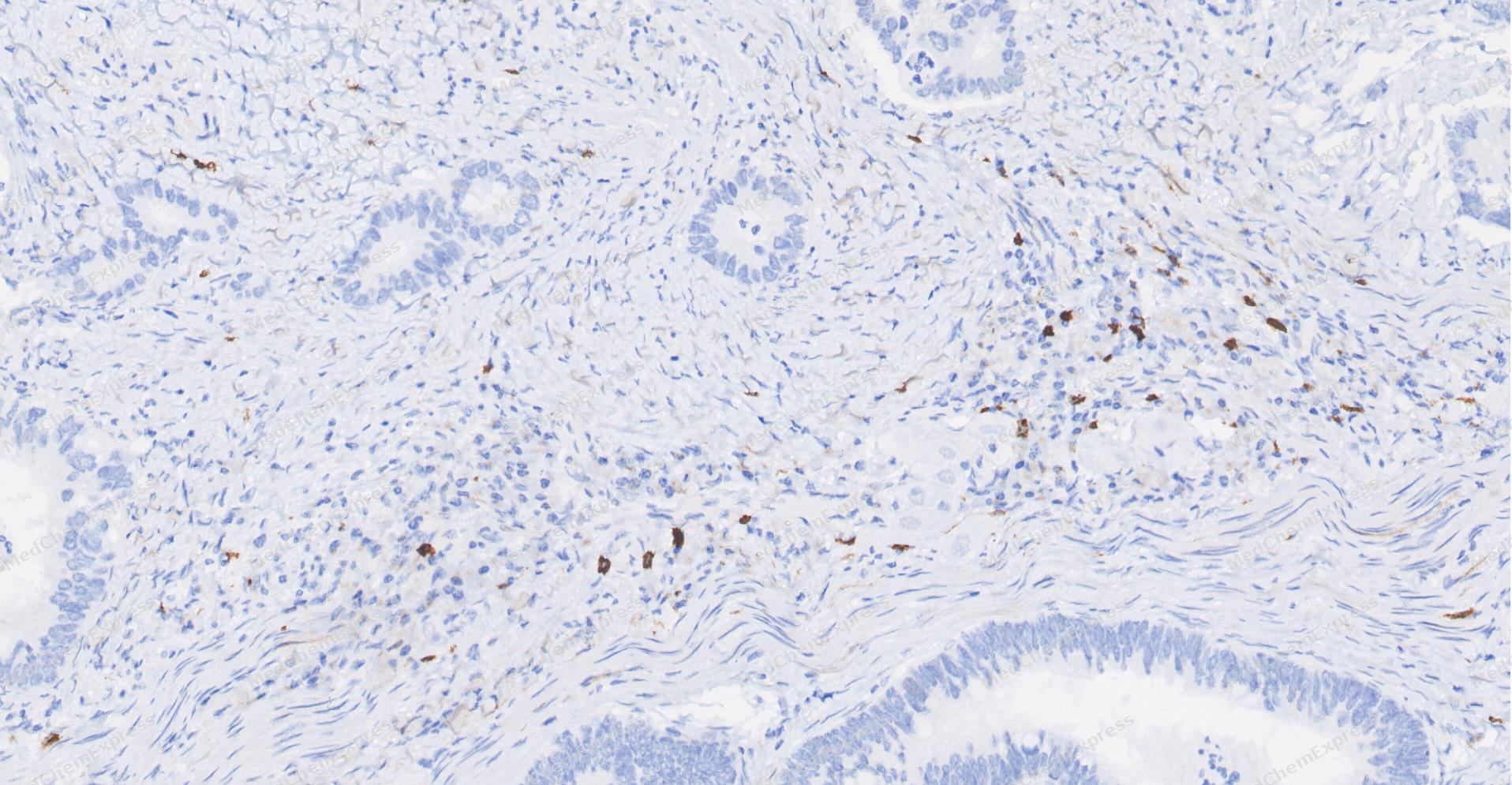 Immunohistochemical analysis of paraffin-embedded human Colon cancer tissue using c-Kit antibody was performed. The section was pretreated using high-temperature and high-pressure mediated EDTA antigen retrieval buffer (pH 9.0), for 5 minutes. The tissues were incubated with primary antibody (HY-P80085, 1:100 dilution) at room temperature for 60 minutes. Detection was performed using an HRP conjugated compact polymer system. DAB was used as the chromogen. The tissues were counterstained with hematoxylin and mounted with neutral balsam mounting medium.
 -
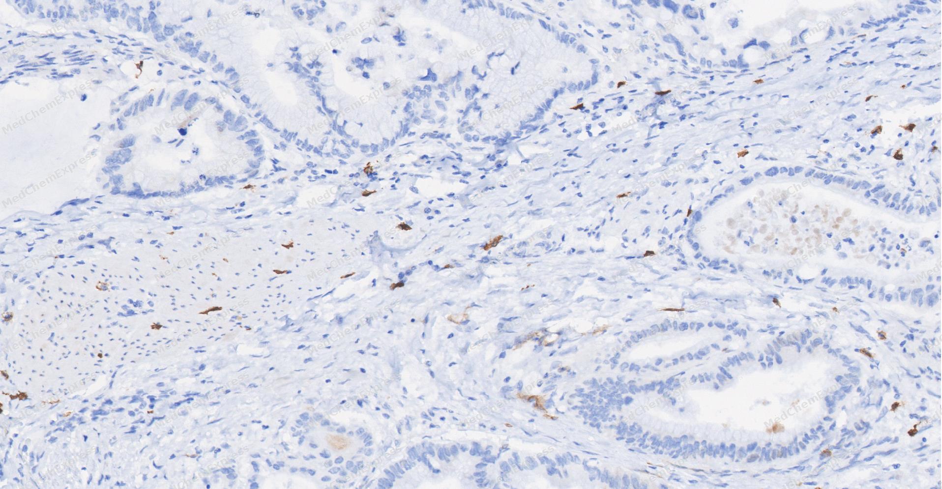 Immunohistochemical analysis of paraffin-embedded human Gastric Cancer tissue using c-Kit antibody was performed. The section was pretreated using high-temperature and high-pressure mediated EDTA antigen retrieval buffer (pH 9.0), for 5 minutes. The tissues were incubated with primary antibody (HY-P80085, 1:100 dilution) at room temperature for 60 minutes. Detection was performed using an HRP conjugated compact polymer system. DAB was used as the chromogen. The tissues were counterstained with hematoxylin and mounted with neutral balsam mounting medium.
 -
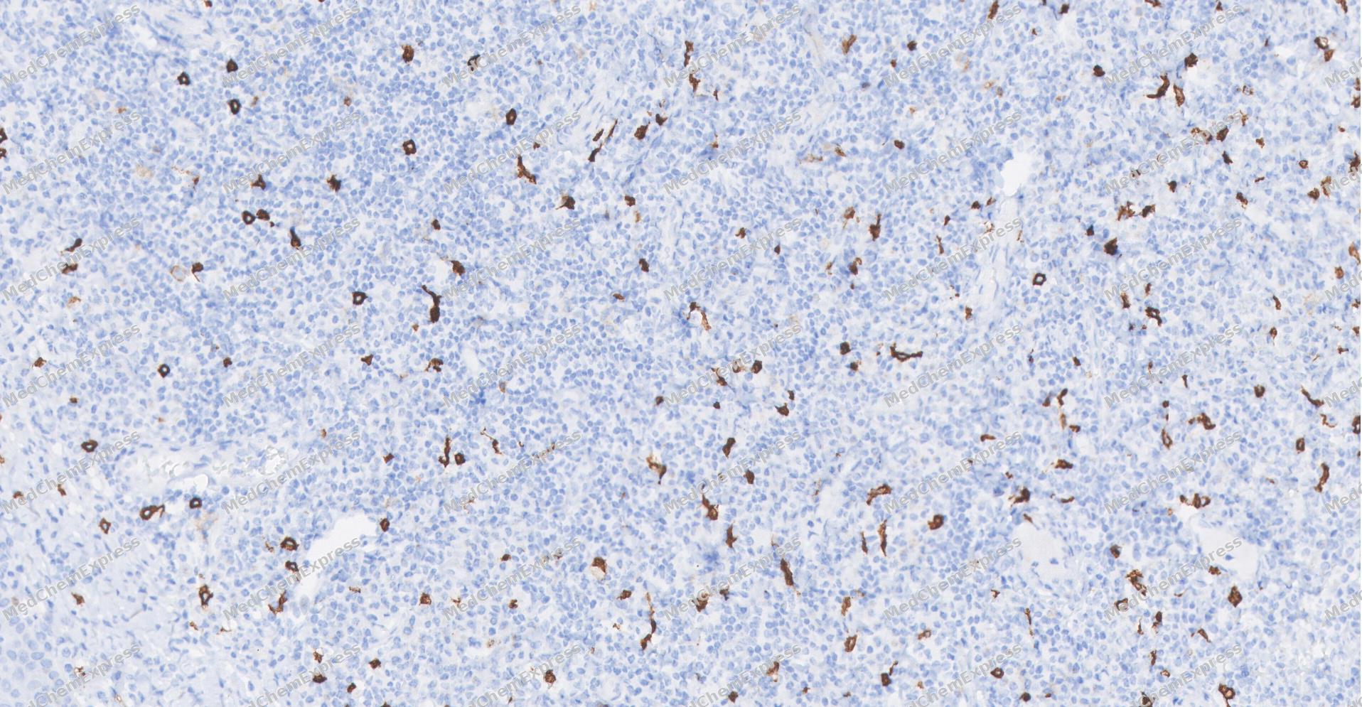 Immunohistochemical analysis of paraffin-embedded human Tonsil tissue using c-Kit antibody was performed. The section was pretreated using high-temperature and high-pressure mediated EDTA antigen retrieval buffer (pH 9.0), for 5 minutes. The tissues were incubated with primary antibody (HY-P80085, 1:100 dilution) at room temperature for 60 minutes. Detection was performed using an HRP conjugated compact polymer system. DAB was used as the chromogen. The tissues were counterstained with hematoxylin and mounted with neutral balsam mounting medium.
 -
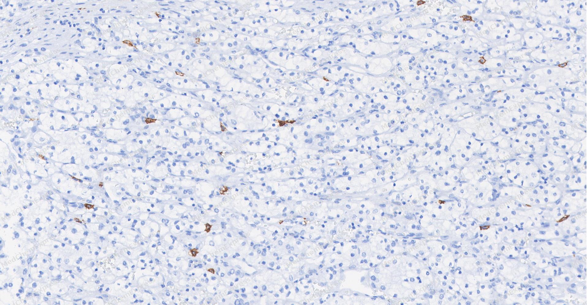 Immunohistochemical analysis of paraffin-embedded human Kidney cancer tissue using c-Kit antibody was performed. The section was pretreated using high-temperature and high-pressure mediated EDTA antigen retrieval buffer (pH 9.0), for 5 minutes. The tissues were incubated with primary antibody (HY-P80085, 1:100 dilution) at room temperature for 60 minutes. Detection was performed using an HRP conjugated compact polymer system. DAB was used as the chromogen. The tissues were counterstained with hematoxylin and mounted with neutral balsam mounting medium.
 -
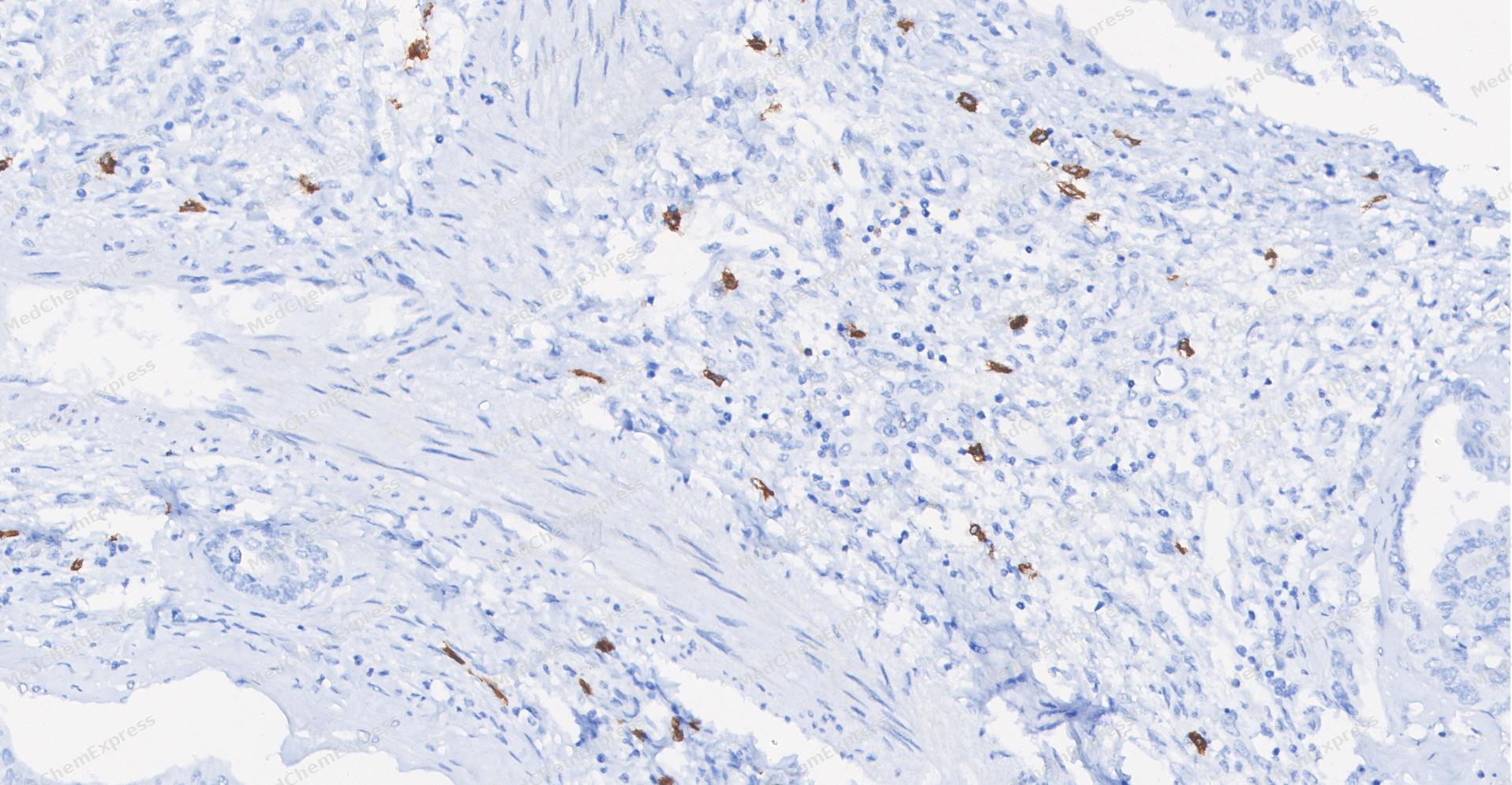 Immunohistochemical analysis of paraffin-embedded human cholangiocarcinoma tissue using c-Kit antibody was performed. The section was pretreated using high-temperature and high-pressure mediated EDTA antigen retrieval buffer (pH 9.0), for 5 minutes. The tissues were incubated with primary antibody (HY-P80085, 1:100 dilution) at room temperature for 60 minutes. Detection was performed using an HRP conjugated compact polymer system. DAB was used as the chromogen. The tissues were counterstained with hematoxylin and mounted with neutral balsam mounting medium.
 -
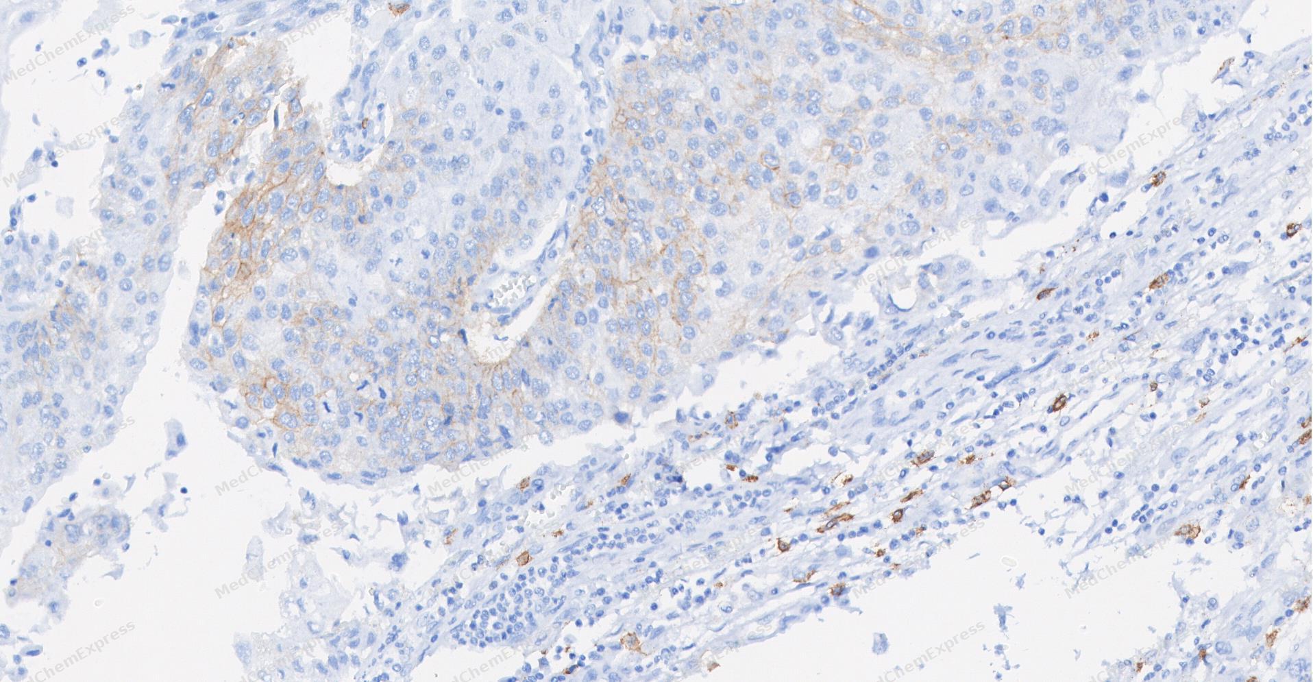 Immunohistochemical analysis of paraffin-embedded human Bladder cancer tissue using c-Kit antibody was performed. The section was pretreated using high-temperature and high-pressure mediated EDTA antigen retrieval buffer (pH 9.0), for 5 minutes. The tissues were incubated with primary antibody (HY-P80085, 1:100 dilution) at room temperature for 60 minutes. Detection was performed using an HRP conjugated compact polymer system. DAB was used as the chromogen. The tissues were counterstained with hematoxylin and mounted with neutral balsam mounting medium.


Background
-
Function
c-Kit (Mast/stem cell growth factor receptor, SCFR or CD117) is a proteinthat in humans is encoded by the KIT gene. c-Kit (CD117) is an important cell surface marker used to identify certain types of hematopoietic (blood) progenitors in the bone marrow. c-Kit is a cytokine receptor expressed on the surface of hematopoietic stem cells as well as other cell types. Altered forms of this receptor may be associated with some types of cancer. c-Kit is a receptor tyrosine kinase type III, which binds to stem cell factor. When c-Kit binds to stem cell factor (SCF) it forms adimer that activates its intrinsic tyrosine kinase activity, that in turn phosphorylates and activates signal transduction molecules that propagate the signal in the cell. Signalling through c-Kit plays a role in cell survival, proliferation, and differentiation.
-
Subcellular Localization
Cell membrane; Single-pass type I membrane protein; Cell membrane; Single-pass type I membrane protein; Cytoplasm
-
Expression
Tissue_specificity:In the testes, this protein was detected in spermatogonia and Leydig cells in the basal layer, but not in Sertoli cells or spermatocytes in the seminiferous tubules (protein level) (PubMed:20601678) . It is also maintained in ejaculated sperm (protein level) (PubMed:20601678) .
Induction:Up-regulated by cis-retinoic acid in neuroblastoma cell lines -
Isoforms & Post-Translational Modification
P10721 has 3 isomers: P10721-1: 109865 Da (predicted); P10721-2: 109451 Da (predicted); P10721-4: 29087 Da (predicted).
Ubiquitinated by SOCS6. KIT is rapidly ubiquitinated after autophosphorylation induced by KITLG/SCF binding, leading to internalization and degradation;Autophosphorylated on tyrosine residues. KITLG/SCF binding enhances autophosphorylation. Isoform 1 shows low levels of tyrosine phosphorylation in the absence of added KITLG/SCF (in vitro). Kinase activity is down-regulated by phosphorylation on serine residues by protein kinase C family members. Phosphorylation at Tyr-568 is required for interaction with PTPN11/SHP-2, CRK (isoform Crk-II) and members of the SRC tyrosine-protein kinase family. Phosphorylation at Tyr-570 is required for interaction with PTPN6/SHP-1. Phosphorylation at Tyr-703, Tyr-823 and Tyr-936 is important for interaction with GRB2. Phosphorylation at Tyr-721 is important for interaction with PIK3R1. Phosphorylation at Tyr-823 and Tyr-936 is important for interaction with GRB7 -
Subunit
Monomer in the absence of bound KITLG/SCF. Homodimer in the presence of bound KITLG/SCF, forming a heterotetramer with two KITLG/SCF molecules. Interacts (via phosphorylated tyrosine residues) with the adapter proteins GRB2 and GRB7 (via SH2 domain), and SH2B2/APS. Interacts (via C-terminus) with MPDZ (via the tenth PDZ domain). Interacts (via phosphorylated tyrosine residues) with PIK3R1 and PIK3 catalytic subunit. Interacts (via phosphorylated tyrosine) with CRK (isoform Crk-II), FYN, SHC1 and MATK/CHK (via SH2 domain). Interacts with LYN and FES/FPS. Interacts (via phosphorylated tyrosine residues) with the protein phosphatases PTPN6/SHP-1 (via SH2 domain), PTPN11/SHP-2 (via SH2 domain) and PTPRU. Interacts with PLCG1. Interacts with DOK1 and TEC. Interacts (KITLG/SCF-bound) with IL1RL1. Interacts with IL1RAP (independent of stimulation with KITLG/SCF). A mast cell-specific KITLG/SCF-induced interleukin-33 signaling complex contains IL1RL1, IL1RAP, KIT and MYD88
-
SwissProt ID
-
Synonyms
CD117, Sl, Kit, Mast/stem cell growth factor receptor Kit, SCFR, Proto-oncogene c-Kit, Tyrosine-protein kinase Kit
-
Research Field
Immunology


Documentation
-
Data Sheet (234 KB)
-
SDS (251 KB)
- English - EN (251 KB)
- Français - FR (251 KB)
- Deutsch - DE (251 KB)
- Norwegian - NO (251 KB)
- Español - ES (251 KB)
- Swedish - SV (251 KB)
- Italian - IT (251 KB)
- Korean - KR (251 KB)
- Portuguese - PT (251 KB)
-
User Guide for Antibodies (1077 KB)

 Powered by Bioz
