



Casein Kinase 2 beta Antibody (YA563)
(Synonyms: CSNK2B; CK2N; G5A; Casein kinase II subunit beta; CK II beta; Phosvitin; Protein G5a)
Based on 1 Customer Validation
Casein Kinase 2 beta Antibody (YA563) is a Rabbit-derived and non-conjugated IgG monoclonal antibody, targeting to Casein Kinase 2 beta.
-
Host:
Rabbit
-
Isotype:
IgG
-
Application:
WB, IHC-F, IHC-P, ICC/IF
-
Reactivity :
Human, Mouse, Rat
-
Formulation:
Supplied in 50 mM Tris-Glycine (pH 7.4), 0.15 M NaCl, 40% Glycerol and 0.05% BSA. Preservative: 0.01% Sodium azide
-
Conjugation:
Non-conjugated


To place orders, for customer services and technical support, please contact: MedChemExpress USA
Tel: 609-228-6898 E-mail: [email protected] [email protected]





Applications
| Application |
WB

WB: Western Blot
|
IHC-P
IHC-P: Immunohistochemistry-Paraffin
|
IHC-F
IHC-F: Immunohistochemistry-Frozen
|
ICC/IF
ICC/IF: Immunocytochemistry/
Immunofluorescence |
|---|---|---|---|---|
| Dilution Ratio | 1:500-1:1000 | 1:50-1:100 | 1:50-1:100 | 1:50-1:200 |


Product Details
Casein Kinase 2 beta Antibody (YA563) is a Rabbit-derived and non-conjugated IgG monoclonal antibody, targeting to Casein Kinase 2 beta.
-
Host Rabbit

-
Clonality Recombinant,Monoclonal

-
Species ReactivityHuman, Mouse, Rat
-
Observed Molecular WeightObserved band size: 25 kDa
Note: Due to possible protein modifications or aggregation, the molecular weight should be confirmed by actual measurement, and the predicted value is for reference only.

-
Calculated Molecular Weight Predicted band size: 25 kDa

Entrez Gene: 1460 Human ; 13001 Mouse ; 81650 Rat
SwissProt: P67870 Human ; P67871 Mouse ; P67874 Rat
OMIM: 618732 Human
Synthetic peptide corresponding to Human Casein Kinase 2 beta.The exact sequence is proprietary to MCE.
Endogenous
affinity purified
Non-conjugated
Unmodified
IgG


Product Properties
-
Appearance
Solution
-
Formulation
Supplied in 50 mM Tris-Glycine (pH 7.4), 0.15 M NaCl, 40% Glycerol and 0.05% BSA. Preservative: 0.01% Sodium azide
-
Concentration
Batch-dependent, Please check the COA for the concentration of each lot. Check Lot Concentration
-
Storage & Stability
Stored at -20°C for 1 year. Avoid repeated freeze / thaw cycles.
-
Shipping
Shipping with blue ice.


Verification Images
-
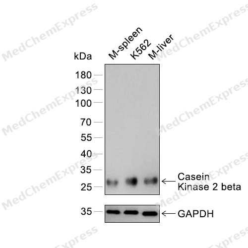 Western blot analysis of extracts from M-spleen (lane 1) and K562 (lane 2) and M-liver (lane 3) using Casein Kinase 2 beta antibody. Proteins were transferred to a PVDF membrane and blocked with 5% nonfat powdered milk in PBST for 2 hour at room temperature. The primary antibody (1/1000) and loading control antibody (GAPDH, 1/3000) was diluted with 5% nonfat powdered milk in PBST at 4°C overnight. Goat Anti-Rabbit IgG-HRP Secondary Antibody (1/8,000) was incubated for 45min at room temperature.
 -
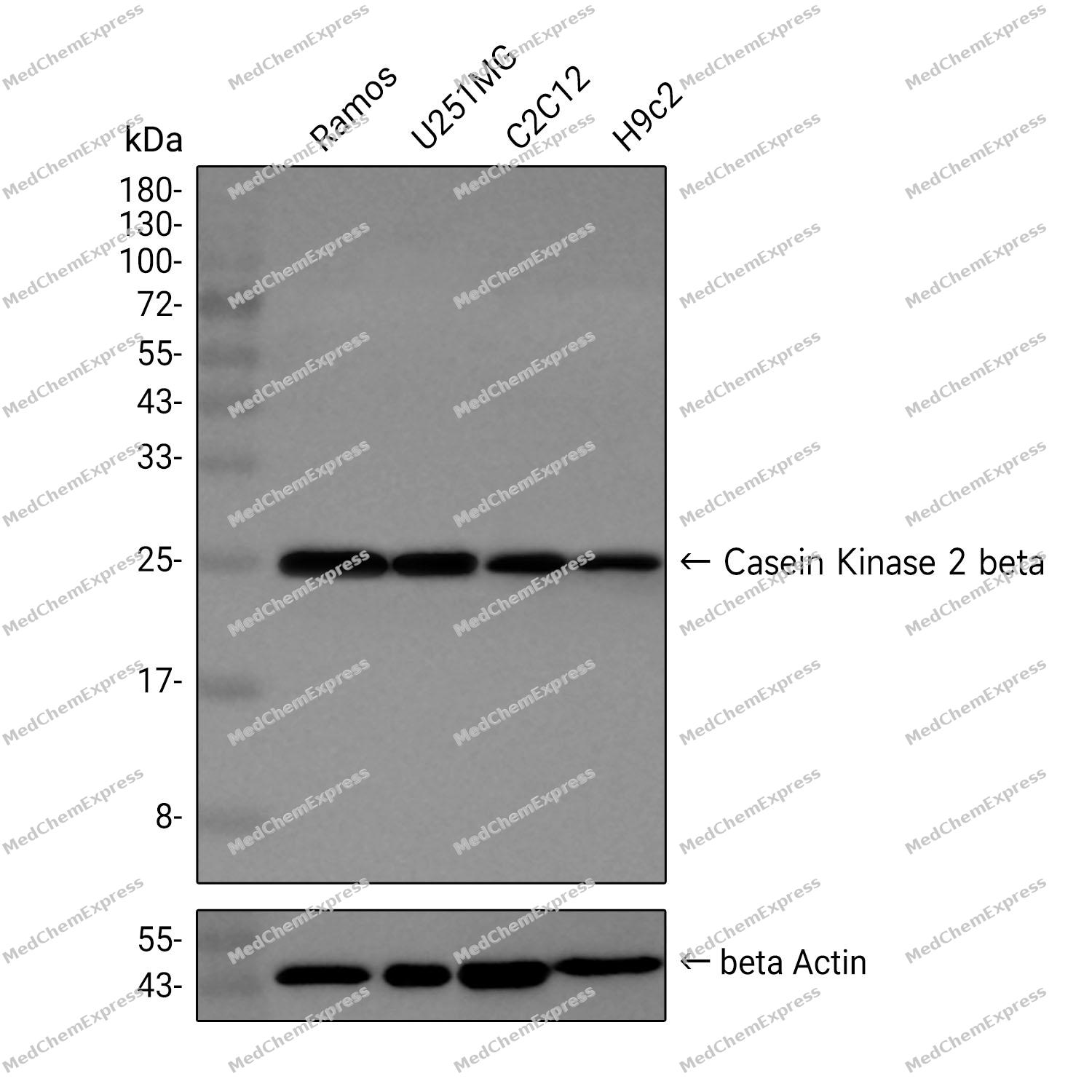 Western blot analysis was performed on extracts from Ramos (lane 1, 15 μg), U251MG (lane 2, 15 μg), C2C12 (lane 3, 15 μg), and H9c2 (lane 4, 15 μg) using Casein Kinase 2 beta Rabbit mAb.Proteins were transferred to a PVDF membrane and blocked with 5% non-fat milk in TBST at 4°C overnight.The primary antibody (1:1000 dilution) and the loading control antibody (beta-Actin, HY-P80438, 1:5000 dilution) was incubated in 5% non-fat milk in TBST for 1 hour at 37°C.Goat Anti-Rabbit IgG-HRP Secondary Antibody (1:40000 dilution) was then applied for 40 minutes at 37°C.
 -
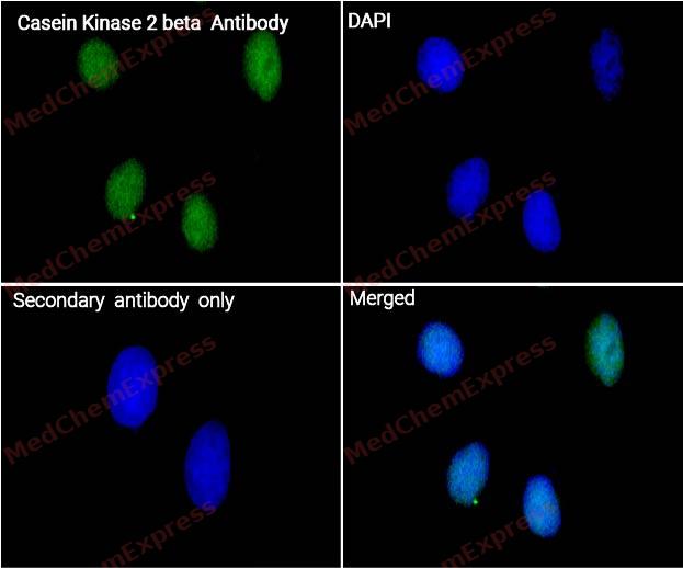 Immunocytochemistry analysis of Hela cells labeling Casein Kinase 2 beta with Casein Kinase 2 beta Antibody (HY-P80586) at 1/100 dilution . Cells were fixed in 4% paraformaldehyde for 15 minutes at room temperature, permeabilized with 0.1% Triton X-100 in PBS for 15 minutes at room temperature, then blocked with quick block buffer for 10 minutes at room temperature. Cells were then incubated with Casein Kinase 2 beta Antibody (HY-P80586) at 1/100 dilution in quick block buffer overnight at 4 ℃. AF488-conjugated Goat Anti-Rabbit IgG H&L(HY-P8002, Green) was used as the secondary antibody at 1/1,000 dilution. PBS instead of the primary antibody was used as the secondary antibody only control. The Nuclear counterstain was DAPI (Blue).
 -
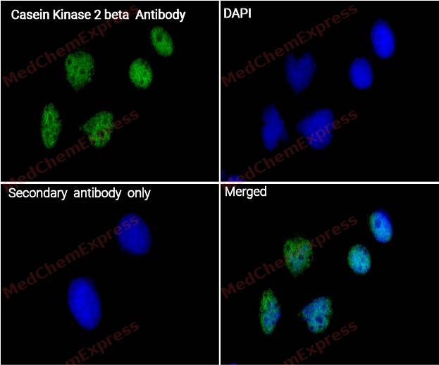 Immunocytochemistry analysis of HePG2 cells labeling Casein Kinase 2 beta with Casein Kinase 2 beta Antibody (HY-P80586) at 1/100 dilution . Cells were fixed in 4% paraformaldehyde for 15 minutes at room temperature, permeabilized with 0.1% Triton X-100 in PBS for 15 minutes at room temperature, then blocked with quick block buffer for 10 minutes at room temperature. Cells were then incubated with Casein Kinase 2 beta Antibody (HY-P80586) at 1/100 dilution in quick block buffer overnight at 4 ℃. AF488-conjugated Goat Anti-Rabbit IgG H&L(HY-P8002, Green) was used as the secondary antibody at 1/1,000 dilution. PBS instead of the primary antibody was used as the secondary antibody only control. The Nuclear counterstain was DAPI (Blue).


Background
-
Function
Casein Kinase 2 beta is a Regulatory subunit of casein kinase II/CK2. As part of the kinase complex regulates the basal catalytic activity of the alpha subunit a constitutively active serine/threonine-protein kinase that phosphorylates a large number of substrates containing acidic residues C-terminal to the phosphorylated serine or threonine. Participates in Wnt signaling; (Microbial infection) Upon infection with Epstein-Barr virus (EBV), the interaction with viral EBNA1 increases the association of CK2 with PML proteins, which increases PML phosphorylation by CK2, triggering the polyubiquitylation and degradation of PML. Seems to also suppress EBV reactivation by mediating ARK2N and JUN at the Z promoter which inhibits BZLF1 transcrition[1][2][3][4][5].
-
Subcellular Localization
Nucleus
-
Subunit
Casein kinase II/CK2 is a tetramer composed of an alpha subunit, an alpha' subunit and two beta subunits. The beta subunit dimerization is mediated by zinc ions. Interacts with DYNLT2 (By similarity). Interacts with CD163. Also a component of a CK2-SPT16-SSRP1 complex composed of SSRP1, SUPT16H, CSNK2A1, CSNK2A2 and CSNK2B, the complex associating following UV irradiation. Interacts with MUSK; mediates phosphorylation of MUSK by CK2. Interacts with FGF1; this interaction is increased in the presence of FIBP, suggesting a possible cooperative interaction between CSNKB and FIBP in binding to FGF1. Interacts (via KSSR motif) with ARK2N (PubMed:24216761). Interacts with JUN and ARK2N; mediates the interaction between ARK2N and JUN (PubMed:31341047)
-
SwissProt ID
-
Synonyms
CSNK2B; CK2N; G5A; Casein kinase II subunit beta; CK II beta; Phosvitin; Protein G5a
-
Research Field
Cell Biology


Documentation
-
Data Sheet (261 KB)
-
SDS (251 KB)
- English - EN (251 KB)
- Français - FR (251 KB)
- Deutsch - DE (251 KB)
- Norwegian - NO (251 KB)
- Español - ES (251 KB)
- Swedish - SV (251 KB)
- Italian - IT (251 KB)
- Korean - KR (251 KB)
- Portuguese - PT (251 KB)
-
User Guide for Antibodies (1077 KB)
References
[1]. Keller DM, et al. A DNA damage-induced p53 serine 392 kinase complex contains CK2, hSpt16, and SSRP1. Mol Cell. 2001 Feb;7(2):283-92. [Content Brief]
[2]. Cheusova T, et al. Casein kinase 2-dependent serine phosphorylation of MuSK regulates acetylcholine receptor aggregation at the neuromuscular junction. Genes Dev. 2006 Jul 1;20(13):1800-16. [Content Brief]
[3]. Sivachandran N, et al. Epstein-Barr virus nuclear antigen 1 Hijacks the host kinase CK2 to disrupt PML nuclear bodies. J Virol. 2010 Nov;84(21):11113-23. [Content Brief]
[4]. Cao JY, et al. Identification of a novel protein interaction motif in the regulatory subunit of casein kinase 2. Mol Cell Biol. 2014 Jan;34(2):246-58. [Content Brief]
[5]. Siddiqi UZ, et al. Identification of ARKL1 as a Negative Regulator of Epstein-Barr Virus Reactivation. J Virol. 2019 Oct 15;93(20):. [Content Brief]

 Powered by Bioz
