



Ephrin Receptor B1 Antibody (YA2176)
(Synonyms: EPHB1; ELK; EPHT2; HEK6; NET; Ephrin type-B receptor 1; ELK; EPH tyrosine kinase 2; EPH-like kinase 6; EK6; hEK6; Neuronally-expressed EPH-related tyrosine kinase; NET; Tyrosine-protein kinase receptor EPH-2)
Based on 1 Customer Validation
Ephrin Receptor B1 Antibody (YA2176) is a Rabbit-derived and non-conjugated IgG monoclonal antibody, targeting to Ephrin Receptor B1.
-
Host:
Rabbit
-
Isotype:
IgG
-
Application:
WB, IP
-
Reactivity :
Human, Mouse, Rat
-
Formulation:
Supplied in rabbit IgG in 10mM PBS, pH 7.4, 150mM sodium chloride, 0.05% BSA, 0.02% sodium azide and 50% glycerol.
-
Conjugation:
Non-conjugated


To place orders, for customer services and technical support, please contact: MedChemExpress USA
Tel: 609-228-6898 E-mail: [email protected] [email protected]





Applications
| Application |
WB

WB: Western Blot
|
IP
IP: Immunoprecipitation
|
|---|---|---|
| Dilution Ratio | 1:500-1:1000 | 1:50 |


Product Details
Ephrin Receptor B1 Antibody (YA2176) is a Rabbit-derived and non-conjugated IgG monoclonal antibody, targeting to Ephrin Receptor B1.
-
Host Rabbit

-
Clonality Recombinant, Monoclonal

-
Species ReactivityHuman, Mouse, Rat
-
Observed Molecular WeightObserved band size: 120 kDa
Note: Due to possible protein modifications or aggregation, the molecular weight should be confirmed by actual measurement, and the predicted value is for reference only.

-
Calculated Molecular Weight Predicted band size: 110 kDa

Entrez Gene: 2047 Human ; 270190 Mouse ; 24338 Rat
SwissProt: P54762 Human ; Q8CBF3 Mouse ; P09759 Rat
OMIM: 600600 Human
A synthesized peptide derived from human Eph receptor B1 aa575-625/984.
Endogenous
Affinity Chromatography
Non-conjugated
Unmodified
IgG


Product Properties
-
Appearance
Liquid
-
Formulation
Supplied in rabbit IgG in 10mM PBS, pH 7.4, 150mM sodium chloride, 0.05% BSA, 0.02% sodium azide and 50% glycerol.
-
Concentration
Batch-dependent, Please check the COA for the concentration of each lot. Check Lot Concentration
-
Storage & Stability
Stored at -20°C for 1 year. Avoid repeated freeze / thaw cycles.
-
Shipping
Shipping with blue ice.


Verification Images
-
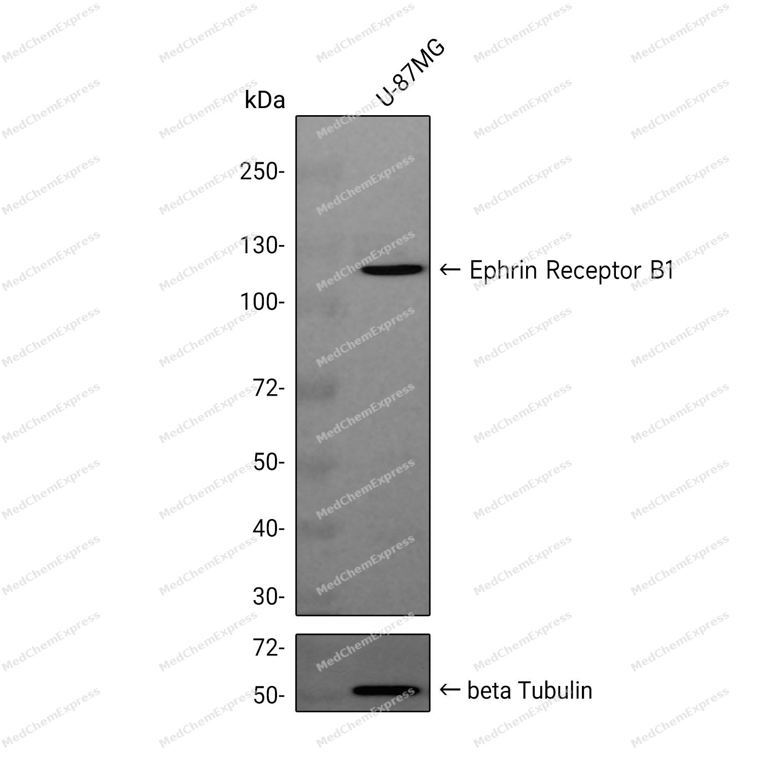 Western blot analysis was performed on extracts from U-87MG (lane 1, 15 μg) using Ephrin Receptor B1 Rabbit mAb.Proteins were transferred to a PVDF membrane and blocked with 5% non-fat milk in TBST at 4°C overnight.The primary antibody (1:1000 dilution) and the loading control antibody (beta-Tubulin, HY-P80955, 1:5000 dilution) was incubated in 5% non-fat milk in TBST for 1 hour at 37°C.Goat Anti-Rabbit IgG-HRP Secondary Antibody (1:40000 dilution) was then applied for 40 minutes at 37°C.


Background
-
Function
Ephrin Receptor B1 receptor tyrosine kinase which binds promiscuously transmembrane ephrin-B family ligands residing on adjacent cells, leading to contact-dependent bidirectional signaling into neighboring cells. The signaling pathway downstream of the receptor is referred to as forward signaling while the signaling pathway downstream of the ephrin ligand is referred to as reverse signaling. Cognate/functional ephrin ligands for this receptor include EFNB1, EFNB2 and EFNB3. During nervous system development, regulates retinal axon guidance redirecting ipsilaterally ventrotemporal retinal ganglion cells axons at the optic chiasm midline. This probably requires repulsive interaction with EFNB2. In the adult nervous system together with EFNB3, regulates chemotaxis, proliferation and polarity of the hippocampus neural progenitors. In addition to its role in axon guidance also plays an important redundant role with other ephrin-B receptors in development and maturation of dendritic spines and synapse formation. May also regulate angiogenesis. More generally, may play a role in targeted cell migration and adhesion. Upon activation by EFNB1 and probably other ephrin-B ligands activates the MAPK/ERK and the JNK signaling cascades to regulate cell migration and adhesion respectively. Involved in the maintenance of the pool of satellite cells (muscle stem cells) by promoting their self-renewal and reducing their activation and differentiation
-
Subcellular Localization
Cell membrane; Single-pass type I membrane protein; Early endosome membrane; Cell projection, dendrite
-
Expression
Tissue_specificity:Primarily expressed in the brain -
Isoforms & Post-Translational Modification
P54762 has 3 isomers: P54762-1: 109885 Da (predicted); P54762-5: 61401 Da (predicted); P54762-6: 26906 Da (predicted).
Phosphorylated. Autophosphorylation is stimulated by the ligand EFNB1. Required for interaction with SH2 domain-containing interactors, for activation of the MAPK/ERK and JUN signaling cascades and for ubiquitination by CBL;Ubiquitinated; (EFNB1)ligand-induced poly- and/or multi-ubiquitination by CBL is regulated by SRC and leads to lysosomal degradation -
Subunit
Heterotetramer upon binding of the ligand. The heterotetramer is composed of an ephrin dimer and a receptor dimer. Oligomerization is probably required to induce biological responses (By similarity). Interacts with EPHB6; transphosphorylates EPHB6 to form an active signaling complex. Interacts with PICK1 (By similarity). Interacts (through Tyr-594) with NCK1 (via SH2 domain); activates the JUN cascade to regulate cell adhesion (By similarity). The ligand-activated form interacts (through Tyr-928) with GRB7 and GRB10 (via SH2 domains). The ligand-activated form interacts (residues within the catalytic domain) with GRB2 (via SH2 domain). Interacts with GRB2, SHC1 and SRC; activates the MAPK/ERK cascade to regulate cell migration. Interacts with CBL; regulates receptor degradation through ubiquitination. Interacts with ACP1
-
SwissProt ID
-
Synonyms
EPHB1; ELK; EPHT2; HEK6; NET; Ephrin type-B receptor 1; ELK; EPH tyrosine kinase 2; EPH-like kinase 6; EK6; hEK6; Neuronally-expressed EPH-related tyrosine kinase; NET; Tyrosine-protein kinase receptor EPH-2
-
Research Field
Cardiovascular


Documentation
-
Data Sheet (234 KB)
-
SDS (252 KB)
- English - EN (252 KB)
- Français - FR (252 KB)
- Deutsch - DE (252 KB)
- Norwegian - NO (252 KB)
- Español - ES (252 KB)
- Swedish - SV (252 KB)
- Italian - IT (252 KB)
- Korean - KR (252 KB)
- Portuguese - PT (252 KB)
-
User Guide for Antibodies (1077 KB)

 Powered by Bioz
