



Heparanase 1 Antibody (YA1601)
(Synonyms: HEP; Heparanase; Heparanase1; Hpa 1; HPA; Hpa1 HPR1; HPSE1; HSE1)
Based on 1 Customer Validation
Heparanase 1 Antibody (YA1601) is a Rabbit-derived and non-conjugated IgG monoclonal antibody, targeting to Heparanase 1.
-
Host:
Rabbit
-
Isotype:
IgG
-
Application:
WB, IP
-
Reactivity :
Human, Mouse, Rat
-
Formulation:
Supplied in rabbit IgG in 10mM PBS, pH 7.4, 150mM sodium chloride, 0.05% BSA, 0.02% sodium azide and 50% glycerol.
-
Conjugation:
Non-conjugated


To place orders, for customer services and technical support, please contact: MedChemExpress USA
Tel: 609-228-6898 E-mail: [email protected] [email protected]





Applications
| Application |
WB

WB: Western Blot
|
IP
IP: Immunoprecipitation
|
|---|---|---|
| Dilution Ratio | 1:500-1:1000 | 1:50 |


Product Details
Heparanase 1 Antibody (YA1601) is a Rabbit-derived and non-conjugated IgG monoclonal antibody, targeting to Heparanase 1.
-
Host Rabbit

-
Clonality Recombinant,Monoclonal

-
Species ReactivityHuman, Mouse, Rat
-
Observed Molecular WeightObserved band size: 65 kDa
Note: Due to possible protein modifications or aggregation, the molecular weight should be confirmed by actual measurement, and the predicted value is for reference only.

-
Calculated Molecular Weight Predicted band size: 61 kDa

Entrez Gene: 10855 Human ; 15442 Mouse ; 64537 Rat
SwissProt: Q9Y251 Human ; Q6YGZ1 Mouse ; Q71RP1 Rat
OMIM: 604724 Human
A synthesized peptide derived from human Heparanase 1 aa1-60/543.
Endogenous
Affinity Chromatography
Non-conjugated
Unmodified
IgG


Product Properties
-
Appearance
Liquid
-
Formulation
Supplied in rabbit IgG in 10mM PBS, pH 7.4, 150mM sodium chloride, 0.05% BSA, 0.02% sodium azide and 50% glycerol.
-
Concentration
Batch-dependent, Please check the COA for the concentration of each lot. Check Lot Concentration
-
Storage & Stability
Stored at -20°C for 1 year. Avoid repeated freeze / thaw cycles.
-
Shipping
Shipping with blue ice.


Verification Images
-
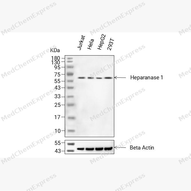 Western blot analysis of extracts from Jurkat (lane 2(20μg), HeLa (lane 3(20μg), HepG2 (lane 4(20μg) and 293T (lane 5(20μg) using Heparanase 1 Antibody (HY-P81856) Rabbit mAb. Proteins were transferred to a PVDF membrane and blocked with 5% non-fat milk in TBST for 2 hour at room temperature. The primary antibody (1/1000)and Loading control antibody (Beta Actin, HY-P80993, 1/10000) was used in 5% non-fat milk in TBST at 4°C overnight. Goat Anti-Rabbit IgG-HRP Secondary Antibody (1/10000) was used for 1 hour at room temperature.


Background
-
Function
Heparanase 1 is an Endoglycosidase that cleaves heparan sulfate proteoglycans (HSPGs) into heparan sulfate side chains and core proteoglycans. Participates in extracellular matrix (ECM) degradation and remodeling. Selectively cleaves the linkage between a glucuronic acid unit and an N-sulfo glucosamine unit carrying either a 3-O-sulfo or a 6-O-sulfo group. Can also cleave the linkage between a glucuronic acid unit and an N-sulfo glucosamine unit carrying a 2-O-sulfo group, but not linkages between a glucuronic acid unit and a 2-O-sulfated iduronic acid moiety. It is essentially inactive at neutral pH but becomes active under acidic conditions such as during tumor invasion and in inflammatory processes. Facilitates cell migration associated with metastasis, wound healing and inflammation. Enhances shedding of syndecans, and increases endothelial invasion and angiogenesis in myelomas. Acts as a procoagulant by increasing the generation of activation factor X in the presence of tissue factor and activation factor VII. Increases cell adhesion to the extracellular matrix (ECM), independent of its enzymatic activity. Induces AKT1/PKB phosphorylation via lipid rafts increasing cell mobility and invasion. Heparin increases this AKT1/PKB activation. Regulates osteogenesis. Enhances angiogenesis through up-regulation of SRC-mediated activation of VEGF. Implicated in hair follicle inner root sheath differentiation and hair homeostasis
-
Subcellular Localization
Lysosome membrane; Peripheral membrane protein; Secreted; Nucleus
-
Expression
Tissue_specificity:Highly expressed in placenta and spleen and weakly expressed in lymph node, thymus, peripheral blood leukocytes, bone marrow, endothelial cells, fetal liver and tumor tissues. Also expressed in hair follicles, specifically in both Henle's and Huxley's layers of inner the root sheath (IRS) at anagen phase -
Isoforms & Post-Translational Modification
Q9Y251 has 4 isomers: Q9Y251-1: 61149 Da (predicted); Q9Y251-2: 54734 Da (predicted); Q9Y251-3: 53161 Da (predicted); Q9Y251-4: 42791 Da (predicted).
Proteolytically processed. The cleavage of the 65 kDa form leads to the generation of a linker peptide, and 8 kDa and 50 kDa products. The active form, the 8/50 kDa heterodimer, is resistant to degradation. Complete removal of the linker peptide appears to be a prerequisite to the complete activation of the enzyme;N-glycosylated. Glycosylation of the 50 kDa subunit appears to be essential for its solubility -
Subunit
Heterodimer; heterodimer formation between the 8 kDa and the 50 kDa subunits is required for enzyme activity. Interacts with TF; the interaction, inhibited by heparin, enhances the generation of activated factor X and activates coagulation. Interacts with HRG; the interaction is enhanced at acidic pH, partially inhibits binding of HPSE to cell surface receptors and modulates its enzymatic activity. Interacts with SDC1; the interaction enhances the shedding of SDC1. Interacts with HPSE2
-
SwissProt ID
-
Synonyms
HEP; Heparanase; Heparanase1; Hpa 1; HPA; Hpa1 HPR1; HPSE1; HSE1
-
Research Field
Tags & Cell Markers


Documentation
-
Data Sheet (234 KB)
-
SDS (251 KB)
- English - EN (251 KB)
- Français - FR (251 KB)
- Deutsch - DE (251 KB)
- Norwegian - NO (251 KB)
- Español - ES (251 KB)
- Swedish - SV (251 KB)
- Italian - IT (251 KB)
- Korean - KR (251 KB)
- Portuguese - PT (251 KB)
-
User Guide for Antibodies (1077 KB)

 Powered by Bioz
