



Phospho-p53 (Ser6) Antibody (YA166)
(Synonyms: TP53; P53; Cellular tumor antigen p53; Antigen NY-CO-13; Phosphoprotein p53; Tumor suppressor p53)
Based on 1 Customer Validation
Phospho-p53 (Ser6) Antibody (YA166) is a Rabbit-derived and non-conjugated IgG monoclonal antibody, targeting to Phospho-p53 (Ser6).
-
Host:
Rabbit
-
Isotype:
IgG
-
Application:
WB, IHC-P, IP
-
Reactivity :
Human
-
Formulation:
Supplied in 50 mM Tris-Glycine (pH 7.4), 0.15 M NaCl, 40% Glycerol and 0.05% BSA. Preservative: 0.01% Sodium azide
-
Conjugation:
Non-conjugated


To place orders, for customer services and technical support, please contact: MedChemExpress USA
Tel: 609-228-6898 E-mail: [email protected] [email protected]





Applications
| Application |
WB

WB: Western Blot
|
IHC-P
IHC-P: Immunohistochemistry-Paraffin
|
IP
IP: Immunoprecipitation
|
|---|---|---|---|
| Dilution Ratio | 1:500-1:1000 | 1:50-1:100 | 1:20 |


Product Details
Phospho-p53 (Ser6) Antibody (YA166) is a Rabbit-derived and non-conjugated IgG monoclonal antibody, targeting to Phospho-p53 (Ser6).
-
Host Rabbit

-
Clonality Recombinant,Monoclonal

-
Species ReactivityHuman
-
Observed Molecular WeightObserved band size: 53 kDa
Note: Due to possible protein modifications or aggregation, the molecular weight should be confirmed by actual measurement, and the predicted value is for reference only.

-
Calculated Molecular Weight Predicted band size: 44 kDa

Entrez Gene: 7157 Human ; 22059 Mouse ; 24842 Rat
SwissProt: P04637 Human ; P02340 Mouse ; P10361 Rat
OMIM: 133239 Human
Synthetic phosphopeptide corresponding to residues surrounding Ser6 of Human p53.The exact sequence is proprietary to MCE.
Endogenous
affinity purified
Non-conjugated
Phosphorylated
IgG


Product Properties
-
Appearance
Solution
-
Formulation
Supplied in 50 mM Tris-Glycine (pH 7.4), 0.15 M NaCl, 40% Glycerol and 0.05% BSA. Preservative: 0.01% Sodium azide
-
Concentration
Batch-dependent, Please check the COA for the concentration of each lot. Check Lot Concentration
-
Storage & Stability
Stored at -20°C for 1 year. Avoid repeated freeze / thaw cycles.
-
Shipping
Shipping with blue ice.


Verification Images
-
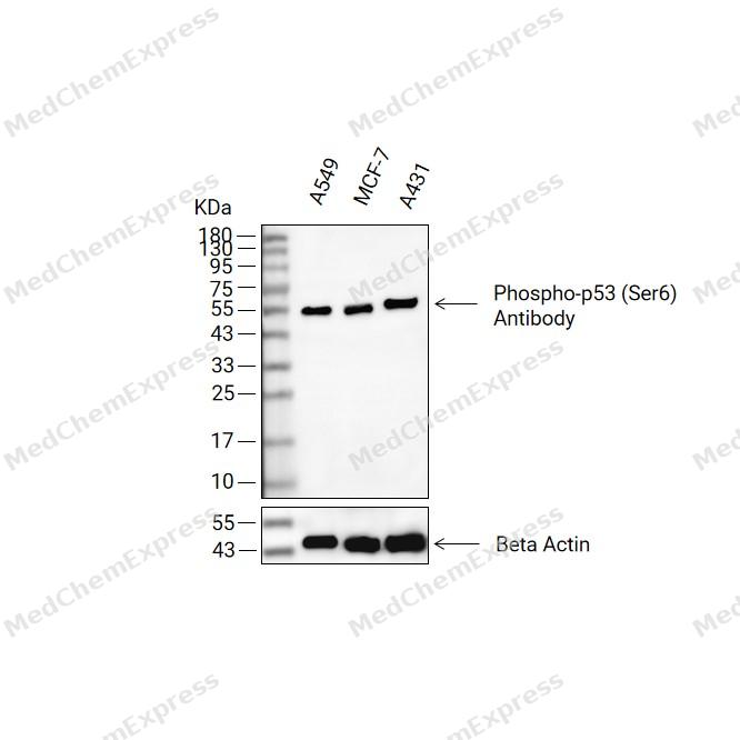 Western blot analysis of extracts from A549(lane 2(20ug) , MCF-7(lane 3(20ug) and A431(lane 4(20ug) using Phospho-p53(Ser6) Antibody (HY-P80844) Rabbit mAb. Proteins were transferred to a PVDF membrane and blocked with 5% non-fat milk in TBST for 2 hour at room temperature. The primary antibody (1/1000) and Loading control antibody (Beta Actin, HY-P83730, 1/10000) was used in 5% non-fat milk in TBST at 4°C overnight. Goat Anti-Mouse/Rabbit IgG-HRP Secondary Antibody (1/10000) was used for 1 hour at room temperature.


Background
-
Function
p53 multifunctional transcription factor that induces cell cycle arrest, DNA repair or apoptosis upon binding to its target DNA sequence. Acts as a tumor suppressor in many tumor types; induces growth arrest or apoptosis depending on the physiological circumstances and cell type. Negatively regulates cell division by controlling expression of a set of genes required for this process. One of the activated genes is an inhibitor of cyclin-dependent kinases. Apoptosis induction seems to be mediated either by stimulation of BAX and FAS antigen expression, or by repression of Bcl-2 expression. Its pro-apoptotic activity is activated via its interaction with PPP1R13B/ASPP1 or TP53BP2/ASPP2. However, this activity is inhibited when the interaction with PPP1R13B/ASPP1 or TP53BP2/ASPP2 is displaced by PPP1R13L/iASPP. In cooperation with mitochondrial PPIF is involved in activating oxidative stress-induced necrosis; the function is largely independent of transcription. Induces the transcription of long intergenic non-coding RNA p21 (lincRNA-p21) and lincRNA-Mkln1. LincRNA-p21 participates in TP53-dependent transcriptional repression leading to apoptosis and seems to have an effect on cell-cycle regulation. Implicated in Notch signaling cross-over. Prevents CDK7 kinase activity when associated to CAK complex in response to DNA damage, thus stopping cell cycle progression. Isoform 2 enhances the transactivation activity of isoform 1 from some but not all TP53-inducible promoters. Isoform 4 suppresses transactivation activity and impairs growth suppression mediated by isoform 1. Isoform 7 inhibits isoform 1-mediated apoptosis. Regulates the circadian clock by repressing CLOCK-BMAL1-mediated transcriptional activation of PER2[1][2][3][4][5][6][7][8][9][10][11][12][13][14][15][16][17][18].
-
Subcellular Localization
Cytoplasm; Nucleus; Nucleus, PML body; Endoplasmic reticulum; Mitochondrion matrix; Cytoplasm, cytoskeleton, microtubule organizing center, centrosome; Nucleus; Cytoplasm; Nucleus; Cytoplasm; Nucleus; Cytoplasm; Nucleus; Cytoplasm; Nucleus; Cytoplasm; Nucleus; Cytoplasm; Cytoplasm
-
Expression
Tissue_specificity:These subtypes are ubiquitous. They are expressed in a variety of normal tissues, but the mode of expression is tissue-dependent. Subtype 2 is expressed in most normal tissues, but not detected in the brain, lungs, prostate, muscle, fetal brain, spinal cord, and fetal liver. Subtype 3 is expressed in most normal tissues, but not detected in the lungs, spleen, testes, fetal brain, spinal cord, and fetal liver. Subtype 7 is expressed in most normal tissues, but not detected in the prostate, uterus, skeletal muscle, and breast. Subtype 8 is detected only in the colon, bone marrow, testes, fetal brain, and intestines. Subtype 9 is expressed in most normal tissues, but not detected in the brain, heart, lungs, fetal liver, salivary glands, breast, or intestines.
Induction:Up-regulated in response to DNA damage. Isoform 2 is not induced in tumor cells in response to stress -
Isoforms & Post-Translational Modification
p53 has 9 isomers: P04637-1: 43653 Da (predicted); P04637-2: 37826 Da (predicted); P04637-3: 38501 Da (predicted); P04637-4: 39320 Da (predicted); P04637-5: 33493 Da (predicted); P04637-6: 34168 Da (predicted); P04637-7: 29553 Da (Predicted); P04637-8: 23726 Da (predicted). P04637-9: 24401 Da (predicted).
CREBBP acetylation of Lys-382 enhances transcriptional activity. EP300 acetylation of Lys-382. SIRT1 deacetylation of Lys-382 weakens its ability to induce apoptosis and regulate cellular senescence. SIRT2 deacetylation of Lys-382 weakens its ability to induce transcriptional activation in an AKT-dependent manner. Acetylation of Lys-381 increases its stability. SIRT6 deacetylation of Lys-381 reduces its stability, thereby regulating cellular senescence. KAT5, KAT6A, and KAT8 acetylation of Lys-120 regulates its ability to induce apoptosis. Lactation of AARS1 prevents liquid-liquid phase separation (LLPS), thereby inhibiting transcription factor activity. Phosphorylation of Ser residues mediates transcriptional activation. Phosphorylation of Ser-9 by HIPK4 enhances its repressive activity against the BIRC5 promoter. VRK1 phosphorylates the Thr-18 site. Following DNA damage, CHEK2 phosphorylates the Ser-20 site, thereby preventing MDM2 ubiquitination. Phosphorylation of the Ser-46 site is essential for CREBBP acetylation. Both isoform 1 and isoform 2 of VRK2 phosphorylate the Thr-18 site. Phosphorylation of the Thr-18 site by VRK2 isoform 2 leads to reduced MDM2-mediated ubiquitination and increased EP300-mediated acetylation. p53/TP53 is stabilized in genotoxic and oxidative stress responses via CDK5-mediated phosphorylation at Ser-15, Ser-33, and Ser-46 sites, leading to p53/TP53 accumulation, particularly in the nucleus, which in turn induces transcriptional activation of p53/TP53 target genes. Phosphorylation at Ser-392 modulates the ability of TP53/p53 aggregates to undergo liquid-liquid phase separation by increasing their fluidity; it is dephosphorylated at Thr-55 by the PP2A-PPP2R5C holoenzyme. The SV40 small T antigen inhibits dephosphorylation of the PP2A AC form; O-glycosylation may occur in the C-terminal basic region. Studies have been conducted in the EB-1 cell line; it is ubiquitinated by MDM2 and SYVN1, leading to proteasome degradation. p53/TP53 can be ubiquitinated by RFWD3, which works synergistically with MDM2. p53/TP53 can be ubiquitinated by MKRN1 at Lys-291 and Lys-292, leading to proteasomal degradation. p53/TP53 can be deubiquitinated by USP10, resulting in its stability. p53/TP53 can be ubiquitinated by TRIM24, RFFL, RNF34, and RNF125, leading to proteasomal degradation. TOPORS-mediated ubiquitination leads to degradation. USP7-mediated deubiquitination leads to stability. Isomer 4 undergoes monoubiquitination in an MDM2-independent manner. COP1-mediated ubiquitination leads to proteasomal degradation. Ubiquitination and subsequent proteasomal degradation are negatively regulated by CCAR2. Polyubiquitination at Lys-24 by MUL1 leads to proteasomal degradation. Deubiquitination at USP3 leads to stability. Ubiquitination at MSL2 promotes its cytoplasmic localization. Furthermore, it can be ubiquitinated by the SCF (FBXO22)-KDMA4A complex, leading to proteasome degradation; monomethylation at Lys-372 by SETD7 stabilizes it and enhances transcriptional activity. SMYD2 monomethylation at Lys-370 reduces DNA binding activity, thus affecting transcriptional regulation. Monomethylation at Lys-372 prevents its interaction with SMYD2 and inhibits subsequent monomethylation at Lys-370. EHMT1 and EHMT2 dimethylate Lys-373. KMT5A monomethylates Lys-382, promoting its interaction with L3MBTL1 and thus inhibiting transcriptional activity. Dimethylation at Lys-370 and Lys-382 reduces p53 ubiquitination levels by stabilizing the binding of p53 to the methyl readout protein PHF20. UBC9 performs SUMO modification on Lys-386. -
Subunit
Forms homodimers and homotetramers. Binds DNA as a homotetramer. Interacts with AXIN1. Interacts with histone acetyltransferases EP300 and methyltransferases HRMT1L2 and CARM1, and recruits them to promoters. Interacts (via C-terminus) with TAF1; when TAF1 is part of the TFIID complex. Interacts with ING4; this interaction may be indirect. Found in a complex with CABLES1 and TP73. Interacts with HIPK1, HIPK2, and TP53INP1. Interacts with WWOX. May interact with HCV core protein. Interacts with USP7 and SYVN1. Interacts with HSP90AB1. Interacts with ARMC10, CDKN2AIP, NUAK1, STK11/LKB1, UHRF2 and E4F1. Interacts with YWHAZ; the interaction enhances TP53 transcriptional activity. Phosphorylation of YWHAZ on 'Ser-58' inhibits this interaction. Interacts (via DNA-binding domain) with MAML1 (via N-terminus). Interacts with MKRN1. Interacts with PML (via C-terminus). Interacts with MDM2; leading to ubiquitination and proteasomal degradation of TP53. Directly interacts with FBXO42; leading to ubiquitination and degradation of TP53.
-
SwissProt ID
-
Synonyms
TP53; P53; Cellular tumor antigen p53; Antigen NY-CO-13; Phosphoprotein p53; Tumor suppressor p53
-
Research Field
Cell Biology


Documentation
-
Data Sheet (263 KB)
-
SDS (252 KB)
- English - EN (252 KB)
- Français - FR (252 KB)
- Deutsch - DE (252 KB)
- Norwegian - NO (252 KB)
- Español - ES (252 KB)
- Swedish - SV (252 KB)
- Italian - IT (252 KB)
- Korean - KR (252 KB)
- Portuguese - PT (252 KB)
-
User Guide for Antibodies (1077 KB)
References
[1]. Guo A, et al. The function of PML in p53-dependent apoptosis. Nat Cell Biol. 2000 Oct;2(10):730-6. [Content Brief]
[2]. Bergamaschi D, et al. iASPP oncoprotein is a key inhibitor of p53 conserved from worm to human. Nat Genet. 2003 Feb;33(2):162-7. [Content Brief]
[3]. Louria-Hayon I, et al. The promyelocytic leukemia protein protects p53 from Mdm2-mediated inhibition and degradation. J Biol Chem. 2003 Aug 29;278(35):33134-41. [Content Brief]
[4]. An W, et al. Ordered cooperative functions of PRMT1, p300, and CARM1 in transcriptional activation by p53. Cell. 2004 Jun 11;117(6):735-48. [Content Brief]
[5]. Ghosh A, et al. Regulation of human p53 activity and cell localization by alternative splicing. Mol Cell Biol. 2004 Sep;24(18):7987-97. [Content Brief]
[6]. Zhao Y, et al. The notch regulator MAML1 interacts with p53 and functions as a coactivator. J Biol Chem. 2007 Apr 20;282(16):11969-81. [Content Brief]
[7]. Taira N, et al. DYRK2 is targeted to the nucleus and controls p53 via Ser46 phosphorylation in the apoptotic response to DNA damage. Mol Cell. 2007 Mar 9;25(5):725-38. [Content Brief]
[8]. Allton K, et al. Trim24 targets endogenous p53 for degradation. Proc Natl Acad Sci U S A. 2009 Jul 14;106(28):11612-6. [Content Brief]
[9]. Huarte M, et al. A large intergenic noncoding RNA induced by p53 mediates global gene repression in the p53 response. Cell. 2010 Aug 6;142(3):409-19. [Content Brief]
[10]. Wu L, et al. Aurora B interacts with NIR-p53, leading to p53 phosphorylation in its DNA-binding domain and subsequent functional suppression. J Biol Chem. 2011 Jan 21;286(3):2236-44. [Content Brief]
[11]. Vaseva AV, et al. p53 opens the mitochondrial permeability transition pore to trigger necrosis. Cell. 2012 Jun 22;149(7):1536-48. [Content Brief]
[12]. Miki T, et al. p53 regulates Period2 expression and the circadian clock. Nat Commun. 2013;4:2444. [Content Brief]
[13]. Polato F, et al. DRAGO (KIAA0247), a new DNA damage-responsive, p53-inducible gene that cooperates with p53 as oncosuppressor. [Corrected]. J Natl Cancer Inst. 2014 Apr;106(4):dju053. [Content Brief]
[14]. Dai Z, et al. Ser392 phosphorylation modulated a switch between p53 and transcriptional condensates. Biochim Biophys Acta Gene Regul Mech. 2022 May;1865(4):194827. [Content Brief]
[15]. Chen Q, et al. Phosphorylation and specific DNA improved the incorporation ability of p53 into functional condensates. Int J Biol Macromol. 2023 Mar 1;230:123221. [Content Brief]
[16]. Zong Z, et al. Alanyl-tRNA synthetase, AARS1, is a lactate sensor and lactyltransferase that lactylates p53 and contributes to tumorigenesis. Cell. 2024 May 9;187(10):2375-2392.e33. [Content Brief]
[17]. Schneider E, et al. Regulation of CAK kinase activity by p53. Oncogene. 1998 Nov 26;17(21):2733-41. [Content Brief]
[18]. Sykes SM, et al. Acetylation of the p53 DNA-binding domain regulates apoptosis induction. Mol Cell. 2006 Dec 28;24(6):841-51. [Content Brief]

 Powered by Bioz
