



phospho-PI3 Kinase p110 beta (Ser1070) Antibody
(Synonyms: PI3-kinase p110 subunit beta; p-PI3Kβ(Ser1070); PI 3-kinase C2β; p110 BETA; p110Beta; Phosphatidylinositol 3 kinase catalytic beta polypeptide; Phosphatidylinositol 4 5 bisphosphate 3 kinase 110 kDa catalytic subunit beta; Phosphatidylinositol 4 5 bisphosphate 3 kinase catalytic subunit beta isoform; Phosphatidylinositol-4; Phosphoinositide 3 kinase catalytic beta polypeptide; PI3 kinase p110 subunit beta; PI3-kinase subunit beta; PI3K; PI3K beta; PI3K-beta; PI3Kbeta; PI3KCB; PIK3C1; Pik3cb; PK3CB_HUMAN; PtdIns 3 kinase p110; PtdIns-3-kinase subunit beta; PtdIns-3-kinase subunit p110-beta; 5-bisphosphate 3-kinase 110 kDa catalytic subunit beta; 5-bisphosphate 3-kinase catalytic subunit beta isoform.)Based on 7 publication(s) in Google Scholar
phospho-PI3 Kinase p110 beta (Ser1070) Antibody is a Rabbit-derived and non-conjugated IgG polyclonal antibody, targeting to phospho-PI3 Kinase p110 beta (Ser1070).
-
Host:
Rabbit
-
Isotype:
IgG
-
Application:
WB, IHC-P, IHC-F, ICC/IF
-
Reactivity :
Human, Mouse, Rat
-
Formulation:
Supplied in 0.01M TBS(pH7.4) with 1% BSA, 0.03% Proclin300 and 50% Glycerol.
-
Conjugation:
Non-conjugated


To place orders, for customer services and technical support, please contact: MedChemExpress USA
Tel: 609-228-6898 E-mail: [email protected] [email protected]



-

HY-P80535
AKT1+AKT2+AKT3 Antibody (YA635) -
HY-P80276
Phospho-AKT1(Ser473) Antibody (YA227) -
HY-P80788
Phospho-AKT1 (Ser473) Antibody (YA228) -
HY-P80789
Phospho-AKT1/2/3 (Thr308) Antibody -
HY-P80231
mTOR Antibody (YA281) -

HY-K0022
Phosphatase Inhibitor Cocktail Ⅱ (100× in ddH2O) -

HY-K0021
Phosphatase Inhibitor Cocktail I (100× in DMSO) -
HY-P80837
Phospho-mTOR (Ser2448) Antibody (YA171) -
HY-P80846
Phospho-PI3 Kinase p85/p55 (Tyr467/Tyr199) Antibody -
HY-P80008
AKT1 Antibody (YA634) -
HY-P80009
AKT1/2/3 Antibody (YA633) -
HY-P80029
Bcl-2 Antibody (YA589)
Publications Citing Use of MedChemExpress (MCE) phospho-PI3 Kinase p110 beta (Ser1070) Antibody
More- Phytomedicine. 2025 Jul 25:143:156788. [Abstract]
- J Mater Chem B. 2025 Oct 22;13(41):13405-13422. [Abstract]
- Drug Des Devel Ther. 2025 Jan 17:19:349-368. [Abstract]
- Biochim Biophys Acta Mol Basis Dis. 2026 Aug;1872(6):168242. [Abstract]
- Sci Rep. 2025 Jan 28;15(1):3533. [Abstract]
- Toxicol Appl Pharmacol. 2025 Dec:505:117545. [Abstract]
- Arch Biochem Biophys. 2024 May:755:109983. [Abstract]

 7 Publications Citing Use of MCE phospho-PI3 Kinase p110 beta (Ser1070) Antibody (8)
-
WB
-
WB
-
WB
-
WB
-
WB


Applications
| Application |
WB

WB: Western Blot
|
IHC-P
IHC-P: Immunohistochemistry-Paraffin
|
IHC-F
IHC-F: Immunohistochemistry-Frozen
|
ICC/IF
ICC/IF: Immunocytochemistry/
Immunofluorescence |
|---|---|---|---|---|
| Dilution Ratio | 1:500-2000 | 1:100-500 | 1:100-500 | 1:100-500 |


Product Details
phospho-PI3 Kinase p110 beta (Ser1070) Antibody is a Rabbit-derived and non-conjugated IgG polyclonal antibody, targeting to phospho-PI3 Kinase p110 beta (Ser1070).
-
Host Rabbit

-
Clonality Polyclonal

-
Species ReactivityHuman, Mouse, Rat Predicted Reactivity: Chicken,Dog,Pig,Cow,Horse,GuineaPig
Note: The predicted reactivity is for reference only and should not be considered a guarantee of product performance.
-
Observed Molecular WeightObserved band size: 110 kDa
Note: Due to possible protein modifications or aggregation, the molecular weight should be confirmed by actual measurement, and the predicted value is for reference only.

-
Calculated Molecular Weight Predicted band size: 110-123 kDa

Entrez Gene: 5291 Human ; 74769 Mouse ;
SwissProt: P42338 Human ; Q3U4Q1 Mouse ; Q8BTI9 Mouse ;
OMIM: 602925 Human
KLH conjugated synthesised phosphopeptide derived from human PI3 Kinase p110 beta around the phosphorylation site of Ser1070: YR(p-S)
Endogenous
affinity purified
Non-conjugated
Phosphorylated
IgG


Product Properties
-
Appearance
Solution
-
Formulation
Supplied in 0.01M TBS(pH7.4) with 1% BSA, 0.03% Proclin300 and 50% Glycerol.
-
Concentration
Batch-dependent, Please check the COA for the concentration of each lot. Check Lot Concentration
-
Storage & Stability
Stored at -20°C for 1 year. Avoid repeated freeze / thaw cycles.
-
Shipping
Shipping with blue ice.


Publications (7)
-
Journal Impact Factor
-
Most Recent
-
Phytomedicine
Halofuginone targets Serine/Glycine synthesis to reverse epidermal growth factor receptor Tyrosine Kinase inhibitor resistance in lung adenocarcinoma. [Abstract]2025 Jul 25:143:156788. PMID: 40354707 -
J Mater Chem B
A metal-organic framework-based co-delivery system for atherosclerosis therapy via macrophage regulation. [Abstract]2025 Oct 22;13(41):13405-13422. PMID: 41002166 -
Drug Des Devel Ther
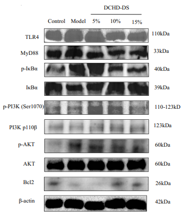
Exploring the Anti-PANoptosis Mechanism of Dachaihu Decoction Against Sepsis-Induced Acute Lung Injury: Network Pharmacology, Bioinformatics, and Experimental Validation. [Abstract]2025 Jan 17:19:349-368. PMID: 39839500
phospho-PI3 Kinase p110 beta (Ser1070) Antibody purchased from MedChemExpress. Usage Cited in: Drug Des Devel Ther. 2025 Jan 17:19:349-368. [Abstract]
DCHD inhibited PANoptosis by regulating the PI3K/AKT/NF-κB signaling pathway.Analysis and quantification of TLR4, Myd88, p-IκBα, p-PI3K, p-AKT, and Bcl2 expression using Western blotting in RAW 264.7 Macrophages.
-
Biochim Biophys Acta Mol Basis Dis
KAT2B inhibits cholangiocarcinoma proliferation by binding with histone demethylase KDM6B. [Abstract]2026 Aug;1872(6):168242. PMID: 41951013 -
Toxicol Appl Pharmacol
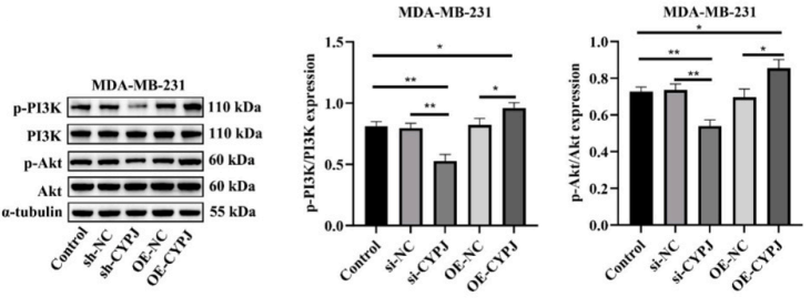
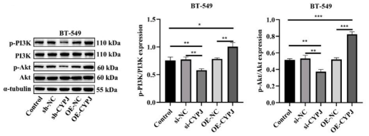
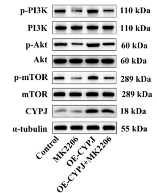
Cyclophilin J inhibited oxidative stress and apoptosis in triple-negative breast cancer cell through activating the PI3K/Akt signaling pathway. [Abstract]2025 Dec:505:117545. PMID: 40915586
phospho-PI3 Kinase p110 beta (Ser1070) Antibody purchased from MedChemExpress. Usage Cited in: Toxicol Appl Pharmacol. 2025 Dec:505:117545. [Abstract]
The proteins of the PI3K/Akt pathway were detected through western blot in MDA-MB-231 cells.
phospho-PI3 Kinase p110 beta (Ser1070) Antibody purchased from MedChemExpress. Usage Cited in: Toxicol Appl Pharmacol. 2025 Dec:505:117545. [Abstract]
The proteins of the PI3K/Akt pathway were detected through western blot in BT-549 cells.
phospho-PI3 Kinase p110 beta (Ser1070) Antibody purchased from MedChemExpress. Usage Cited in: Toxicol Appl Pharmacol. 2025 Dec:505:117545. [Abstract]
The proteins of the PI3K/Akt pathway and CYPJ were detected through Western blot in tumor tissues.
-
Arch Biochem Biophys
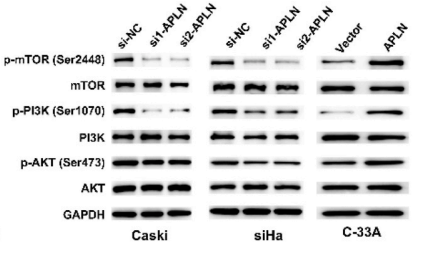
APLN promotes the proliferation, migration, and glycolysis of cervical cancer through the PI3K/AKT/mTOR pathway. [Abstract]2024 May:755:109983. PMID: 38561035
phospho-PI3 Kinase p110 beta (Ser1070) Antibody purchased from MedChemExpress. Usage Cited in: Arch Biochem Biophys. 2024 May:755:109983. [Abstract]
Western blotting of the PI3K/AKT/mTOR pathway-related proteins in caski, siHa, C-33A cells.
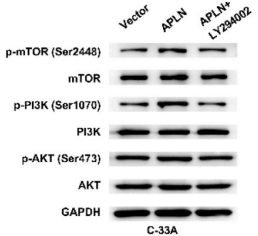
phospho-PI3 Kinase p110 beta (Ser1070) Antibody purchased from MedChemExpress. Usage Cited in: Arch Biochem Biophys. 2024 May:755:109983. [Abstract]
PI3K inhibitor LY294002 was added in APLN overexpressed C-33A cells. Western blotting of the PI3K/AKT/mTOR pathway-related proteins.
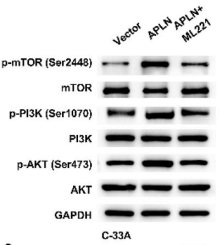
phospho-PI3 Kinase p110 beta (Ser1070) Antibody purchased from MedChemExpress. Usage Cited in: Arch Biochem Biophys. 2024 May:755:109983. [Abstract]
APLN receptor inhibitor ML221 was added in APLN overexpressed C-33A cells. Western blotting of the PI3K/AKT/mTOR pathway-related proteins.
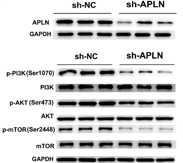
phospho-PI3 Kinase p110 beta (Ser1070) Antibody purchased from MedChemExpress. Usage Cited in: Arch Biochem Biophys. 2024 May:755:109983. [Abstract]
Western blotting of APLN and the PI3K/AKT/mTOR pathwayrelated proteins in APLN knockdown in CaSki cells.


Verification Images
-
 Western blot analysis of extracts from K562 (lane2, 20μg), MCF7 (lane3, 20μg), Jurkat (lane4, 20μg) and Hela (lane5, 20μg) using phospho-PI3 Kinase p110 beta (Ser1070) Antibody (HY-P81211). Proteins were transferred to a PVDF membrane and blocked with 5% BSA in TBST for 2 hour at room temperature. The primary antibody (1/1000) and Loading control antibody (Beta Actin, HY-P80993, 1/10,000) was used in 5% BSA in TBST at 4°C overnight. Goat Anti-Rabbit IgG-HRP Secondary Antibody (HY-P8001,1/10,000) was used for 1 hour at room temperature.
 -
 Immunohistochemical analysis of paraffin-embedded human breast cancer tissue using phospho-PI3 Kinase p110 beta (Ser1070) antibody. The section was pre-treated using heat mediated antigen retrieval with Tris/EDTA buffer (pH 9.0) for 20 minutes. The tissues were probed with the primary antibody (HY-P81211, 1/100) overnight at 4℃. The detection was performed using an HRP conjugated compact polymer system. DAB was used as the chromogen. Tissues were counterstained with hematoxylin and mounted with neutral balsam.
 -
 Immunohistochemical analysis of paraffin-embedded human endometrial cancer tissue using phospho-PI3 Kinase p110 beta (Ser1070) antibody. The section was pre-treated using heat mediated antigen retrieval with Tris/EDTA buffer (pH 9.0) for 20 minutes. The tissues were probed with the primary antibody (HY-P81211, 1/100) overnight at 4℃. The detection was performed using an HRP conjugated compact polymer system. DAB was used as the chromogen. Tissues were counterstained with hematoxylin and mounted with neutral balsam.
 -
 Immunohistochemical analysis of paraffin-embedded human colon cancer tissue using phospho-PI3 Kinase p110 beta (Ser1070) antibody. The section was pre-treated using heat mediated antigen retrieval with Tris/EDTA buffer (pH 9.0) for 20 minutes. The tissues were probed with the primary antibody (HY-P81211, 1/100) overnight at 4℃. The detection was performed using an HRP conjugated compact polymer system. DAB was used as the chromogen. Tissues were counterstained with hematoxylin and mounted with neutral balsam.
 -
 Immunocytochemistry analysis of Hela cells labeling phospho-PI3 Kinase p110 beta (Ser1070) with phospho-PI3 Kinase p110 beta (Ser1070) Antibody (HY-P81211) at 1/100 dilution. Cells were fixed in 4% paraformaldehyde for 15 minutes at room temperature, permeabilized with 0.1% Triton X-100 in PBS for 15 minutes at room temperature, then blocked with quick block buffer for 10 minutes at room temperature. Cells were then incubated with phospho-PI3 Kinase p110 beta (Ser1070) Antibody (HY-P81211) at 1/100 dilution in quick block buffer overnight at 4 ℃. AF488-conjugated Goat Anti-Rabbit IgG H&L (HY-P8002, Green) was used as the secondary antibody at 1/1,000 dilution. PBS instead of the primary antibody was used as the secondary antibody only control. The Nuclear counterstain was DAPI (Blue).
 -
 Immunocytochemistry analysis of Hela cells labeling phospho-PI3 Kinase p110 beta (Ser1070) with phospho-PI3 Kinase p110 beta (Ser1070) Antibody (HY-P81211) at 1/300 dilution. Cells were fixed in 4% paraformaldehyde for 15 minutes at room temperature, permeabilized with 0.1% Triton X-100 in PBS for 15 minutes at room temperature, then blocked with quick block buffer for 10 minutes at room temperature. Cells were then incubated with phospho-PI3 Kinase p110 beta (Ser1070) Antibody (HY-P81211) at 1/300 dilution in quick block buffer overnight at 4 ℃. AF488-conjugated Goat Anti-Rabbit IgG H&L (HY-P8002, Green) was used as the secondary antibody at 1/1,000 dilution. PBS instead of the primary antibody was used as the secondary antibody only control. The Nuclear counterstain was DAPI (Blue).
 -
 Tyramide signaling amplification based immunofluorescence analysis of paraffin-embedded human prostate cancer tissue using phospho-PI3 Kinase p110 beta (Ser1070) antibody. The section was pre-treated using heat mediated antigen retrieval with Tris/EDTA buffer (pH 9.0) for 20 minutes. The tissues were probed with the primary antibody (HY-P81211, 1/300) overnight at 4℃. The detection was performed using an HRP conjugated compact polymer and fluorescent tyramide signal amplification system.Immunostaining was performed using a Bicolor signal amplification fluorescence staining kit. Tissues were counterstained with DAPI (blue) and mounted with Anti-fade fluorescence mounting medium.
 -
 Tyramide signaling amplification based immunofluorescence analysis of paraffin-embedded human breast cancer tissue using phospho-PI3 Kinase p110 beta (Ser1070) antibody. The section was pre-treated using heat mediated antigen retrieval with Tris/EDTA buffer (pH 9.0) for 20 minutes. The tissues were probed with the primary antibody (HY-P81211, 1/500) overnight at 4℃. The detection was performed using an HRP conjugated compact polymer and fluorescent tyramide signal amplification system.Immunostaining was performed with Vari Fluor 594 TSA (200×)(HY-D1835). Tissues were counterstained with DAPI (blue) and mounted with Anti-fade fluorescence mounting medium.
 -
 Tyramide signaling amplification based immunofluorescence analysis of paraffin-embedded human endometrial cancer tissue using phospho-PI3 Kinase p110 beta (Ser1070) antibody. The section was pre-treated using heat mediated antigen retrieval with Tris/EDTA buffer (pH 9.0) for 20 minutes. The tissues were probed with the primary antibody (HY-P81211, 1/500) overnight at 4℃. The detection was performed using an HRP conjugated compact polymer and fluorescent tyramide signal amplification system.Immunostaining was performed with Vari Fluor 594 TSA (200×)(HY-D1835). Tissues were counterstained with DAPI (blue) and mounted with Anti-fade fluorescence mounting medium.
 -
 Tyramide signaling amplification based immunofluorescence analysis of paraffin-embedded human colon cancer tissue using phospho-PI3 Kinase p110 beta (Ser1070) antibody. The section was pre-treated using heat mediated antigen retrieval with Tris/EDTA buffer (pH 9.0) for 20 minutes. The tissues were probed with the primary antibody (HY-P81211, 1/500) overnight at 4℃. The detection was performed using an HRP conjugated compact polymer and fluorescent tyramide signal amplification system.Immunostaining was performed with Vari Fluor 594 TSA (200×)(HY-D1835). Tissues were counterstained with DAPI (blue) and mounted with Anti-fade fluorescence mounting medium.


Background
-
Function
PI3 Kinase p110 beta is a Phosphoinositide-3-kinase (PI3K) phosphorylates phosphatidylinositol derivatives at position 3 of the inositol ring to produce 3-phosphoinositides. Uses ATP and PtdIns(4,5)P2 (phosphatidylinositol 4,5-bisphosphate) to generate phosphatidylinositol 3,4,5-trisphosphate (PIP3). PIP3 plays a key role by recruiting PH domain-containing proteins to the membrane, including AKT1 and PDPK1, activating signaling cascades involved in cell growth, survival, proliferation, motility and morphology. Involved in the activation of AKT1 upon stimulation by G-protein coupled receptors (GPCRs) ligands such as CXCL12, sphingosine 1-phosphate, and lysophosphatidic acid. May also act downstream receptor tyrosine kinases. Required in different signaling pathways for stable platelet adhesion and aggregation. Plays a role in platelet activation signaling triggered by GPCRs, alpha-IIb/beta-3 integrins (ITGA2B/ ITGB3) and ITAM (immunoreceptor tyrosine-based activation motif)-bearing receptors such as GP6. Regulates the strength of adhesion of ITGA2B/ ITGB3 activated receptors necessary for the cellular transmission of contractile forces. Required for platelet aggregation induced by F2 (thrombin) and thromboxane A2 (TXA2). Has a role in cell survival. May have a role in cell migration. Involved in the early stage of autophagosome formation. Modulates the intracellular level of PtdIns3P (phosphatidylinositol 3-phosphate) and activates PIK3C3 kinase activity. May act as a scaffold, independently of its lipid kinase activity to positively regulate autophagy. May have a role in insulin signaling as scaffolding protein in which the lipid kinase activity is not required. May have a kinase-independent function in regulating cell proliferation and in clathrin-mediated endocytosis. Mediator of oncogenic signal in cell lines lacking PTEN. The lipid kinase activity is necessary for its role in oncogenic transformation. Required for the growth of ERBB2 and RAS driven tumors. Also has a protein kinase activity showing autophosphorylation[1][2].
-
Subcellular Localization
Cytoplasm; Nucleus
-
Expression
Tissue_specificity:general expression -
Subunit
Heterodimer of a catalytic subunit PIK3CB and a p85 regulatory subunit (PIK3R1, PIK3R2 or PIK3R3). Interaction with PIK3R2 is required for nuclear localization and nuclear export.
-
SwissProt ID
-
Synonyms
PI3-kinase p110 subunit beta; p-PI3Kβ(Ser1070); PI 3-kinase C2β; p110 BETA; p110Beta; Phosphatidylinositol 3 kinase catalytic beta polypeptide; Phosphatidylinositol 4 5 bisphosphate 3 kinase 110 kDa catalytic subunit beta; Phosphatidylinositol 4 5 bisphosphate 3 kinase catalytic subunit beta isoform; Phosphatidylinositol-4; Phosphoinositide 3 kinase catalytic beta polypeptide; PI3 kinase p110 subunit beta; PI3-kinase subunit beta; PI3K; PI3K beta; PI3K-beta; PI3Kbeta; PI3KCB; PIK3C1; Pik3cb; PK3CB_HUMAN; PtdIns 3 kinase p110; PtdIns-3-kinase subunit beta; PtdIns-3-kinase subunit p110-beta; 5-bisphosphate 3-kinase 110 kDa catalytic subunit beta; 5-bisphosphate 3-kinase catalytic subunit beta isoform.
Documentation
-
Data Sheet (236 KB)
-
SDS (254 KB)
- English - EN (254 KB)
- Français - FR (254 KB)
- Deutsch - DE (254 KB)
- Norwegian - NO (254 KB)
- Español - ES (254 KB)
- Swedish - SV (254 KB)
- Italian - IT (254 KB)
- Korean - KR (254 KB)
- Portuguese - PT (254 KB)
-
User Guide for Antibodies (1077 KB)

 Powered by Bioz
