



AKT1+AKT2+AKT3 Antibody (YA635)
(Synonyms: AKT1,AKT2,AKT3)Based on 6 publication(s) in Google Scholar
AKT1+AKT2+AKT3 Antibody (YA635) is a Rabbit-derived and non-conjugated IgG monoclonal antibody, targeting to AKT1+AKT2+AKT3.
-
Host:
Rabbit
-
Isotype:
IgG
-
Application:
WB, IHC-F, IHC-P, ICC/IF, IP
-
Reactivity :
Human, Mouse, Rat, Hamster
-
Formulation:
Supplied in 50 mM Tris-Glycine (pH 7.4), 0.15 M NaCl, 40% Glycerol and 0.05% BSA. Preservative: 0.01% Sodium azide
-
Conjugation:
Non-conjugated


To place orders, for customer services and technical support, please contact: MedChemExpress USA
Tel: 609-228-6898 E-mail: [email protected] [email protected]



-

HY-P80789
Phospho-AKT1/2/3 (Thr308) Antibody -
HY-P80276
Phospho-AKT1(Ser473) Antibody (YA227) -
HY-P80846
Phospho-PI3 Kinase p85/p55 (Tyr467/Tyr199) Antibody -
HY-P8001
HRP-conjugated Goat Anti-Rabbit IgG H&L -
HY-P80867
PI3 Kinase p85 alpha Antibody (YA689) -
HY-P80788
Phospho-AKT1 (Ser473) Antibody (YA228) -
HY-P81211
phospho-PI3 Kinase p110 beta (Ser1070) Antibody -
HY-P86645
PI3 Kinase p85 alpha Antibody (YA6337) -
HY-P8004
HRP-conjugated Goat Anti-Mouse IgG H&L -
HY-P80231
mTOR Antibody (YA281) -
HY-P80137
GAPDH Antibody (YA418) -
HY-P81162
PI3 Kinase p110 alpha/PIK3CA Antibody (YA3537)
Publications Citing Use of MedChemExpress (MCE) AKT1+AKT2+AKT3 Antibody (YA635)
More
 6 Publications Citing Use of MCE AKT1+AKT2+AKT3 Antibody (YA635) (2)
-
WB
-
WB


Applications
| Application |
WB

WB: Western Blot
|
IHC-P
IHC-P: Immunohistochemistry-Paraffin
|
IHC-F
IHC-F: Immunohistochemistry-Frozen
|
ICC/IF
ICC/IF: Immunocytochemistry/
Immunofluorescence |
IP
IP: Immunoprecipitation
|
|---|---|---|---|---|---|
| Dilution Ratio | 1:500-1:1000 | 1:50-1:100 | 1:50-1:100 | 1:50-1:200 | 1:20 |


Product Details
AKT1+AKT2+AKT3 Antibody (YA635) is a Rabbit-derived and non-conjugated IgG monoclonal antibody, targeting to AKT1+AKT2+AKT3.
-
Host Rabbit

-
Clonality Recombinant,Monoclonal

-
Species ReactivityHuman, Mouse, Rat, Hamster
-
Observed Molecular WeightObserved band size: 56 kDa
Note: Due to possible protein modifications or aggregation, the molecular weight should be confirmed by actual measurement, and the predicted value is for reference only.

-
Calculated Molecular Weight Predicted band size: 56 kDa

Entrez Gene: 207 Human ; 208 Human ; 10000 Human
SwissProt: P31749 Human ; P31751 Human ; Q9Y243 Human ;
OMIM: 114480 Human
Synthetic peptide corresponding to Human AKT1/2/3 aa:281-480.
Endogenous
affinity purified
Non-conjugated
Unmodified
IgG


Product Properties
-
Appearance
Solution
-
Formulation
Supplied in 50 mM Tris-Glycine (pH 7.4), 0.15 M NaCl, 40% Glycerol and 0.05% BSA. Preservative: 0.01% Sodium azide
-
Concentration
Batch-dependent, Please check the COA for the concentration of each lot. Check Lot Concentration
-
Storage & Stability
Stored at -20°C for 1 year. Avoid repeated freeze / thaw cycles.
-
Shipping
Shipping with blue ice.


Publications (6)
-
Journal Impact Factor
-
Most Recent
-
Phytomedicine
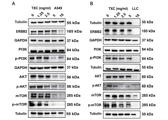
Tougu Xiaotong capsules inhibit lung cancer and gefitinib-resistant cells by blocking ERBB2/PI3K-AKT-mTOR signaling and triggering ferroptosis. [Abstract]2026 Jan:150:157741. PMID: 41477979
AKT1+AKT2+AKT3 Antibody (YA635) purchased from MedChemExpress. Usage Cited in: Phytomedicine. 2026 Jan:150:157741. [Abstract]
A549 and LLC cells were treated with increasing doses of TXC for 24 h, followed by western blotting for ERBB2, PI3K, phospho-PI3K, AKT, phospho-AKT, mTOR, phospho-Mtor.
-
J Mater Chem B
A metal-organic framework-based co-delivery system for atherosclerosis therapy via macrophage regulation. [Abstract]2025 Oct 22;13(41):13405-13422. PMID: 41002166 -
Neuropharmacology
Novel mechanism of hypidone hydrochloride (YL-0919) in Parkinson's disease: inhibiting neuronal ferroptosis by targeting the Sigma1R-PI3K-AKT-ACSL4 axis. [Abstract]2026 Jul 1:292:110950. PMID: 41887568 -
Viruses
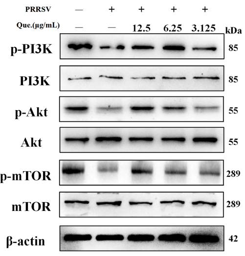
Quercetin Regulates Autophagy to Inhibit PRRSV Replication Through the PI3K/Akt/mTOR Signaling Pathway. [Abstract]2025 Dec 17;17(12):1637. PMID: 41472306
AKT1+AKT2+AKT3 Antibody (YA635) purchased from MedChemExpress. Usage Cited in: Viruses. 2025 Dec 17;17(12):1637. [Abstract]
Quercetin suppresses the PI3K/Akt/mTOR signaling pathway. MARC-145 cells were infected with PRRSV (MOI of 0.8) and treated with quercetin for 48 h.


Verification Images
-
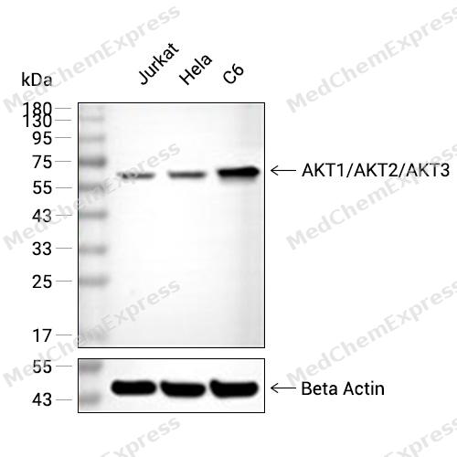 Western blot analysis of extracts from Jurkat (lane 2(20μg), Hela (lane 3(20μg) and C6(lane 4(20μg) using AKT1/AKT2/AKT3 (HY-P80535) Rabbit mAb. Proteins were transferred to a PVDF membrane and blocked with 5% non-fat milk in TBST for 2 hour at room temperature. The primary antibody (1/1000) and Loading control antibody (Beta Actin, HY-P80438, 1/10000) was used in 5% non-fat milk in TBST at 4°C overnight. Goat Anti-Mouse/Rabbit IgG-HRP Secondary Antibody (1/10000) was used for 1 hour at room temperature.
 -
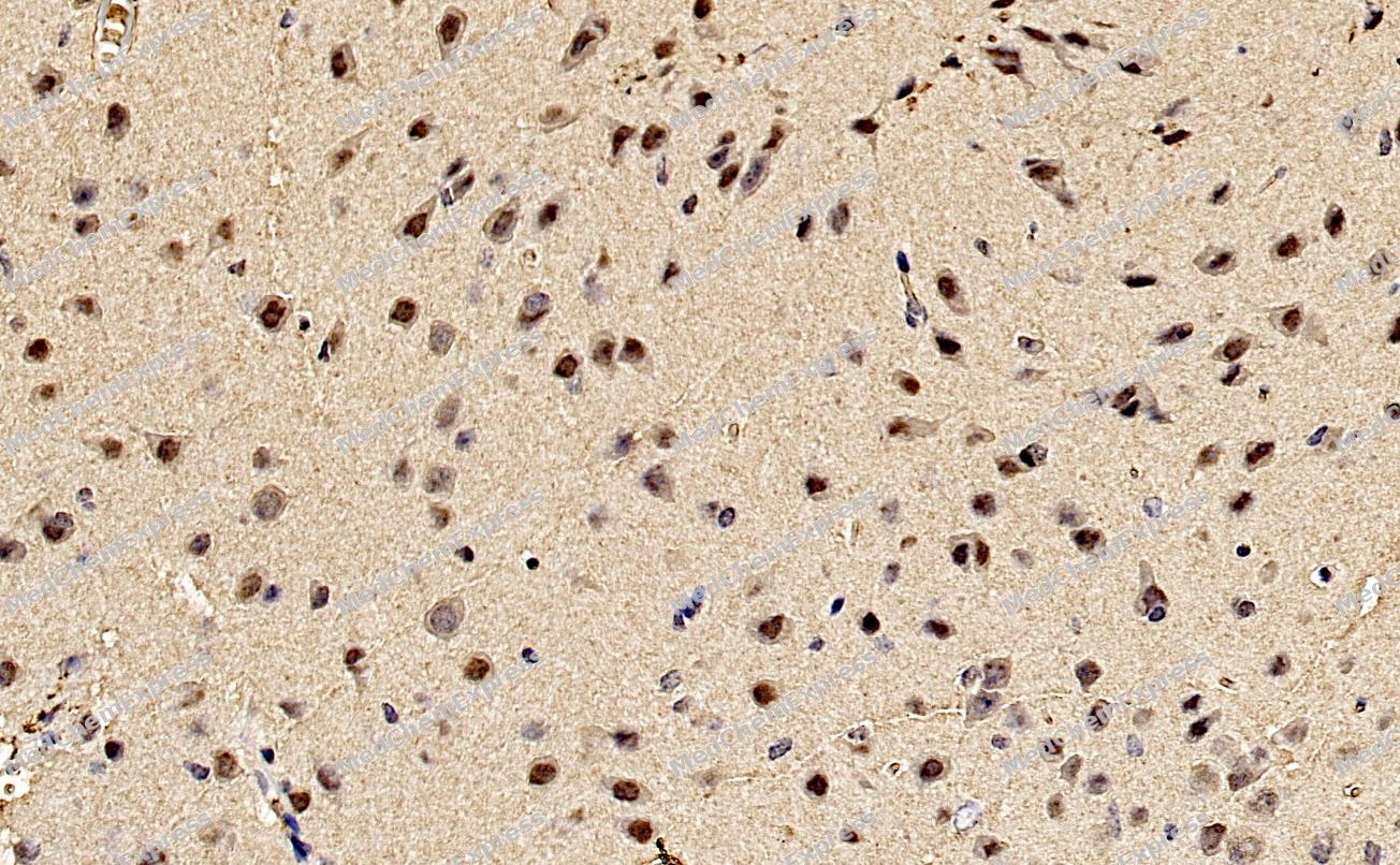 Immunohistochemical analysis of paraffin-embedded Mouse brain tissue using AKT1/AKT2/AKT3 Antibody (YA635). The section was pre-treated using heat mediated antigen retrieval with Tris-EDTA buffer (pH 9.0) for 8 minutes. The tissues were blocked in QuickBlock for 20 minutes at room temperature, washed with ddH2O and PBS, and then probed with the primary antibody (HY-P80535, 1/100) in 4℃ overnight. The detection was performed using an HRP conjugated compact polymer system. DAB was used as the chromogen. Tissues were counterstained with hematoxylin and mounted with DPX.
 -
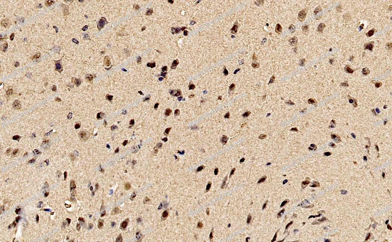 Immunohistochemical analysis of paraffin-embedded Mouse brain tissue using AKT1/AKT2/AKT3 Antibody (YA635). The section was pre-treated using heat mediated antigen retrieval with Tris-EDTA buffer (pH 9.0) for 8 minutes. The tissues were blocked in QuickBlock for 20 minutes at room temperature, washed with ddH2O and PBS, and then probed with the primary antibody (HY-P80535, 1/100) in 4℃ overnight. The detection was performed using an HRP conjugated compact polymer system. DAB was used as the chromogen. Tissues were counterstained with hematoxylin and mounted with DPX.
 -
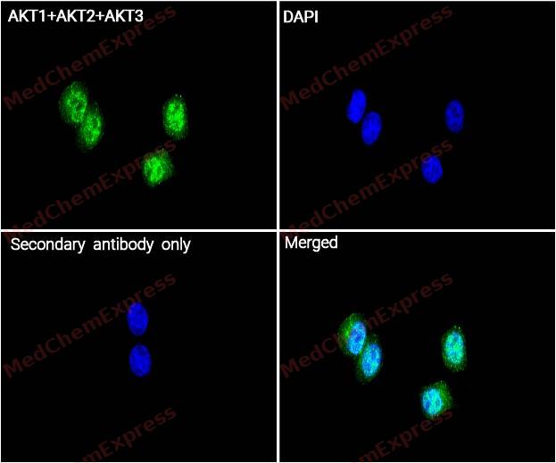 Immunocytochemistry analysis of MCF-7 cells labeling AKT1+AKT2+AKT3 with AKT1+AKT2+AKT3 Antibody (HY-P80535) at 1/100 dilution. Cells were fixed in 4% paraformaldehyde for 15 minutes at room temperature, permeabilized with 0.1% Triton X-100 in PBS for 15 minutes at room temperature, then blocked with quick block buffer for 10 minutes at room temperature. Cells were then incubated with AKT1+AKT2+AKT3 Antibody (HY-P80535) at 1/100 dilution in quick block buffer overnight at 4 ℃. AF488-conjugated Goat Anti-Rabbit IgG H&L(HY-P8002, Green) was used as the secondary antibody at 1/1,000 dilution. PBS instead of the primary antibody was used as the secondary antibody only control. The Nuclear counterstain was DAPI (Blue).
 -
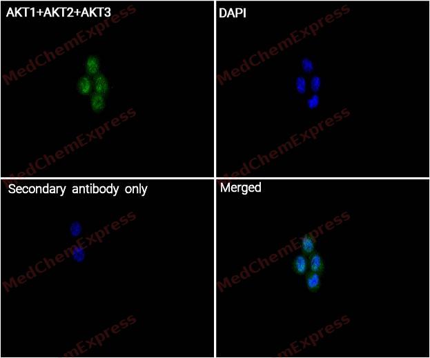 Immunocytochemistry analysis of MCF-7 cells labeling AKT1+AKT2+AKT3 with AKT1+AKT2+AKT3 Antibody (HY-P80535) at 1/200 dilution. Cells were fixed in 4% paraformaldehyde for 15 minutes at room temperature, permeabilized with 0.1% Triton X-100 in PBS for 15 minutes at room temperature, then blocked with quick block buffer for 10 minutes at room temperature. Cells were then incubated with AKT1+AKT2+AKT3 Antibody (HY-P80535) at 1/200 dilution in quick block buffer overnight at 4 ℃. AF488-conjugated Goat Anti-Rabbit IgG H&L(HY-P8002, Green) was used as the secondary antibody at 1/1,000 dilution. PBS instead of the primary antibody was used as the secondary antibody only control. The Nuclear counterstain was DAPI (Blue).


Background
-
Function
AKT1+AKT2+AKT3 is a P31749: AKT1 is one of 3 closely related serine/threonine-protein kinases (AKT1, AKT2 and AKT3) called the AKT kinase, and which regulate many processes including metabolism, proliferation, cell survival, growth and angiogenesis. This is mediated through serine and/or threonine phosphorylation of a range of downstream substrates.
P31751: Serine/threonine kinase closely related to AKT1 and AKT3. All 3 enzymes, AKT1, AKT2 and AKT3, are collectively known as AKT kinase. AKT regulates many processes including metabolism, proliferation, cell survival, growth and angiogenesis, through the phosphorylation of a range of downstream substrates.
Q9Y243: AKT3 is one of 3 closely related serine/threonine-protein kinases (AKT1, AKT2 and AKT3) called the AKT kinase, and which regulate many processes including metabolism, proliferation, cell survival, growth and angiogenesis. This is mediated through serine and/or threonine phosphorylation of a range of downstream substrates.
[1][2][3][4][5][6][7]. -
Subcellular Localization
P31749: Cytoplasm; Nucleus; Cell membrane; Mitochondrion intermembrane space
P31751: Cytoplasm; Nucleus; Cell membrane; Peripheral membrane protein; Early endosome
Q9Y243: Nucleus; Cytoplasm; Membrane; Peripheral membrane protein -
Expression
Tissue_specificity: P31749: Expressed in prostate cancer and levels increase from the normal to the malignant state (at protein level). Expressed in all human cell types so far analyzed. The Tyr-176 phosphorylated form shows a significant increase in expression in breast cancers during the progressive stages i.e. normal to hyperplasia (ADH) , ductal carcinoma in situ (DCIS) , invasive ductal carcinoma (IDC) and lymph node metastatic (LNMM) stages
P31751: Widely expressed. Expressed in myoblasts (PubMed:17565718)
Q9Y243: In adult tissues, it is highly expressed in brain, lung and kidney, but weakly in heart, testis and liver. In fetal tissues, it is highly expressed in heart, liver and brain and not at all in kidney -
Subunit
P31749: Interacts with BTBD10 (By similarity). Interacts with KCTD20 (By similarity). Interacts (via the C-terminus) with CCDC88A (via its C-terminus). Interacts with GRB10.
P31751: Interacts with BTBD10 (By similarity).
Q9Y243: Interacts (via PH domain) with TCL1A; this enhances AKT3 phosphorylation and activation. Interacts with TRAF6. -
Synonyms
AKT1,AKT2,AKT3
-
Research Field
Signal Transduction
Documentation
-
Data Sheet (263 KB)
-
SDS (251 KB)
- English - EN (251 KB)
- Français - FR (251 KB)
- Deutsch - DE (251 KB)
- Norwegian - NO (251 KB)
- Español - ES (251 KB)
- Swedish - SV (251 KB)
- Italian - IT (251 KB)
- Korean - KR (251 KB)
- Portuguese - PT (251 KB)
-
User Guide for Antibodies (1077 KB)
References
[1]. Nicholson KM, et al. The protein kinase B/Akt signalling pathway in human malignancy. Cell Signal. 2002 May;14(5):381-95. [Content Brief]
[2]. Rönnstrand L, et al. Signal transduction via the stem cell factor receptor/c-Kit. Cell Mol Life Sci. 2004 Oct;61(19-20):2535-48. [Content Brief]
[3]. Cartlidge RA, et al. The tRNA methylase METTL1 is phosphorylated and inactivated by PKB and RSK in vitro and in cells. EMBO J. 2005 May 4;24(9):1696-705. [Content Brief]
[4]. Heron-Milhavet L, et al. Akt1 and Akt2: differentiating the aktion. Histol Histopathol. 2011 May;26(5):651-62. [Content Brief]
[5]. Hers I, et al. Akt signalling in health and disease. Cell Signal. 2011 Oct;23(10):1515-27. [Content Brief]
[6]. Walia V, et al. Akt Regulates a Rab11-Effector Switch Required for Ciliogenesis. Dev Cell. 2019 Jul 22;50(2):229-246.e7. [Content Brief]
[7]. Choppara S, et al. Degradation of FBXO31 by APC/C is regulated by AKT- and ATM-mediated phosphorylation. Proc Natl Acad Sci U S A. 2018 Jan 30;115(5):998-1003. [Content Brief]

 Powered by Bioz
