



Arcapillin
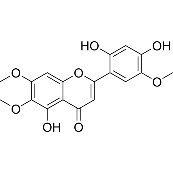
Arcapillin is a flavonoid that can be isolated from Artemisia capillaris Thunb. Arcapillin induces dose-dependent relaxation of ileum and pulmonary artery smooth muscle, causes slight urinary bladder smooth muscle contraction at highest tested concentrations. Arcapillin binds to the active site of SARS-CoV-2 Mpro via interactions with Gln139, His163, and His164, exhibits antiviral activity against SARS-CoV-2 and MERS-CoV. Arcapillin can be used for the research of gastrointestinal disorders, COVID-19, and Middle East respiratory syndrome (MERS).
For research use only. We do not sell to patients.
- CAS No.: 83162-82-7
- Formula: C18H16O8
- Molecular Weight:360.31
-
Storage:
Please store the product under the recommended conditions in the Certificate of Analysis.

To place orders, for customer services and technical support, please contact: MedChemExpress USA
Tel: 609-228-6898 E-mail: [email protected] [email protected]


-
 Biological Activity
-
Chemical Information
- Protocols
- Purity & Documentation
- References
-
 Help & FAQs




Biological Activity
Description
IC50 & Target
PTP1B[1]
In Vitro
Arcapillin binds to the SARS-CoV-2 main protease (PDB: 7L0D) with a docking score of -6.16 kcal mol−1, indicating favorable interactions with the enzyme's active site[2].
Arcapillin (72 h) exhibits antiviral activity against SARS-CoV-2 in Vero E6 cells with an IC50 of 190.8 μg mL−1, and has a low cytotoxic profile in the same cell line with a CC50 of 1228 μg mL−1, yielding a selectivity index of 6.40[2].
Arcapillin (72 h) exhibits potent antiviral activity against MERS-CoV in Vero E6 cells with an IC50 of 16.58 μg mL−1, and has a CC50 of 54.17 μg mL−1 in the same cell line, yielding a selectivity index of 3.26[2].
MedChemExpress (MCE) has not independently confirmed the accuracy of these methods. They are for reference only.


Chemical Information
-
CAS No. 83162-82-7
-
Molecular Weight 360.31
-
Formula C18H16O8
-
SMILES
O=C1C=C(C2=CC(OC)=C(O)C=C2O)OC3=CC(OC)=C(OC)C(O)=C13
-
Structure Classification
-
Shipping
Room temperature in continental US; may vary elsewhere.
-
Storage
Please store the product under the recommended conditions in the Certificate of Analysis.


Purity & Documentation
References

Calculators
Concentration (start) × Volume (start) = Concentration (final) × Volume (final)


Arcapillin Related Classifications
HY-121020 Related Classifications
 Powered by Bioz