



Baquiloprim

Based on 1 Customer Validation
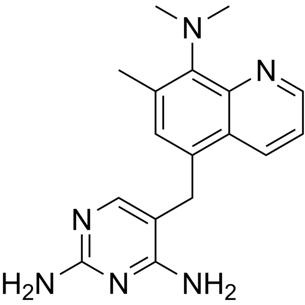
Baquiloprim is an orally active Antibiotic and selective inhibitor of Bacterial Dihydrofolate reductase (DHFR), with an IC50 of 19 nM against E. coli and an IC50 of 190 μM against rat liver dihydrofolate reductase. Baquiloprim is a bacteriostatic broad-spectrum antibacterial agent that acts against Gram-negative and Gram-positive bacteria. Baquiloprim inhibits glucuronidation in cultured hepatocytes. Baquiloprim can be used in research related to Pasteurella haemolytica infection, bacterial infection, Escherichia coli diarrhea, bacterial infection in pigs, and pneumonic pasteurellosis.
For research use only. We do not sell to patients.
- Purity : 99.66%
- CAS No.: 102280-35-3
- Formula: C17H20N6
- Molecular Weight:308.38
-
Storage:
-20°C, sealed storage, away from moisture
* In solvent : -80°C, 6 months; -20°C, 1 month (sealed storage, away from moisture)

To place orders, for customer services and technical support, please contact: MedChemExpress USA
Tel: 609-228-6898 E-mail: [email protected] [email protected]


-
 Biological Activity
-
Chemical Information
- Protocols
- Purity & Documentation
- References
-
 Help & FAQs


All Antibiotic Isoforms
More

Biological Activity
Description
In Vitro
Baquiloprim (24 h) exhibits broad-spectrum in vitro antibacterial activity against cattle, sheep, and pig bacterial isolates, with geometric mean MIC50 ranging from 0.03 mg litre-1 to 0.18 mg litre-1[2].
Baquiloprim (1:100 ratio with sulphadimidine; 24 h) shows high synergistic in vitro antibacterial activity with sulphadimidine against most cattle bacterial pathogens, with FIC indices below 0.5 and reduced MICs for baquiloprim in the 1:100 mixture[2].
Baquiloprim (<0.008-8.0 μg/mL alone; 0.004:0.4-0.5:50 μg/mL in 1:100 ratio with sulphadimidine) alone and in 1:100 combination with sulphadimidine inhibits growth of susceptible porcine bacterial pathogens[4].
Baquiloprim potently inhibits Pasteurella haemolytica in vitro with a mean MIC of 0.09 mg/L, and its activity is synergistically enhanced by sulphadimidine, reducing its mean MIC to 0.02 mg/l[6].
MedChemExpress (MCE) has not independently confirmed the accuracy of these methods. They are for reference only.
Parmacokinetics
In Vivo
Baquiloprim (10 mg/kg; i.m.; once daily; 3 days) achieves mean peak serum concentrations of 0.55 μg/mL and maintains concentrations above the assay limit for at least 24 hours post-dose, with an elimination half-life of 6.3 hours[4].
Baquiloprim (30 mg/kg; i.m.; single dose) achieves a mean peak serum concentration of 0.87 μg/mL and maintains measurable concentrations in most pigs for at least 48 hours, with an extended elimination half-life of 10.6 hours[4].
Baquiloprim (4 mg/kg; oral; single dose 6 hours pre-infection, second dose 48 hours post-infection) combined with sulphadimidine suppresses P. haemolytica-induced pneumonic pasteurellosis in calves, as evidenced by normal body temperatures, minimal signs, negligible lung lesions, and no bacterial recovery[6].
MedChemExpress (MCE) has not independently confirmed the accuracy of these methods. They are for reference only.
-
Animal Model:Dwarf goats (female, healthy, mean bodyweight 30.6 kg)[1]
-
Dosage:8 mg/kg (i.v.); 8 mg/kg (intra-ruminal)
-
Administration:i.v.; single dose; intra-ruminal; single dose via nasogastric catheter
-
Result:Exhibited mean distribution half-life (t1/2α) of 0.89 hours, mean elimination half-life (t1/2β) of 14.0 hours, mean volume of distribution at steady-state (Vdss) of 14.1 litres/kg, mean AUC0-48h of 9.6 μg hours/mL, and mean clearance (CI) of 0.77 litres/kg/hours after intravenous administration.
Reached mean maximum plasma concentration (Cmax) of 0.09 μg/mL, mean time to maximum concentration (Tmax) of 35 hours, mean AUC0-48h of 3.2 μg hours/mL, and mean systemic oral bioavailability of 33.7 per cent after intra-ruminal administration.
-
Animal Model:CD-1 (female, 18 to 20 g, Escherichia coli P855 infection model)[2]
-
Dosage:0.5 mg/kg
-
Administration:p.o.; at 1 and 6 hours post-infection
-
Result:Achieved an average ED50 of 0.5 mg/kg across three experiments.
-
Animal Model:female hybrid (6 weeks old, 13-18 kg)[4]
-
Dosage:10 mg/kg (total formulation)
-
Administration:i.m.; once daily; 3 days
-
Result:Reached mean maximum serum concentration (Cmax) of 0.55 μg/mL at 2.5 hours after the first dose.
Had mean elimination half-life (t½) of 6.3 hours, area under the serum concentration-time curve (AUC∞) of 6.27 μg·mL-1·hour, volume of distribution (Vd(area)/F) of 2.41 litre/kg, and clearance (Clp/F) of 273 mL·hour-1·kg-1.
Achieved mean baquiloprim concentrations of 0.05 μg/mL, 0.08 μg/mL, and 0.11 μg/mL at 24 hours after the first, second, and third doses, respectively.
Detected concentrations ≥0.02 μg/mL in all samples taken 24 hours after each injection.
-
Animal Model:male hybrid (7 weeks old, 23-36 kg)[4]
-
Dosage:30 mg/kg (total formulation)
-
Administration:i.m.; single dose
-
Result:Reached mean maximum serum concentration (Cmax) of 0.87 μg/mL at 3.3 hours post-dose.
Had mean elimination half-life (t½) of 10.6 hours, area under the serum concentration-time curve (AUC∞) of 16.8 μg·mL-1·hour, volume of distribution (Vd(area)/F) of 4.60 litre/kg, and clearance (Clp/F) of 308 mL·hour-1·kg-1.
Detected concentrations ≥0.02 μg/mL in all but two samples taken 48 hours post-dose.
-
Animal Model:Friesian calves (female, 21 weeks old, mean bodyweights 117-121 kg, endobronchial inoculation with Pasteurella haemolytica type A1)[6]
-
Dosage:4 mg/kg (single dose 6 hours pre-infection); 4 mg/kg (second dose 48 hours post-infection)
-
Administration:oral; single dose (6 hours pre-infection); second dose (48 hours post-infection)
-
Result:Maintained mean rectal temperatures close to pre-infection levels (maximum individual temperature 39.3°C).
Achieved a mean score of 1.6 (single dose) or 1.5 (repeat dose), versus 8.6 in untreated controls.
Had post-mortem lung lesion scores of 0.2 (single dose) and 0.25 (repeat dose), compared to 2.4 in untreated controls.
Prevented isolation of P. haemolytica from all treated calves, whereas the organism was isolated from all 5 untreated controls.


Chemical Information
-
CAS No. 102280-35-3
-
Appearance Solid
-
Molecular Weight 308.38
-
Formula C17H20N6
-
Color White to yellow
-
SMILES
NC1=NC=C(CC2=C3C=CC=NC3=C(N(C)C)C(C)=C2)C(N)=N1
-
Shipping
Room temperature in continental US; may vary elsewhere.
-
Storage
-20°C, sealed storage, away from moisture
* In solvent : -80°C, 6 months; -20°C, 1 month (sealed storage, away from moisture)


Purity & Documentation
-
Data Sheet (284 KB)
-
SDS (252 KB)
- English - EN (252 KB)
- Français - FR (252 KB)
- Deutsch - DE (252 KB)
- Norwegian - NO (252 KB)
- Español - ES (252 KB)
- Swedish - SV (252 KB)
- Italian - IT (252 KB)
- Korean - KR (252 KB)
- Portuguese - PT (252 KB)
-
Handling Instructions (2659 KB)
References
[1]. Lewicki J, et al. Oral bioavailability and pharmacokinetics of baquiloprim in dwarf goats. Research in veterinary science. 1995 May;58(3):268-71. [Content Brief]
[2]. White G, et al. Baquiloprim, a new antifolate antibacterial: in vitro activity and pharmacokinetic properties in cattle. Research in veterinary science. 1993 May;54(3):372-8. [Content Brief]
[3]. White DG, et al. Comparison of danofloxacin with baquiloprim/sulphadimidine for the treatment of experimentally induced Escherichia coli diarrhoea in calves. The Veterinary record. 1998 Sep 05;143(10):273-6. [Content Brief]
[4]. Davies AM, et al. Pharmacokinetics of baquiloprim and sulphadimidine in pigs after intramuscular administration. Research in veterinary science. 1994 Jul;57(1):69-74. [Content Brief]
[5]. Sunderland SJ, et al. Efficacy of danofloxacin 18% injectable solution in the treatment of Escherichia coli diarrhoea in young calves in Europe. Research in veterinary science. 2003 Apr;74(2):171-8. [Content Brief]
[6]. Dassanayake L, et al. Administration of a bolus formulation of baquiloprim and sulphadimidine to calves: plasma concentration--time profiles and efficacy in suppressing experimental pneumonic pasteurellosis. Veterinary microbiology. 1994 Jan;38(3):255-62. [Content Brief]

Calculators
Concentration (start) × Volume (start) = Concentration (final) × Volume (final)


Baquiloprim Related Classifications
HY-19581 Related Classifications
 Powered by Bioz
Keywords
- Baquiloprim
- 102280-35-3
- Antibiotic
- Bacterial
- Dihydrofolate reductase (DHFR)
- pneumonic pasteurellosis
- Gram-negative bacteria
- E. coli
- Gram-positive bacteria
- rat liver dihydrofolate reductase
- sulphonamides
- bacterial dihydrofolate reductase
- escherichia coli diarrhoea
- hepatocytes
- pasteurella haemolytica infection
- Inhibitor
- inhibitor
- inhibit