
BChE-IN-16
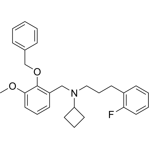
BChE-IN-16 (compound 87) is a highly potent BChE inhibitor with an IC50 of 3.8 nM for hBChE. BChE-IN-16 has low cytotoxicity, potential CNS permeability, unique adaptability and can be used in Alzheimer's disease (AD) research.
For research use only. We do not sell to patients.
- Formula: C28H32FNO2
- Molecular Weight:433.56
-
Storage:
Please store the product under the recommended conditions in the Certificate of Analysis.
Biological Activity
Description
IC50 & Target
|
hBCHE 3.8 nM (IC50) |
Cellular Effect
|
Cell Line
|
Type | Value | Description | References |
|---|---|---|---|---|
| HepG2 | IC50 |
76 μM
Compound: 87
|
Cytotoxicity against human HepG2 cells assessed as inhibition of cell growth incubated for 24 hrs by MTT assay
|
[PMID: 36996715] |
| SH-SY5Y | IC50 |
87 μM
Compound: 87
|
Cytotoxicity against human SH-SY5Y cells assessed as inhibition of cell growth incubated for 24 hrs by MTT assay
|
[PMID: 36996715] |
In Vitro
BChE-IN-16 has zero inhibition of hAChE when the IC50 value is greater than 10 μM[1].
BChE-IN-16 competitively inhibits hBChE with a Ki value of 4.04 nM[1].
BChE-IN-16 is cytotoxic to SH-SY5Y cells with an IC50 value of 87.0 μM[1].
BChE-IN-16 is cytotoxic to HepG2 cells with an IC50 value of 76.0 μM[1].
MedChemExpress (MCE) has not independently confirmed the accuracy of these methods. They are for reference only. Further protocols information, click here.
Chemical Information
-
Molecular Weight 433.56
-
Formula C28H32FNO2
-
SMILES
COC1=CC=CC(CN(C2CCC2)CCCC3=C(C=CC=C3)F)=C1OCC4=CC=CC=C4
-
Shipping
Room temperature in continental US; may vary elsewhere.
-
Storage
Please store the product under the recommended conditions in the Certificate of Analysis.
Protocols
-
Cell Cytotoxicity Assay
Cytotoxicity assays are usually based on the assessment of cell membrane damage, which can also be indirectly detected by measuring cell viability. Detection methods include MTT assay, CKK-8 assay, LDH assay and ATP assay, etc.
-
Mammalian live/dead viability and cytotoxicity staining
Live/dead viability and cytotoxicity staining assays are based on the simultaneous detection of intracellular esterase activity in metabolically active (viable) cells and membrane integrity loss in non-viable cells. In commonly used dual-staining approaches, membrane-permeant fluorogenic substrates are converted by intracellular esterases into fluorescent products in live cells, while impermeant DNA-binding dyes selectively enter cells with compromised plasma membranes and label nucleic acids in dead or dying cells, enabling discrimination between viable and non-viable populations by fluorescence microscopy or flow cytometry.
-
Transepithelial/transendothelial electrical resistance assay
TEER measures electrical resistance across epithelial or endothelial monolayers cultured on permeable supports, and the readout reflects ionic conductance through the cell barrier, especially the paracellular pathway regulated by junctional integrity. TEER can be measured without destroying the monolayer and is commonly used before or during transport, permeability, barrier-disruption, and barrier-maturation experiments. TEER values are influenced by biological maturation and technical conditions; reported factors include temperature, medium formulation, passage number, electrode geometry, membrane properties, and junctional length during early monolayer maturation. Therefore, TEER should be interpreted with blank-insert subtraction, area normalization, repeated readings, and, when possible, orthogonal barrier readouts such as FITC-dextran flux or tight-junction staining.
-
Cell Viability Determination by MTT Colorimetric Assay
The following protocol uses the MTT colorimetric assay as a classic literature-established method for assessing cell viability/metabolic activity in cultured mammalian cells. MTT[3-(4,5-dimethylthiazol-2-yl)-2,5-diphenyltetrazolium bromide] is reduced by metabolically active cells to a colored formazan product; the amount of formazan is quantified spectrophotometrically and provides an indirect measure of metabolically active viable cells. Importantly, MTT reduction reflects cellular oxidoreductase/metabolic activity rather than an absolute direct count of living cells, so changes in cellular metabolism can alter the signal independently of cell number.
-
Alzheimer’s Disease Modeling
Alzheimer’s Disease (AD) is a neurodegenerative disorder characterized by a progressive decline in cognitive functions and loss of specific types of neurons and synapses. Alzheimer's symptoms can be simulated in mice by injecting drugs (such as Aβ) or genetically modified.
Purity & Documentation
References
Calculators
Concentration (start) × Volume (start) = Concentration (final) × Volume (final)

