



Benproperine phosphate
Based on 2 publication(s) in Google Scholar
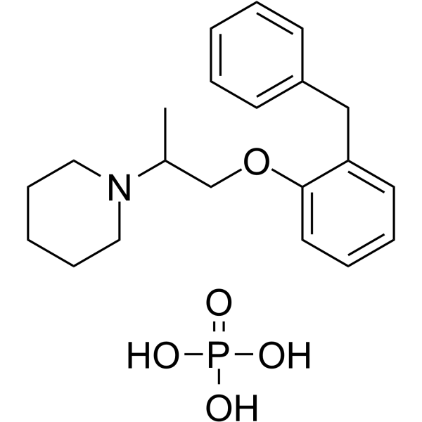
Benproperine phosphate is an orally active, potent actin-related protein 2/3 complex subunit 2 (ARPC2) inhibitor. Benproperine phosphate attenuates the actin polymerization rate of action polymerization nucleation by impairing Arp2/3 function. Benproperine phosphate has the potential for a cough suppressant and suppresses cancer cell migration and tumor metastasis.
Nur für Forschungszwecke. Wir verkaufen nicht an Patienten.
- Reinheit : 99.93%
- CAS. Nr.: 19428-14-9
- Formel: C21H30NO5P
- Molecular Weight:407.44
-
Speicherung:
4°C, sealed storage, away from moisture
* In solvent : -80°C, 2 years; -20°C, 1 year (sealed storage, away from moisture)

To place orders, for customer services and technical support, please contact: MedChemExpress USA
Tel: 609-228-6898 E-mail: [email protected] [email protected]


-
 Biologische Aktivität
-
Chemical Information
-
Lösungsmittel & Löslichkeit
- Protokoll
- Reinheit & Dokumentation
- Verweise
-
 Help & FAQs


-
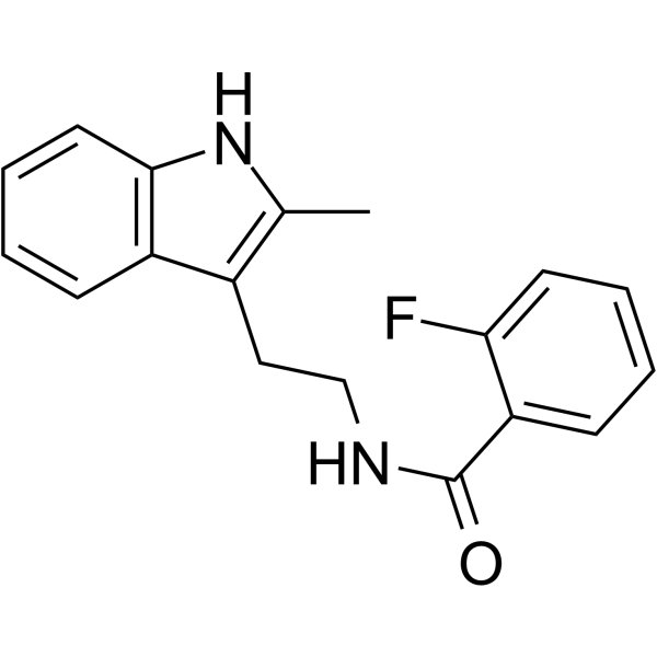
HY-16926
CK-666Arp2/3 Complex Inhibitor -
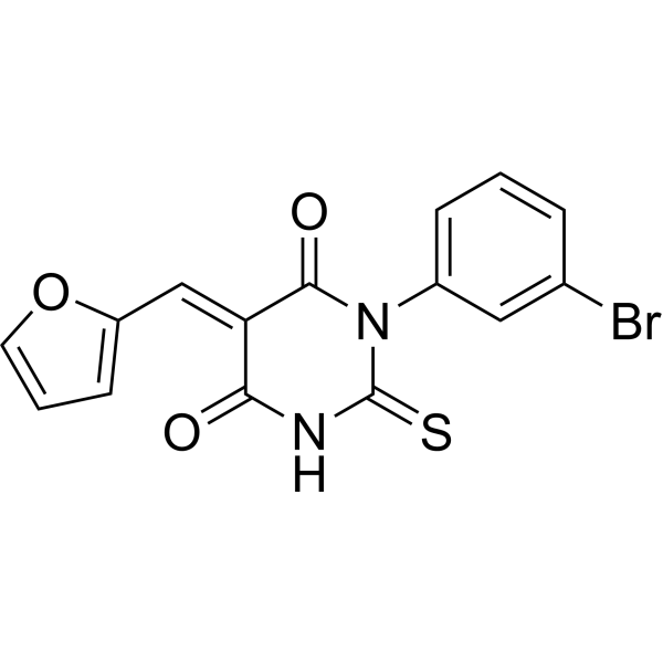
HY-16931
SMIFH2Formin Inhibitor -
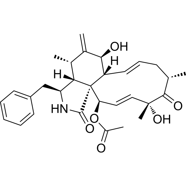
HY-N6682
Cytochalasin DActin Polymerization Inhibitor -
HY-15892
CK-636Arp2/3 Complex Inhibitor -
HY-12762
QS11ARFGAP1 Inhibitor -
HY-16928
Cytochalasin BMycotoxin -
HY-16929
Latrunculin AG-actin Polymerization Inhibitor -
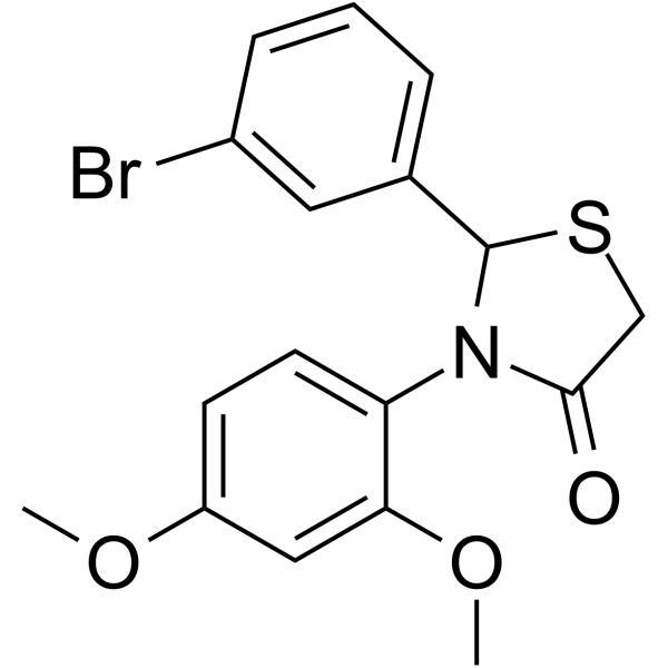
HY-16927
CK-869Arp2/3 Complex Inhibitor -
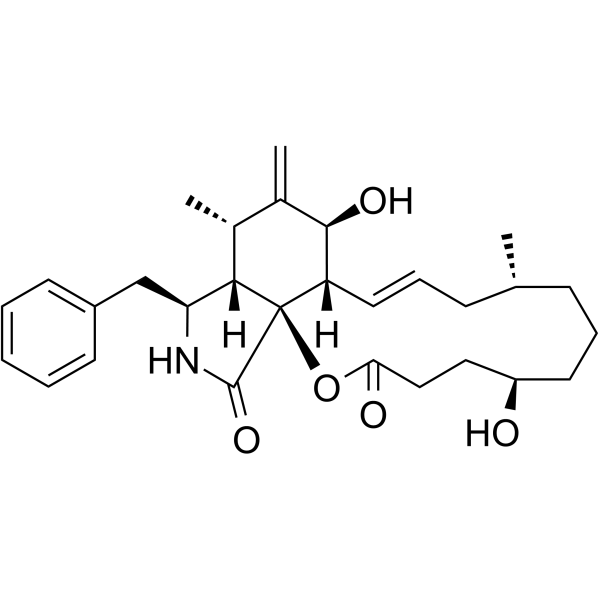
HY-N6701
Dihydrocytochalasin BActin Inhibitor -
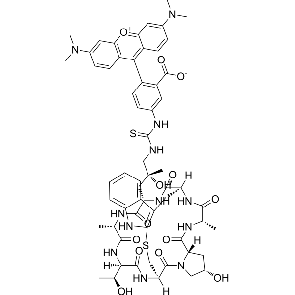
HY-P2270
Phalloidin-TRITCF-actin Probe -
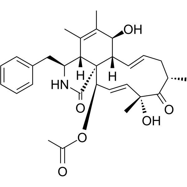
HY-N6774
Cytochalasin C -
HY-119357
TN-16Tubulin Inhibitor
Publications Citing Use of MedChemExpress (MCE) Benproperine phosphate
More

Biologische Aktivität
Beschreibung
In Vitro
Benproperine phosphate (20-120 μM; for 24?hours) inhibits cell viability in a dose-dependent manner[1].
?
Benproperine phosphate (10 μM; for 24?hours) significantly inhibits the migration of various types of cancer cells and inhibits the migration and invasion of DLD-1, AsPC-1 cells with IC50 values of 1-2?μM. Benproperine phosphate (10 μM; for 24?hours) does not affect cortactin-rich lamellipodium in MCF-10A cells[1].
MedChemExpress (MCE) has not independently confirmed the accuracy of these methods. They are for reference only.
-
Cell Line:DLD-1, AsPC-1, CFPAC-1, A375P, A375P, MDA-MB-231, DU145, DU145 cancer cells
-
Concentration:20, 40, 60, 80, 100, 120 μM
-
Incubation Time:For 24 hours
-
Result:Inhibited cell viability in a dose-dependent manner.
In Vivo
? Benproperine phosphate shows a marked decrease in the lung metastasis of AsPC-1 cells (56.1% inhibition) in mouse. Benproperine phosphate significantly suppressed the liver metastasis of HCT-116 cells by 78.9% and DLD-1 cells by 78.2%[1].
MedChemExpress (MCE) has not independently confirmed the accuracy of these methods. They are for reference only.
-
Animal Model:Female BALB/c nude mice of 6-week-old with AsPC-1 cells[1]
-
Dosage:50, 100 mg/kg
-
Administration:Oral gavage; 5 days per week for 4 weeks
-
Result:Inhibited primary pancreatic tumor growth compared to the vehicle control (47.7% inhibition) without body weights change.
- Scratch/Wound-Healing Migration Assay
- Cell migration
- Zigmond Chamber Chemotaxis Assay
- Under-Agarose Cell Migration Assay
- Neural Crest/Neuronal Cell Migration Explant Assay
- Transwell/Boyden Chamber Migration Assay
- 3D Collagen/Hydrogel Matrix Migration Assay
- Cell-Exclusion Zone Migration Assay
- Dunn/Insall Chamber Chemotaxis Assay
- Orthotopic Cell-Line Xenograft
- Phalloidin F-actin cytoskeleton staining


Chemical Information
-
CAS. Nr. 19428-14-9
-
Appearance Solid
-
Molecular Weight 407.44
-
Formel C21H30NO5P
-
Color White to off-white
-
SMILES
CC(N1CCCCC1)COC2=CC=CC=C2CC3=CC=CC=C3.O=P(O)(O)O
-
Versand
Room temperature in continental US; may vary elsewhere.
-
Speicherung
4°C, sealed storage, away from moisture
* In solvent : -80°C, 2 years; -20°C, 1 year (sealed storage, away from moisture)


Publications (2)
-
Journal Impact Factor
-
Most Recent


Lösungsmittel & Löslichkeit
In Vitro:
DMSO : 100 mg/mL (245.43 mM; Need ultrasonic; Hygroscopic DMSO has a significant impact on the solubility of product, please use newly opened DMSO)
H2O : ≥ 100 mg/mL (245.43 mM)
* "≥" means soluble, but saturation unknown.
Please refer to the solubility information to select the appropriate solvent. Once prepared, please aliquot and store the solution to prevent product inactivation from repeated freeze-thaw cycles.
Storage method and period of stock solution: -80°C, 2 years; -20°C, 1 year (sealed storage, away from moisture). When stored at -80°C, please use it within 2 years. When stored at -20°C, please use it within 1 year.
* Note: If you choose water as the stock solution, please dilute it to the working solution, then filter and sterilize it with a 0.22 μm filter before use.
Please refer to the solubility information to select the appropriate solvent. Once prepared, please aliquot and store the solution to prevent product inactivation from repeated freeze-thaw cycles.
Storage method and period of stock solution: -80°C, 2 years; -20°C, 1 year (sealed storage, away from moisture). When stored at -80°C, please use it within 2 years. When stored at -20°C, please use it within 1 year.
* Note: If you choose water as the stock solution, please dilute it to the working solution, then filter and sterilize it with a 0.22 μm filter before use.
Konzentration (Stammlösung) × Volumen (Stammlösung) = Konzentration (Ziellösung) × Volumen (Ziellösung)
In Vivo:
Select the appropriate dissolution method based on your experimental animal and administration route.
- For the following dissolution methods, please ensure to first prepare a clear stock solution using an In Vitro approach and then sequentially add co-solvents:
- To ensure reliable experimental results, the clarified stock solution can be appropriately stored based on storage conditions. As for the working solution for In Vivo experiments, it is recommended to prepare freshly and use it on the same day.
- The percentages shown for the solvents indicate their volumetric ratio in the final prepared solution. If precipitation or phase separation occurs during preparation, heat and/or sonication can be used to aid dissolution.
Add each solvent one by one: 10% DMSO 40% PEG300 5% Tween-80 45% Saline
Solubility: ≥ 2.08 mg/mL (5.11 mM); Clear solution
This protocol yields a clear solution of ≥ 2.08 mg/mL (saturation unknown).
Taking 1 mL working solution as an example, add 100 μL DMSO stock solution (20.8 mg/mL) to 400 μL PEG300, and mix evenly; then add 50 μL Tween-80 and mix evenly; then add 450 μL Saline to adjust the volume to 1 mL.
Preparation of Saline: Dissolve 0.9 g sodium chloride in ddH₂O and dilute to 100 mL to obtain a clear Saline solution.
Add each solvent one by one: 10% DMSO 90% (20% SBE-β-CD in Saline)
Solubility: ≥ 2.08 mg/mL (5.11 mM); Clear solution
This protocol yields a clear solution of ≥ 2.08 mg/mL (saturation unknown).
Taking 1 mL working solution as an example, add 100 μL DMSO stock solution (20.8 mg/mL) to 900 μL 20% SBE-β-CD in Saline, and mix evenly.
Preparation of 20% SBE-β-CD in Saline (4°C, storage for one week): 2 g SBE-β-CD powder is dissolved in 10 mL Saline, completely dissolve until clear.
For the following dissolution methods, please prepare the working solution directly:
It is recommended to prepare fresh solutions and use them promptly within a short period of time.
The percentages shown for the solvents indicate their volumetric ratio in the final prepared solution. If precipitation or phase separation occurs during preparation, heat and/or sonication can be used to aid dissolution.
Add each solvent one by one: Saline
Solubility: ≥ 100 mg/mL (245.43 mM); Clear solution
In Vivo Dissolution Calculator
Please enter the basic information of animal experiments:
-
-
-
-
Recommended: Prepare an additional quantity of animals to account for potential losses during experiments.
Working solution concentration: 0.22 mg/mL
This product has good water solubility, please refer to the measured solubility data in water/PBS/Saline for details.


Reinheit & Dokumentation
-
Data Sheet (278 KB)
-
SDS (393 KB)
- English - EN (393 KB)
- Français - FR (393 KB)
- Deutsch - DE (393 KB)
- Norwegian - NO (393 KB)
- Español - ES (393 KB)
- Swedish - SV (393 KB)
- Italian - IT (393 KB)
- Korean - KR (393 KB)
- Portuguese - PT (393 KB)
-
Handling Instructions (2659 KB)
Verweise


Complete Stock Solution Preparation Table
Please refer to the solubility information to select the appropriate solvent. Once prepared, please aliquot and store the solution to prevent product inactivation from repeated freeze-thaw cycles.
Storage method and period of stock solution: -80°C, 2 years; -20°C, 1 year (sealed storage, away from moisture). When stored at -80°C, please use it within 2 years. When stored at -20°C, please use it within 1 year.
| Optional Solvent | Concentration Solvent Mass | 1 mg | 5 mg | 10 mg | 25 mg |
|---|---|---|---|---|---|
| DMSO / H2O | 1 mM | 2.4543 mL | 12.2717 mL | 24.5435 mL | 61.3587 mL |
| 5 mM | 0.4909 mL | 2.4543 mL | 4.9087 mL | 12.2717 mL | |
| 10 mM | 0.2454 mL | 1.2272 mL | 2.4543 mL | 6.1359 mL | |
| 15 mM | 0.1636 mL | 0.8181 mL | 1.6362 mL | 4.0906 mL | |
| 20 mM | 0.1227 mL | 0.6136 mL | 1.2272 mL | 3.0679 mL | |
| 25 mM | 0.0982 mL | 0.4909 mL | 0.9817 mL | 2.4543 mL | |
| 30 mM | 0.0818 mL | 0.4091 mL | 0.8181 mL | 2.0453 mL | |
| 40 mM | 0.0614 mL | 0.3068 mL | 0.6136 mL | 1.5340 mL | |
| 50 mM | 0.0491 mL | 0.2454 mL | 0.4909 mL | 1.2272 mL | |
| 60 mM | 0.0409 mL | 0.2045 mL | 0.4091 mL | 1.0226 mL | |
| 80 mM | 0.0307 mL | 0.1534 mL | 0.3068 mL | 0.7670 mL | |
| 100 mM | 0.0245 mL | 0.1227 mL | 0.2454 mL | 0.6136 mL |
* Note: If you choose water as the stock solution, please dilute it to the working solution, then filter and sterilize it with a 0.22 μm filter before use.


Benproperine phosphate Related Classifications
HY-114657A Related Classifications
 Powered by Bioz