



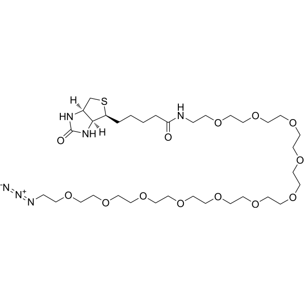
Biotin-PEG11-azide

Based on 1 Customer Validation
Biotin-PEG11-azide is a PEG-based PROTAC linker that can be used in the synthesis of PROTACs. Biotin-PEG11-azide is a click chemistry reagent, it contains an Azide group and can undergo copper-catalyzed azide-alkyne cycloaddition reaction (CuAAc) with molecules containing Alkyne groups. It can also undergo strain-promoted alkyne-azide cycloaddition (SPAAC) reactions with molecules containing DBCO or BCN groups.
For research use only. We do not sell to patients.
- Purity : 98.0%
- CAS No.: 2276672-04-7
- Formula: C34H64N6O13S
- Molecular Weight:796.97
-
Storage:Powder -20°C, 3 years ; In solvent -80°C, 6 months , -20°C, 1 month

To place orders, for customer services and technical support, please contact: MedChemExpress USA
Tel: 609-228-6898 E-mail: [email protected] [email protected]


-
 Biological Activity
-
Chemical Information
- Protocols
- Purity & Documentation
- References
-
 Help & FAQs


-
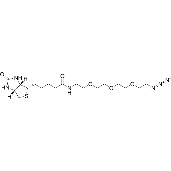
HY-130143
Biotin-PEG3-azidePROTAC Linker -
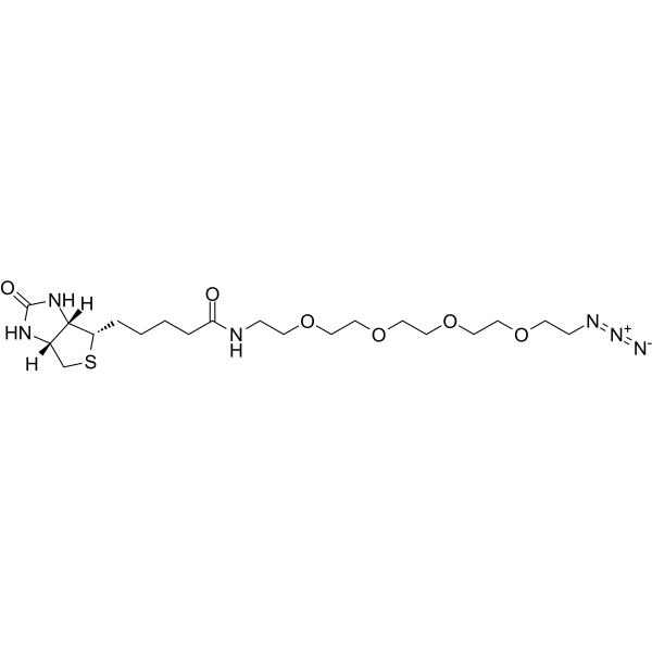
HY-140910
Biotin-PEG4-azidePROTAC Linker -
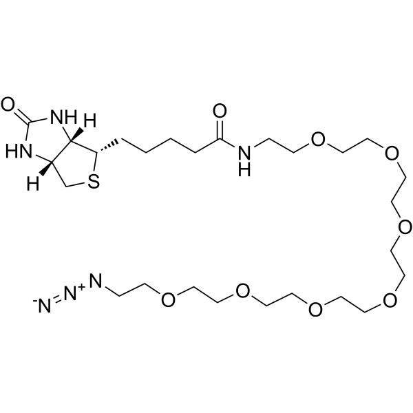
HY-133175
Biotin-PEG7-azidePROTAC linker -
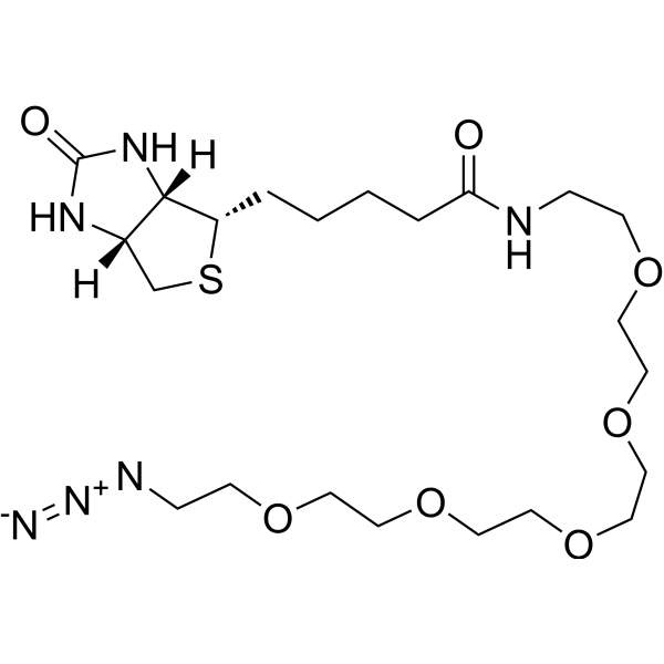
HY-133174
Biotin-PEG5-azidePROTAC linker -
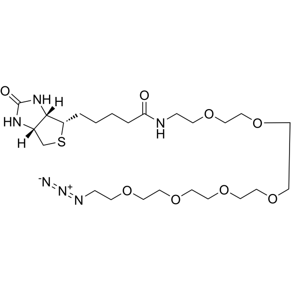
HY-140911
Biotin-PEG6-azidePROTAC Linkers -
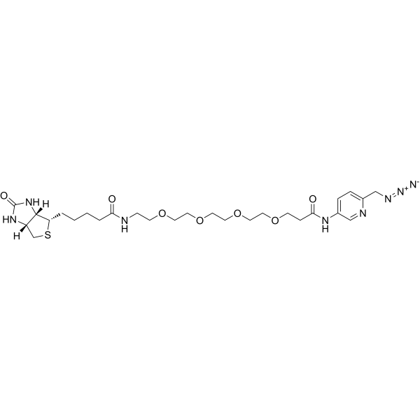
HY-140915
Biotin-PEG4-Picolyl azidePROTAC Linkers -
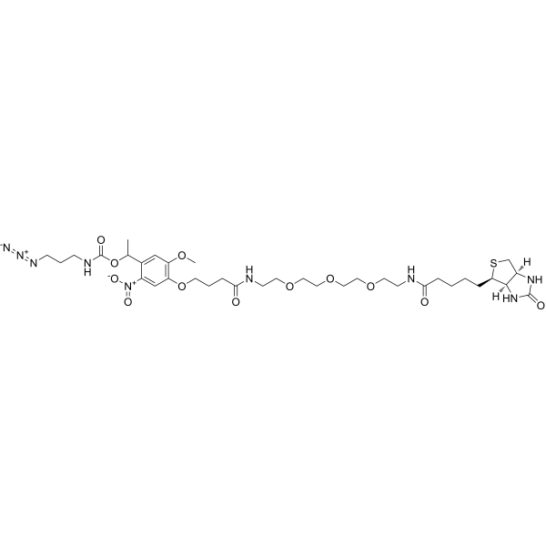
HY-140132
PC Biotin-PEG3-azideADC Linker -
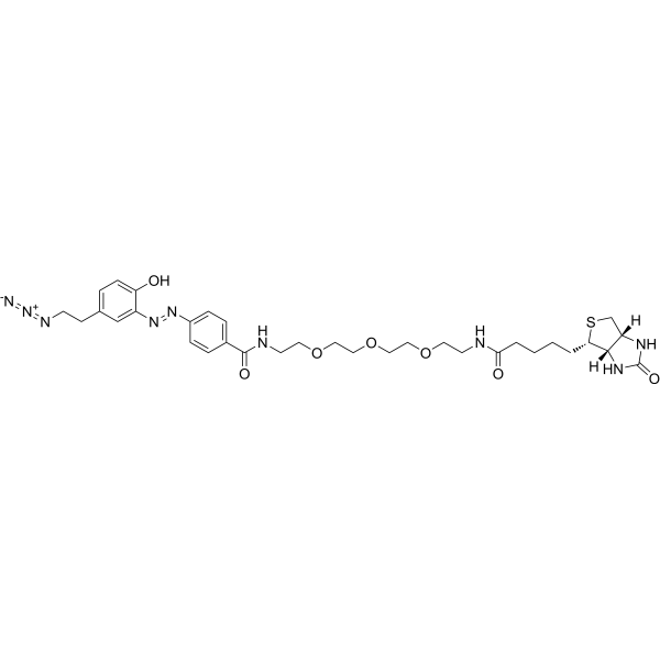
HY-140918
Diazo Biotin-PEG3-azidePROTAC Linker -
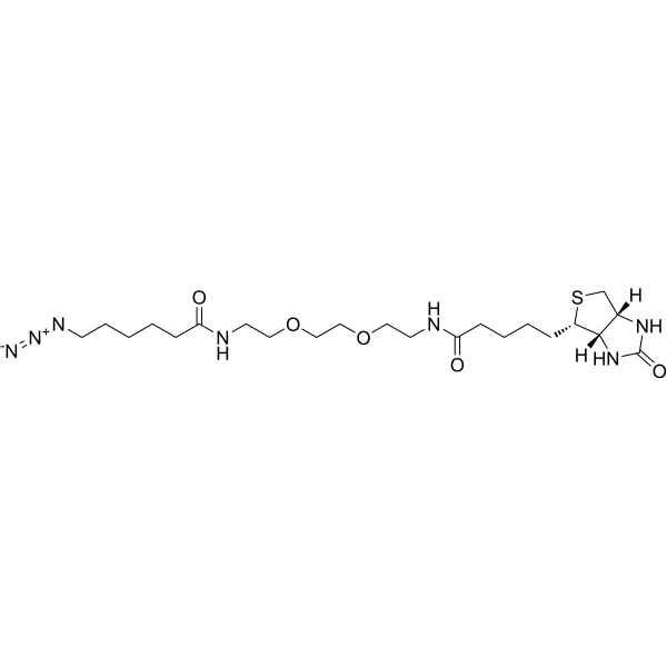
HY-117042
Biotin-PEG2-C6-azidePROTAC Linker -
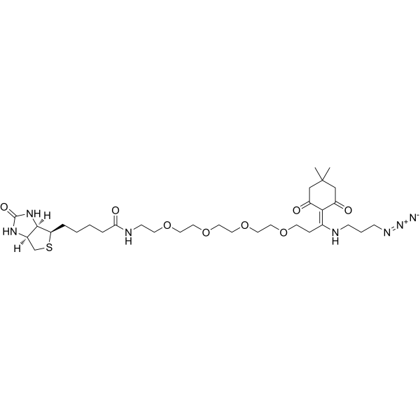
HY-140916
Dde Biotin-PEG4-azidePROTAC Linker -
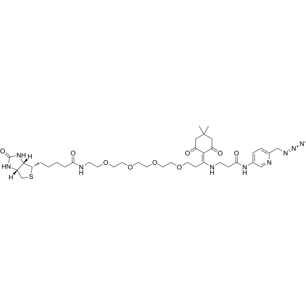
HY-140917
Dde Biotin-PEG4-Picolyl azidePROTAC Linker
All PROTAC Linkers Isoforms
More

Biological Activity
Description
IC50 & Target
|
PEGs |
In Vitro
PROTACs contain two different ligands connected by a linker; one is a ligand for an E3 ubiquitin ligase and the other is for the target protein. PROTACs exploit the intracellular ubiquitin-proteasome system to selectively degrade target proteins[1].
MedChemExpress (MCE) has not independently confirmed the accuracy of these methods. They are for reference only. Further protocols information, click here.


Chemical Information
-
CAS No. 2276672-04-7
-
Appearance Solid
-
Molecular Weight 796.97
-
Formula C34H64N6O13S
-
Color White to off-white
-
SMILES
O=C(NCCOCCOCCOCCOCCOCCOCCOCCOCCOCCOCCOCCN=[N+]=[N-])CCCC[C@@H]1SC[C@]([C@]1([H])N2)([H])NC2=O
-
Shipping
Room temperature in continental US; may vary elsewhere.
-
Storage
Powder -20°C 3 years In solvent -80°C 6 months -20°C 1 month

Protocols
-
EdU Incorporation Assay (Click Chemistry-Based DNA Synthesis Measurement)
The EdU incorporation assay measures DNA synthesis by adding the thymidine analog 5-ethynyl-2′-deoxyuridine to cells or tissues, where it is incorporated into newly synthesized DNA during S phase. Incorporated EdU is detected by copper-catalyzed azide-alkyne cycloaddition, in which a fluorescent azide covalently reacts with the ethynyl group on EdU, allowing S-phase cells to be detected by fluorescence microscopy, flow cytometry, or high-content imaging. EdU detection does not require DNA denaturation or anti-BrdU antibody access, which preserves sample structure and improves compatibility with immunostaining and multiparameter cytometry compared with BrdU-based detection. EdU can be cytotoxic in a cell-type- and exposure-dependent manner, so pulse duration, concentration, and continuous-labeling designs should be validated for each cell type.


Purity & Documentation
-
Data Sheet (267 KB)
-
SDS (252 KB)
- English - EN (252 KB)
- Français - FR (252 KB)
- Deutsch - DE (252 KB)
- Norwegian - NO (252 KB)
- Español - ES (252 KB)
- Swedish - SV (252 KB)
- Italian - IT (252 KB)
- Korean - KR (252 KB)
- Portuguese - PT (252 KB)
-
Handling Instructions (2659 KB)
References

Calculators
Concentration (start) × Volume (start) = Concentration (final) × Volume (final)


Biotin-PEG11-azide Related Classifications
HY-140912 Related Classifications
 Powered by Bioz