



Bintrafusp alfa

Based on 1 Customer Validation
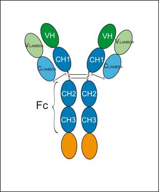
Bintrafusp alfa (M 7824) is a first-in-class bifunctional fusion protein composed of the extracellular domain of TGF-βRII fused to a human IgG1 mAb blocking programmed cell death ligand. Bintrafusp alfa can be used for the research of cancer.
For research use only. We do not sell to patients.
- Purity : 97.96%
- CAS No.: 1918149-01-5
- Molecular Weight:177.21 kDa
-
Storage:
Please store the product under the recommended conditions in the Certificate of Analysis.


To place orders, for customer services and technical support, please contact: MedChemExpress USA
Tel: 609-228-6898 E-mail: [email protected] [email protected]


-
 Biological Activity
-
Chemical Information
- Protocols
- Purity & Documentation
- References
-
 Help & FAQs


-

HY-P99020
FresolimumabTGF-β Inhibitor -
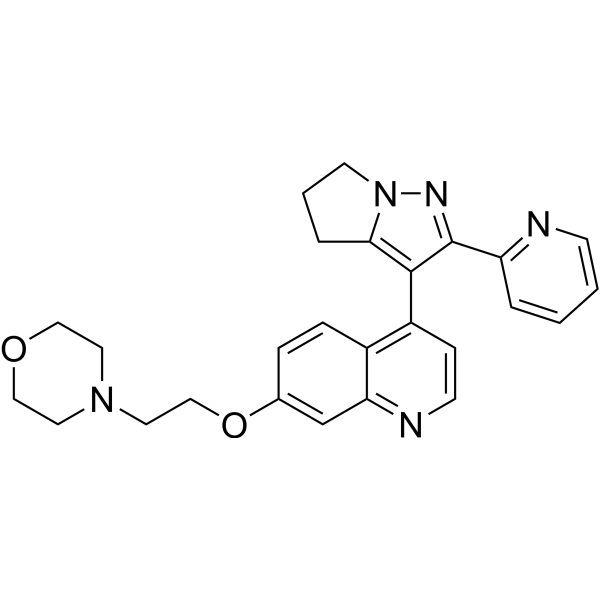
HY-12075
LY2109761TGF-βR I/II Inhibitor -
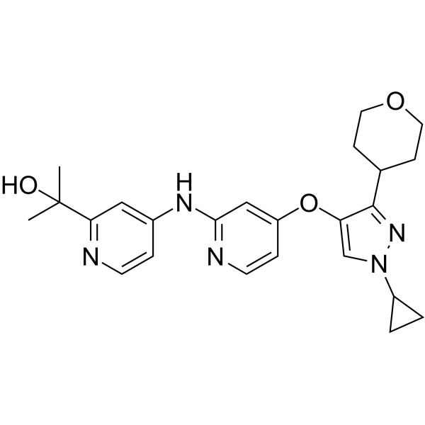
HY-103021
LY3200882ALK5 Inhibitor -

HY-10431
SB-431542TGF-β Receptor Kinase Inhibitor -
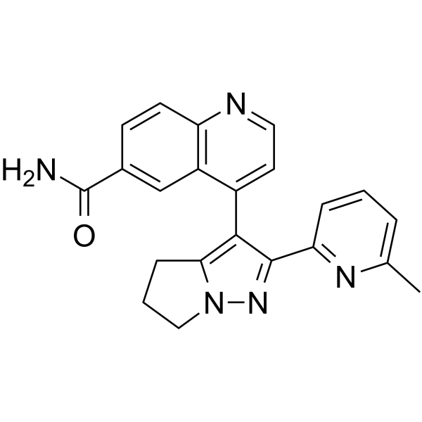
HY-13226
GalunisertibTGFβRI Inhibitor -
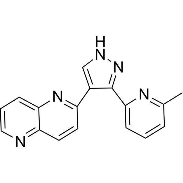
HY-13012
RepSoxTGFβR-1/ALK5 Inhibitor -
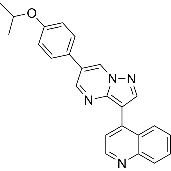
HY-12273
DMH-1BMP Inhibitor -
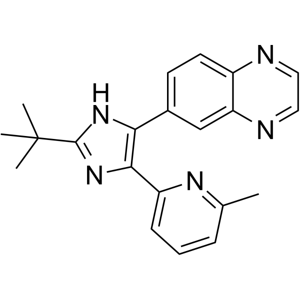
HY-12043
SB 525334ALK5 Inhibitor -
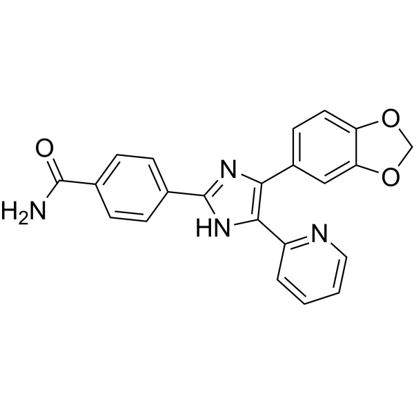
HY-10431G
SB-431542 (GMP)TGF-β Receptor Kinase Inhibitor -
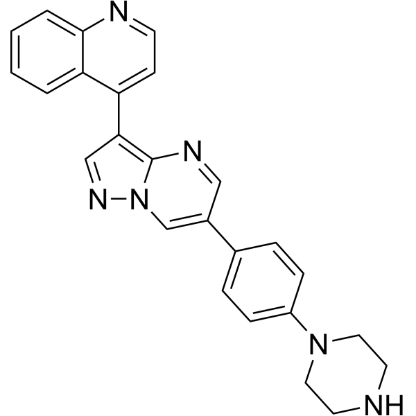
HY-12071
LDN193189ALK2/3 Inhibitor -
HY-13012G
RepSox (GMP)TGF-β-RI/ALK5 Inhibitor (GMP) -

HY-P990957
Ficerafusp alfaAnti-EGFR/TGFβ Antibody


Biological Activity
Description
Isotype
Human IgG1 lambda
Recommend Isotype Controls
In Vitro
Bintrafusp alfa can confer long-term protective immunity, decrease regulatory T-cell function, substantially increase CD8+ T cell and natural killer cell infiltration, and decrease myeloid-derived suppressor cell infiltration within tumors[1].
MedChemExpress (MCE) has not independently confirmed the accuracy of these methods. They are for reference only. Further protocols information, click here.
Clinical Trial
| NCT Number | Sponsor | Condition | Start Date |
Phase

|
|---|---|---|---|---|
| NCT01329991 | Plexxikon| | 2011-05 | PHASE1 |
Conjugated
Unconjugated
Reconsititution
The product can be reconstituted/diluted with sterile PBS or saline.
Format
-
Human IgG1 lambda
Application
ELISA, FACS, Functional assay


Chemical Information
-
CAS No. 1918149-01-5
-
Appearance Liquid
-
Molecular Weight 177.21 kDa
-
Color Colorless to light yellow
-
SMILES
[Bintrafusp alfa]
-
Synonyms
M7824; MSB0011359C
-
Shipping
Shipping with dry ice.
-
Formulation
Please refer to the lot-specific COA for specific buffer information.
-
Storage
Please store the product under the recommended conditions in the Certificate of Analysis.

Protocols
-
Apoptosis
Apoptosis, also called programmed cell death, is generally characterized by distinct morphological characteristics.
-
Cell Cytotoxicity Assay
Cytotoxicity assays are usually based on the assessment of cell membrane damage, which can also be indirectly detected by measuring cell viability. Detection methods include MTT assay, CKK-8 assay, LDH assay and ATP assay, etc.
-
Mammalian live/dead viability and cytotoxicity staining
Live/dead viability and cytotoxicity staining assays are based on the simultaneous detection of intracellular esterase activity in metabolically active (viable) cells and membrane integrity loss in non-viable cells. In commonly used dual-staining approaches, membrane-permeant fluorogenic substrates are converted by intracellular esterases into fluorescent products in live cells, while impermeant DNA-binding dyes selectively enter cells with compromised plasma membranes and label nucleic acids in dead or dying cells, enabling discrimination between viable and non-viable populations by fluorescence microscopy or flow cytometry.
-
Apoptosis Solutions
Apoptosis is a regulated, generally non-lytic cell-death pathway that removes unwanted, damaged, infected, or abnormal cells through coordinated morphological changes, caspase activation, DNA fragmentation, and membrane remodeling. The intrinsic apoptosis pathway is controlled mainly by mitochondrial outer membrane permeabilization, BCL-2 family proteins, cytochrome c release, apoptosome formation, caspase-9 activation, and downstream executioner caspase-3/7 activation. The extrinsic apoptosis pathway is initiated by death receptors such as Fas, TNFR, and TRAIL receptors, which recruit adaptor proteins and activate caspase-8 before engaging executioner caspases or mitochondrial amplification through BID cleavage. Apoptosis is linked to many phenotypes, including cancer cell killing, tissue homeostasis, immune regulation, neurodegeneration, infection response, and treatment-induced cytotoxicity; unresolved questions include how apoptosis interacts with necroptosis, pyroptosis, ferroptos


Purity & Documentation
-
Data Sheet (256 KB)
-
SDS (252 KB)
- English - EN (252 KB)
- Français - FR (252 KB)
- Deutsch - DE (252 KB)
- Norwegian - NO (252 KB)
- Español - ES (252 KB)
- Swedish - SV (252 KB)
- Italian - IT (252 KB)
- Korean - KR (252 KB)
- Portuguese - PT (252 KB)
-
Inhibitory Antibodies User Guide (603 KB)
References

Calculators
Concentration (start) × Volume (start) = Concentration (final) × Volume (final)


Bintrafusp alfa Related Classifications
HY-P99480 Related Classifications
 Powered by Bioz