



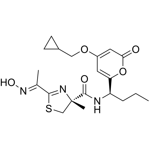
PM534
PM534 is a potent tubulin inhibitor (KD = 20 nM). PM534 targets and binds to the entire colchicine-binding domain (CBD) of tubulin, thereby inhibiting tubulin assembly; this leads to cell cycle arrest at the G2-M phase and induces multinucleation and apoptosis. PM534 exhibits microtubule-destabilizing agent (MDA) activity, potent broad-spectrum antitumor activity, and anti-angiogenic effects. PM534 can be used in research concerning human non-small cell lung cancer (NSCLC), breast cancer, ovarian cancer, and prostate cancer.
For research use only. We do not sell to patients.
- CAS No.: 2446376-25-4
- Formula: C20H27N3O5S
- Molecular Weight:421.51
-
Storage:
Please store the product under the recommended conditions in the Certificate of Analysis.

To place orders, for customer services and technical support, please contact: MedChemExpress USA
Tel: 609-228-6898 E-mail: [email protected] [email protected]


-
 Biological Activity
-
Chemical Information
- Protocols
- Purity & Documentation
- References
-
 Help & FAQs




Biological Activity
Description
Cellular Effect
|
Cell Line

|
Type | Value | Description | References |
|---|---|---|---|---|
| A549 | IC50 |
1.5 nM
Compound: PM534
|
Antiproliferative activity against human A549 cells assessed as inhibition of cell growth incubated for 48 hrs by MTT assay
Antiproliferative activity against human A549 cells assessed as inhibition of cell growth incubated for 48 hrs by MTT assay
|
38294341 |
| HeLa | IC50 |
1.3 nM
Compound: PM534
|
Antiproliferative activity against human HeLa cells expressing tubulin betaIII assessed as inhibition of cell growth incubated for 48 hrs by MTT assay
Antiproliferative activity against human HeLa cells expressing tubulin betaIII assessed as inhibition of cell growth incubated for 48 hrs by MTT assay
|
38294341 |
| A2780 | IC50 |
1.4 nM
Compound: PM534
|
Antiproliferative activity against human A2780 cells assessed as inhibition of cell growth incubated for 48 hrs by MTT assay
Antiproliferative activity against human A2780 cells assessed as inhibition of cell growth incubated for 48 hrs by MTT assay
|
38294341 |
| HeLa | IC50 |
1.6 nM
Compound: PM534
|
Antiproliferative activity against human HeLa cells assessed as inhibition of cell growth incubated for 48 hrs by MTT assay
Antiproliferative activity against human HeLa cells assessed as inhibition of cell growth incubated for 48 hrs by MTT assay
|
38294341 |
| BT-474 | GI50 |
2.3 nM
|
Significantly inhibits cell activity.
Significantly inhibits cell activity.
|
41481906 |
| HCC1937 | GI50 |
1.8 nM
|
Significantly inhibits cell activity.
Significantly inhibits cell activity.
|
41481906 |
| HCC1954 | GI50 |
22.4 nM
|
Significantly inhibits cell activity.
Significantly inhibits cell activity.
|
41481906 |
| MCF7 | GI50 |
2.1 nM
|
Significantly inhibits cell activity.
Significantly inhibits cell activity.
|
41481906 |
| MDA-MB-231 | GI50 |
2.2 nM
|
Significantly inhibits cell activity.
Significantly inhibits cell activity.
|
41481906 |
| MDA-MB-436 | ED50 |
2.6 nM
|
Significantly inhibits cell activity.
Significantly inhibits cell activity.
|
41481906 |
| A2780 | GI50 |
1.1 nM
|
Significantly inhibits cell activity.
Significantly inhibits cell activity.
|
41481906 |
| ES-2 | GI50 |
2.5 nM
|
Significantly inhibits cell activity.
Significantly inhibits cell activity.
|
41481906 |
| IGROV-1 | GI50 |
2.8 nM
|
Significantly inhibits cell activity.
Significantly inhibits cell activity.
|
41481906 |
| SK-OV-3 | GI50 |
2.5 nM
|
Significantly inhibits cell activity.
Significantly inhibits cell activity.
|
41481906 |
| LNCaP | GI50 |
3.0 nM
|
Significantly inhibits cell activity.
Significantly inhibits cell activity.
|
41481906 |
| PC-3 | GI50 |
1.7 nM
|
Significantly inhibits cell activity.
Significantly inhibits cell activity.
|
41481906 |
| VCaP | GI50 |
2.7 nM
|
Significantly inhibits cell activity.
Significantly inhibits cell activity.
|
41481906 |
| LoVo | GI50 |
1.9 nM
|
Overcomes drug resistance and inhibits cell growth.
Overcomes drug resistance and inhibits cell growth.
|
41481906 |
| HeLa | GI50 |
1.1 nM
|
Overcomes drug resistance and inhibits cell growth.
Overcomes drug resistance and inhibits cell growth.
|
41481906 |
In Vitro
PM534 (0.625-5 μM) inhibits tubulin assembly activity in a concentration-dependent manner in a cell-free system[1].
PM534 (1-2 nM; 24 h) induces complete depolymerization of the microtubule cytoskeleton and disrupts mitotic spindle formation in A549 cells[1].
PM534 (0.06-240 nM; 72 h) inhibits cell proliferation in A549 (IC50 = 1.5 nM), Calu-6, NCI-H23, NCI-H460, A2780 (IC50 = 1.4 nM), A2780AD (IC50 = 3.2 nM), HeLa (IC50 = 1.6 nM), and HeLa βIII (IC50 = 1.3 nM) cells, with a GI50 value of 2.2 nM[1].
PM534 (0.06-240 nM; 72 h) inhibits the growth of BT-474 (GI50 = 2.3 nM), HCC1937 (GI50 = 1.8 nM), HCC1954 (GI50 = 22.4 nM), MCF7 (GI50 = 2.1 nM), MDA-MB-231 (GI50 = 2.2 nM), MDA-MB-436 (GI50 = 2.6 nM), A2780 (GI50 = 1.1 nM), ES-2 (GI50 = 2.5 nM), IGROV-1 (GI50 = 2.8 nM), SK-OV-3 (GI50 = 2.5 nM), 22Rv1 (GI50 = 2.1 nM), LN-Cap (GI50 = 3.0 nM), PC-3 (GI50 = 1.7 nM), and VCaP (GI50 = 2.7 nM) cells[2].
PM534 (10 nM; 1-24 h) induces irreversible anti-proliferative effects in MDA-MB-231 and MDA-MB-436 cells[2].
PM534 (10-1000 nM; 2 h) competitively binds to the colchicine site and displaces EBI in cell lysates[2].
PM534 (10 nM; 24 h) induces G2-M phase cell cycle arrest, upregulates Cyclin B1 and pH3 expression, and causes multinucleation in MDA-MB-231, A2780, and 22Rv1 cells[2].
PM534 (0.5-10 nM; 30 min) inhibits cell adhesion activity in HUVEC cells (IC50 = 2.3 nM)[2].
PM534 (0.6-10 nM; 24 h) inhibits cell migration in HUVEC cells[2].
PM534 (0.5-10 nM; 24 h) inhibits cell invasion, disrupts capillary-like tube formation, and disrupts established networks in HUVEC cells[2].
PM534 (0.06-240 nM; 72 h) exhibits activity in overcoming drug resistance and inhibiting cell growth in LoVo (GI50 = 1.9 nM), LoVo-DOX (GI50 = 5.8 nM), HeLa wt (GI50 = 1.1 nM), and HeLa βIII (GI50 = 1 nM) cells[2].
MedChemExpress (MCE) has not independently confirmed the accuracy of these methods. They are for reference only.
-
Cell Line:A549
-
Concentration:1 nM, 2 nM
-
Incubation Time:24 h
-
Result:Induced complete disorganization of the tubulin cytoskeleton during interphase.
Abrogated the formation of the mitotic spindle.
Generated a cell phenotype of hypercondensed chromatin and aberrant chromosome distribution.
-
Cell Line:OV-3, 22Rv1, LN-Cap, PC-3, VCaP, LoVo, LoVo-DOX, HeLa, HeLa βIII
-
Concentration:Perform serial dilutions starting from 240 nM down to 0.06 nM using a 1:2.5 dilution factor
-
Incubation Time:72 h
-
Result:BT-474, HCC1937, HCC1954, MCF7, MDA-MB-231, MDA-MB-436, A2780, ES-2, IGROV-1, SK- Inhibited cell proliferation and growth in a dose-dependent manner.
-
Cell Line:MDA-MB-231, A2780, 22Rv1
-
Concentration:10 nM
-
Incubation Time:24 h
-
Result:Induced the formation of multinucleated and aberrantly divided cells.
-
Cell Line:MDA-MB-231, A2780, 22Rv1
-
Concentration:10 nM
-
Incubation Time:24 h
-
Result:Caused a pronounced accumulation of cells in the G2-M phase.
-
Cell Line:MDA-MB-231, A2780, 22Rv
-
Concentration:10 nM
-
Incubation Time:6, 15, 24, 48 h
-
Result:Upregulated the expression of cyclin B1 and phosphorylated histone H3 (pH3).
-
Cell Line:HUVEC
-
Concentration:0.6, 1.6, 4, 10 nM
-
Incubation Time:24 h
-
Result:Significantly delayed wound closure at lower concentrations and completely inhibited cell migration at concentrations above 4 nM.
-
Cell Line:HUVEC
-
Concentration:0.5, 1.0, 2.5, 5, 10 nM
-
Incubation Time:24 h
-
Result:Concentration-dependently inhibited invasion and completely abrogated cell invasion at concentrations of 5 nM and 10 nM.
In Vivo
PM534 (2.5 or 5 mg/kg; i.v.; once weekly; 3 weeks) reduces tumor volume, prolongs survival, induces apoptosis and necrosis, and disrupts blood vessels in A2780, ES-2, MDA-MB-231, and HCC1937 xenograft mouse models[2].
PM534 (2.5 or 3.75 mg/kg; i.v.; once weekly; 3 weeks) reduces tumor volume and prolongs survival in 22Rv1 and VCaP xenograft mouse models[2].
PM534 (5 mg/kg; i.v.; single injection; 1 week) overcomes multidrug resistance and significantly reduces tumor volume in LoVo, LoVo-DOX, HeLa wt, and HeLa βIII xenograft mouse models[2].
MedChemExpress (MCE) has not independently confirmed the accuracy of these methods. They are for reference only.
-
Animal Model:Athymic nu/nu mice (Female, 4-6 weeks old) were subcutaneously injected with 5 x 10⁶ NCI-H460 cells[1]
-
Dosage:0.75, 1.1, 1.7, 2.5 mg/kg
-
Administration:i.v.; once a week; 2 weeks
-
Result:Inhibited tumor growth and improved survival rates.
-
Animal Model:Athymic nude-Foxn1nu/nu mice (Female, 4-6 weeks old) were subcutaneously injected with 5 x 10⁶ A2780, ES-2, MDA-MB-231, HCC1937 cells[2]
-
Dosage:2.5 mg/kg (ES-2, HCC1937), 5 mg/kg (A2780, MDA-MB-231)
-
Administration:i.v.; once a week; 3 weeks
-
Result:Reduced tumor volume.
Prolonged survival.
Induced apoptosis and necrosis.
Disrupted blood vessels.
-
Animal Model:Athymic nude-Foxn1nu/nu and CB17/Icr-Prkdecid mice (male, 4-6 weeks old) were subcutaneously injected with 5 x 10⁶ 22Rv1 and VCaP cells, respectively[2]
-
Dosage:2.5 mg/kg (VCaP), 3.75 mg/kg (22Rv1)
-
Administration:i.v.; once a week; 3 weeks
-
Result:Reduce tumor volume and prolong survival.
-
Animal Model:Athymic nude-Foxn1nu/nu mice (Female, 4-6 weeks old) were subcutaneously injected with 5 x 10⁶ LoVo, LoVo-DOX, HeLa wt, HeLa βIII cells[2]
-
Dosage:5 mg/kg
-
Administration:i.v.; single dose; 1 week
-
Result:Overcame multidrug resistance and significantly reduced tumor volume.
- Detection of Apoptosis
- TUNEL staining for apoptotic DNA fragmentation
- Flow cytometric DNA-content cell-cycle staining
- Annexin V plus membrane-impermeant dye apoptosis staining
- BrdU Incorporation Assay
- Apoptosis Solutions
- Protocol for Cell Cycle
- Subcutaneous Cell-Line-Derived Xenograft
- Breast Cancer Modeling
Clinical Trial
| NCT Number | Sponsor | Condition | Start Date |
Phase
|
|---|---|---|---|---|
| NCT01329991 | Plexxikon| | 2011-05 | PHASE1 |


Chemical Information
-
CAS No. 2446376-25-4
-
Molecular Weight 421.51
-
Formula C20H27N3O5S
-
SMILES
O=C([C@]1(N=C(SC1)/C(C)=N/O)C)N[C@@H](C2=CC(OCC3CC3)=CC(O2)=O)CCC
-
Shipping
Room temperature in continental US; may vary elsewhere.
-
Storage
Please store the product under the recommended conditions in the Certificate of Analysis.


Purity & Documentation
References
[1]. Lucena-Agell D,et al. PM534, an Optimized Target-Protein Interaction Strategy through the Colchicine Site of Tubulin. J Med Chem. 2024 Feb 22;67(4):2619-2630. [Content Brief]
[2]. Aviles P, et al. PM534, a Novel Colchicine Site Tubulin Inhibitor with Broad-Spectrum and Resistance-Overcoming Antitumor Activity. Mol Cancer Ther. 2026 Jun 1;25(6):935-947. [Content Brief]

Calculators
Concentration (start) × Volume (start) = Concentration (final) × Volume (final)


PM534 Related Classifications
HY-163270 Related Classifications
 Powered by Bioz