



EHop-016
Based on 9 publication(s) in Google Scholar
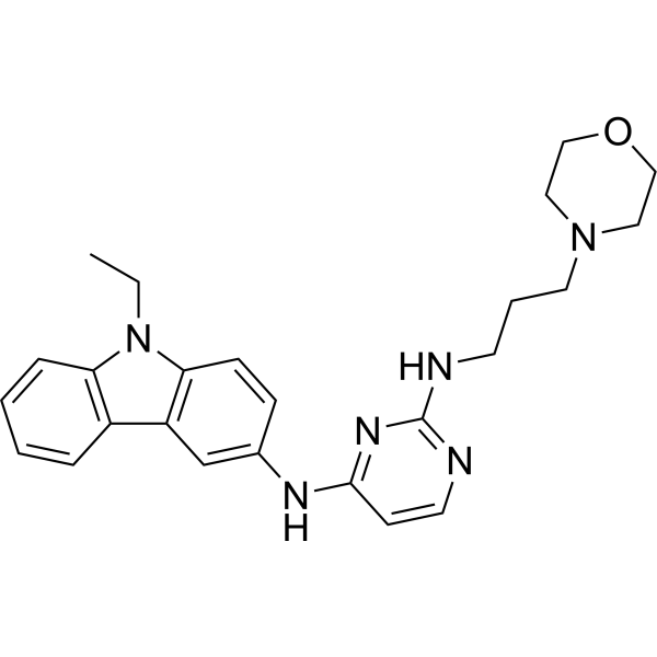
EHop-016 is a potent and selective Rac GTPase Rac1 and Rac3 inhibitor. EHop-016 inhibits Rac1 activity with an IC50 of 1.1 μM in MDA-MB-435 cells. EHop-016 inhibits Vav2 interaction with Rac, Rac-activated PAK1, lamellipodia formation, and cell migration.
Nos produits utilisent uniquement pour la recherche. Nous ne vendons pas aux patients.
- Pureté : 99.38%
- CAS No.: 1380432-32-5
- Formule: C25H30N6O
- Masse moléculaire:430.55
-
Stockage:Powder -20°C, 3 years , 4°C, 2 years ; In solvent -80°C, 2 years , -20°C, 1 year

To place orders, for customer services and technical support, please contact: MedChemExpress USA
Tel: 609-228-6898 E-mail: [email protected] [email protected]


-
 Activité biologique
-
Chemical Information
-
Solvant et solubilité
- Protocole
- Pureté et documentation
- Références
-
 Help & FAQs


-
HY-15723A
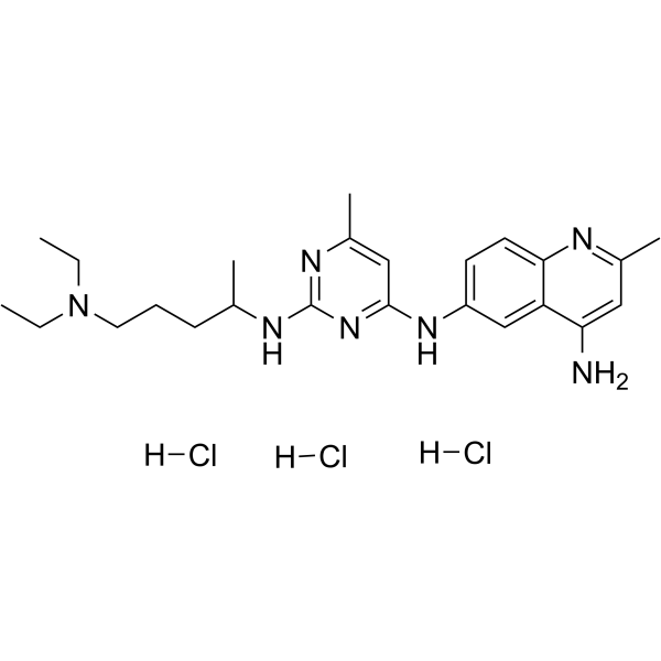
NSC 23766 trihydrochlorideRac Inhibitor -
HY-16659
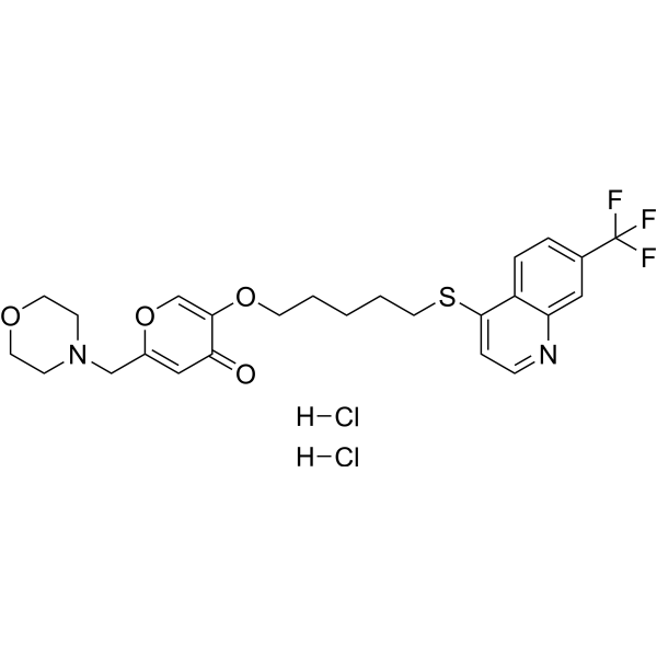
EHT 1864Rac Family Inhibitor -
HY-15723
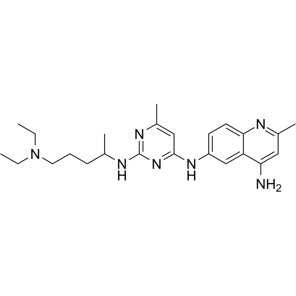
NSC 23766Ras Inhibitor -
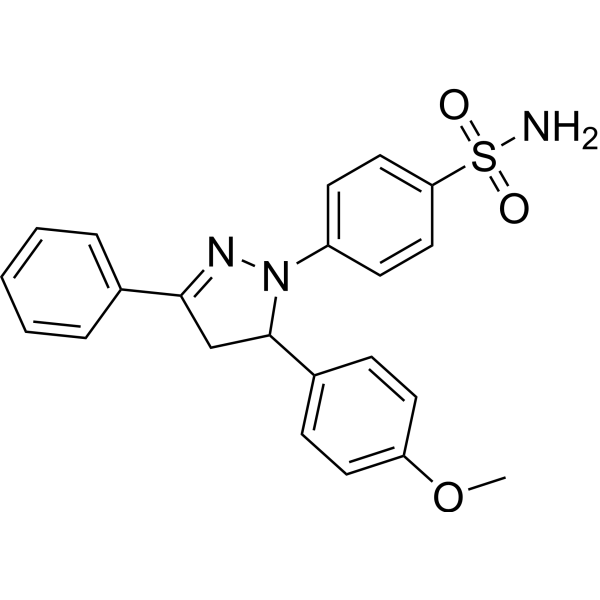
HY-12755
ML141Cdc42 GTPase Inhibitor -
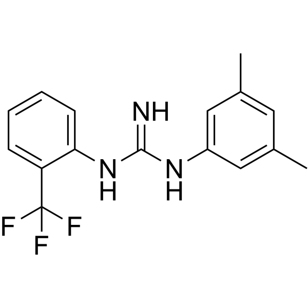
HY-104064
1A-116Rac1 Inhibitor -
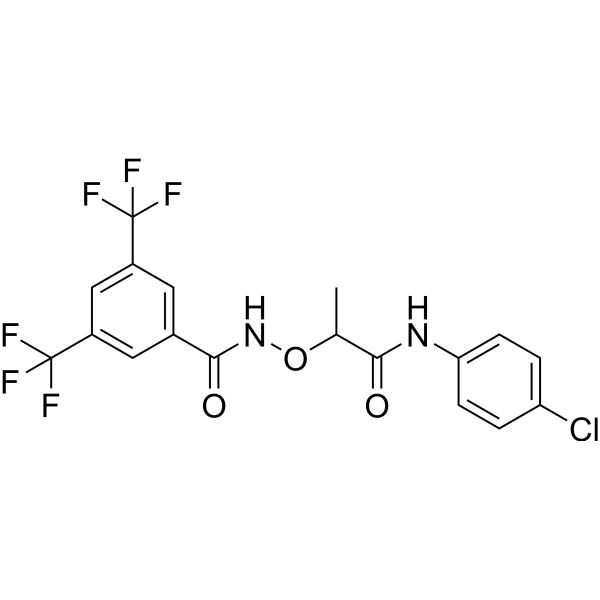
HY-13991
CCG-1423Rho/MRTF/SRF Pathway Inhibitor -
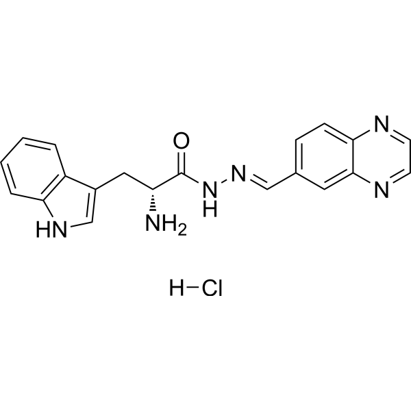
HY-12646
Rhosin hydrochlorideRho GTPases Inhibitor -

HY-10071
Y-27632ROCK Inhibitor -
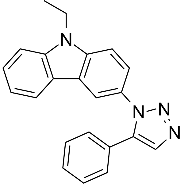
HY-112842
MBQ-167Rac/Cdc42 Inhibitor -
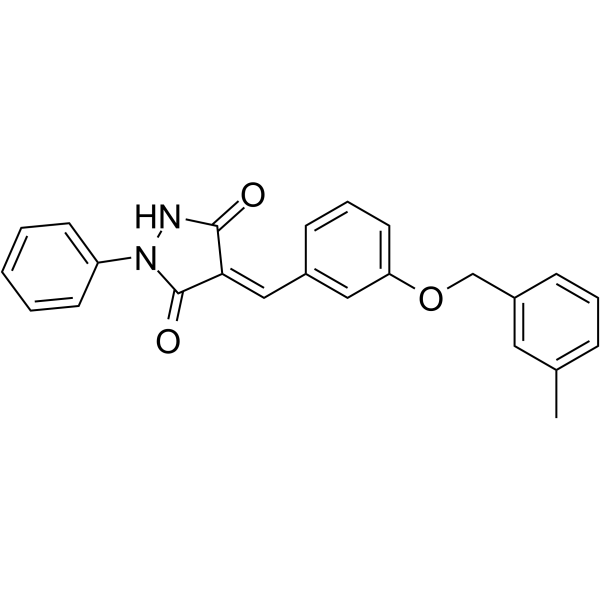
HY-12649
Y16GEF-Rho GTPase Inhibitor -
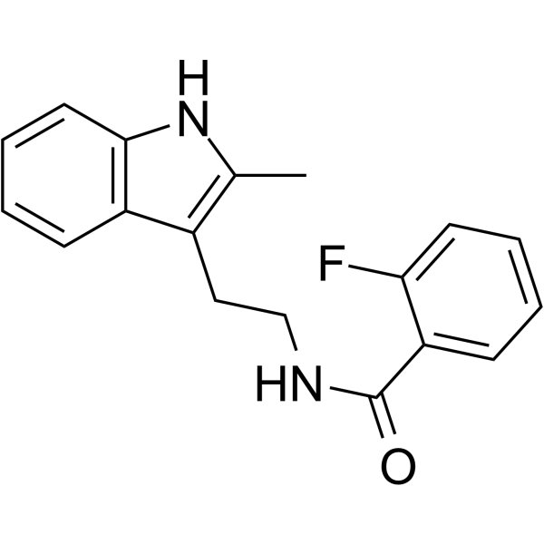
HY-16926
CK-666Arp2/3 Complex Inhibitor -
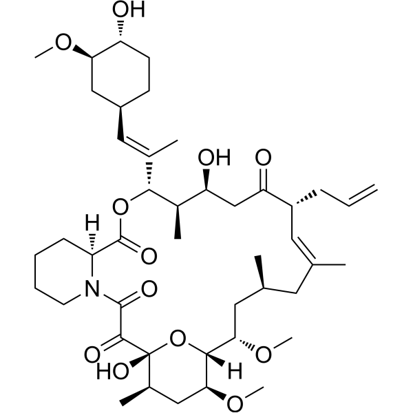
HY-13756
TacrolimusCalcineurin Inhibitor
Publications Citing Use of MedChemExpress (MCE) EHop-016
More- Pharmacol Res. 2026 Jan:223:108072. [Abstract]
- J Exp Med. 2023 Mar 6;220(3):e20221316. [Abstract]
- Biochem Pharmacol. 2021 Feb:184:114399. [Abstract]
- Biochim Biophys Acta Mol Basis Dis. 2026 Apr 15;1872(6):168261. [Abstract]
- Cancers (Basel). 2025 Jan 23;17(3):361. [Abstract]
- Appl Microbiol Biotechnol. 2018 Jul;102(14):5965-5975. [Abstract]
- Mol Oncol. 2019 Sep;13(9):2010-2030. [Abstract]
- J Immunol. 2026 Aug 4;215(8):vkag222.
- bioRxiv. 2025 January 15.
 Customer Validation & Images

-
Cell Proliferation/Viability Assay

-
Bio/Physico-chemical Assay

-
Cell Imaging/Staining

-
RT-PCR

-
ELISA


Activité biologique
Description
Cellular Effect
|
Cell Line

|
Type | Value | Description | References |
|---|---|---|---|---|
| MCF7 | GI50 |
14 μM
Compound: EHop-016
|
Growth inhibition of human MCF7 cells after 24 hrs by SRB assay
Growth inhibition of human MCF7 cells after 24 hrs by SRB assay
|
[PMID: 29358027] |
| MDA-MB-231 | GI50 |
15 μM
Compound: EHop-016
|
Growth inhibition of human MDA-MB-231 cells after 24 hrs by SRB assay
Growth inhibition of human MDA-MB-231 cells after 24 hrs by SRB assay
|
[PMID: 29358027] |
In Vitro
EHop-016 (1-10 μM; 24 hours; MDA-MB-435 cells) treatment inhibits Rac1 and Rac3 activity. At higher concentrations, EHop-016 inhibits the close homolog Cdc42. In MDA-MB-435 cells that demonstrate high active levels of the Rac GEF Vav2, EHop-016 inhibits the association of Vav2 with a nucleotide-free Rac1(G15A) [1].
EHop-016 also inhibits the Rac activity of MDA-MB-231 metastatic breast cancer cells and reduces Rac-directed lamellipodia formation in both cell lines. EHop-016 decreases Rac downstream effects of PAK1 (p21-activated kinase 1) activity and directed migration of metastatic cancer cells[1].
EHop-016 affectes cell viability by down-regulating Akt and Jun kinase activities and c-Myc and Cyclin D expression, as well as increasing caspase 3/7 activities in metastatic cancer cells[2].
MedChemExpress (MCE) has not independently confirmed the accuracy of these methods. They are for reference only.
-
Cell Line:MDA-MB-435 cells
-
Concentration:1 μM, 2 μM, 4 μM, 5 μM, 10 μM
-
Incubation Time:24 hours
-
Result:The activity Rac3 was inhibited by 58%.
In Vivo
MedChemExpress (MCE) has not independently confirmed the accuracy of these methods. They are for reference only.
-
Animal Model:Female athymic nu/nu mice (4-5 weeks old) injected with GFP-MDA-MB-435 cells[2]
-
Dosage:10 mg/kg, 25 mg/kg
-
Administration:Intraperitoneal injection; 3 times a week; for 55 days
-
Result:Significantly reduced mammary fat pad tumor growth, metastasis, and angiogenesis.
- Scratch/Wound-Healing Migration Assay
- Cell migration
- Zigmond Chamber Chemotaxis Assay
- Under-Agarose Cell Migration Assay
- Neural Crest/Neuronal Cell Migration Explant Assay
- Transwell/Boyden Chamber Migration Assay
- 3D Collagen/Hydrogel Matrix Migration Assay
- Cell-Exclusion Zone Migration Assay
- Dunn/Insall Chamber Chemotaxis Assay


Chemical Information
-
CAS No. 1380432-32-5
-
Appearance Solid
-
Masse moléculaire 430.55
-
Formule C25H30N6O
-
Color Light yellow to yellow
-
SMILES
CCN1C2=C(C3=C1C=CC=C3)C=C(NC4=NC(NCCCN5CCOCC5)=NC=C4)C=C2
-
Livraison
Room temperature in continental US; may vary elsewhere.
-
Stockage
Powder -20°C 3 years 4°C 2 years In solvent -80°C 2 years -20°C 1 year


Publications (9)
-
Journal Impact Factor
-
Most Recent
-
Pharmacol Res
Functional, synaptoproteomic and structural adaptations underlying sex-dependent traumatic stress susceptibility/resilience in the hippocampus. [Abstract]2026 Jan:223:108072. PMID: 41453670 -
J Exp Med
Repurposing a tricyclic antidepressant in tumor and metabolism disease treatment through fatty acid uptake inhibition. [Abstract]2023 Mar 6;220(3):e20221316. PMID: 36520461
EHop-016 purchased from MedChemExpress. Usage Cited in: J Exp Med. 2023 Mar 6;220(3):e20221316. [Abstract]
EHop-016 (0, 1, 2, 3, μM; 4 days) did not affect cell proliferation in MDAMB-231 cells.
EHop-016 purchased from MedChemExpress. Usage Cited in: J Exp Med. 2023 Mar 6;220(3):e20221316. [Abstract]
EHop-016 (2 μM; 24 h and incubated with BODIPY-C16 (2 μM) for 2 h or TMR-Dextran (1 mg/ml) for 1 h) reduced the uptake of BODIPY-C16 and TMR-Dextran in MDAMB-231 cells.
EHop-016 purchased from MedChemExpress. Usage Cited in: J Exp Med. 2023 Mar 6;220(3):e20221316. [Abstract]
EHop-016 (2 μM; 24 h and then incubated with BODIPY-C16 (5 μM) for 30 min prior to TMR-Dextran (1 mg/ml) treatment for another 30 min) reduced the uptake of BODIPY-C16 and TMR-Dextran in MDAMB-231 cells.
-
Biochem Pharmacol
DOCK2 contributes to endotoxemia-induced acute lung injury in mice by activating proinflammatory macrophages. [Abstract]2021 Feb:184:114399. PMID: 33382969
EHop-016 purchased from MedChemExpress. Usage Cited in: Biochem Pharmacol. 2021 Feb:184:114399. [Abstract]
EHop-016 (1 μM; 24 h) costimulated with LPS exhibited remarkably suppressed LPS-induced IL-1β and TNF-α in RAW264.7 cells.
EHop-016 purchased from MedChemExpress. Usage Cited in: Biochem Pharmacol. 2021 Feb:184:114399. [Abstract]
EHop-016 (1 μM; 24 h) costimulated with LPS exhibited remarkably suppressed LPS-induced IL-1β and TNF-α, IL-6 in RAW264.7 cells.

EHop-016 purchased from MedChemExpress. Usage Cited in: Biochem Pharmacol. 2021 Feb:184:114399. [Abstract]
EHop-016 (1 μM; 15 min) costimulated with LPS exhibited remarkably inhibited LPS-induced IKK-β phosphorylation in RAW264.7 cells.
-
Biochim Biophys Acta Mol Basis Dis
2026 Apr 15;1872(6):168261. PMID: 41990488 -
Cancers (Basel)
RAC1-Amplified and RAC1-A159V Hotspot-Mutated Head and Neck Cancer Sensitive to the Rac Inhibitor EHop-016 In Vivo: A Proof-of-Concept Study. [Abstract]2025 Jan 23;17(3):361. PMID: 39941730 -
Appl Microbiol Biotechnol
Deacetylmycoepoxydiene is an agonist of Rac1, and simultaneously induces autophagy and apoptosis. [Abstract]2018 Jul;102(14):5965-5975. PMID: 29740674 -
Mol Oncol
RAC1 inhibition reverses cisplatin resistance in esophageal squamous cell carcinoma and induces downregulation of glycolytic enzymes. [Abstract]2019 Sep;13(9):2010-2030. PMID: 31314174 -
-


Solvant et solubilité
In Vitro:
DMSO : ≥ 32 mg/mL (74.32 mM; Hygroscopic DMSO has a significant impact on the solubility of product, please use newly opened DMSO)
* "≥" means soluble, but saturation unknown.
Please refer to the solubility information to select the appropriate solvent. Once prepared, please aliquot and store the solution to prevent product inactivation from repeated freeze-thaw cycles.
Storage method and period of stock solution: -80°C, 2 years; -20°C, 1 year. When stored at -80°C, please use it within 2 years. When stored at -20°C, please use it within 1 year.
Please refer to the solubility information to select the appropriate solvent. Once prepared, please aliquot and store the solution to prevent product inactivation from repeated freeze-thaw cycles.
Storage method and period of stock solution: -80°C, 2 years; -20°C, 1 year. When stored at -80°C, please use it within 2 years. When stored at -20°C, please use it within 1 year.
Concentration (start) × Volume (start) = Concentration (final) × Volume (final)
In Vivo:
Select the appropriate dissolution method based on your experimental animal and administration route.
- For the following dissolution methods, please ensure to first prepare a clear stock solution using an In Vitro approach and then sequentially add co-solvents:
- To ensure reliable experimental results, the clarified stock solution can be appropriately stored based on storage conditions. As for the working solution for In Vivo experiments, it is recommended to prepare freshly and use it on the same day.
- The percentages shown for the solvents indicate their volumetric ratio in the final prepared solution. If precipitation or phase separation occurs during preparation, heat and/or sonication can be used to aid dissolution.
Add each solvent one by one: 10% DMSO 40% PEG300 5% Tween-80 45% Saline
Solubility: ≥ 2.5 mg/mL (5.81 mM); Clear solution
This protocol yields a clear solution of ≥ 2.5 mg/mL (saturation unknown).
Taking 1 mL working solution as an example, add 100 μL DMSO stock solution (25.0 mg/mL) to 400 μL PEG300, and mix evenly; then add 50 μL Tween-80 and mix evenly; then add 450 μL Saline to adjust the volume to 1 mL.
Preparation of Saline: Dissolve 0.9 g sodium chloride in ddH₂O and dilute to 100 mL to obtain a clear Saline solution.
Add each solvent one by one: 10% DMSO 90% (20% SBE-β-CD in Saline)
Solubility: ≥ 2.5 mg/mL (5.81 mM); Clear solution
This protocol yields a clear solution of ≥ 2.5 mg/mL (saturation unknown).
Taking 1 mL working solution as an example, add 100 μL DMSO stock solution (25.0 mg/mL) to 900 μL 20% SBE-β-CD in Saline, and mix evenly.
Preparation of 20% SBE-β-CD in Saline (4°C, storage for one week): 2 g SBE-β-CD powder is dissolved in 10 mL Saline, completely dissolve until clear.
For the following dissolution methods, please prepare the working solution directly:
It is recommended to prepare fresh solutions and use them promptly within a short period of time.
The percentages shown for the solvents indicate their volumetric ratio in the final prepared solution. If precipitation or phase separation occurs during preparation, heat and/or sonication can be used to aid dissolution.
Add each solvent one by one: 50% PEG300 50% Saline
Solubility: 10 mg/mL (23.23 mM); Suspended solution; Need ultrasonic
In Vivo Dissolution Calculator
Please enter the basic information of animal experiments:
-
-
-
-
Recommended: Prepare an additional quantity of animals to account for potential losses during experiments.
Please enter your animal formula composition:
-
%DMSO +
Recommended: Keep the proportion of DMSO in working solution below 2% if your animal is weak.
-
%+
-
+%Tween-80 + +
-
%Saline +
The co-solvents required include: DMSO, . All of co-solvents are available by MedChemExpress (MCE). , Tween 80. All of co-solvents are available by MedChemExpress (MCE).
Working solution concentration: 0.22 mg/mL
Method for preparing stock solution: mg drug dissolved in μL DMSO. Stock solution concentration: mg/mL.
1. Take μL DMSO stock solution;
2. Add μL .
μL , mix evenly;
3. Then add μL Tween 80, mix evenly;
4. Then add μL
Please ensure that the stock solution in the first step is dissolved to a clear state, and add co-solvents in sequence. You can use ultrasonic heating (ultrasonic cleaner, recommended frequency 20-40 kHz), vortexing, etc. to assist dissolution.


Pureté et documentation
-
Fiche technique (279 KB)
-
SDS (394 KB)
- English - EN (394 KB)
- Français - FR (394 KB)
- Deutsch - DE (394 KB)
- Norwegian - NO (394 KB)
- Español - ES (394 KB)
- Swedish - SV (394 KB)
- Italian - IT (394 KB)
- Korean - KR (394 KB)
- Portuguese - PT (394 KB)
-
Instruction de manipulation (2659 KB)
Références
[1]. Montalvo-Ortiz BL, et al. Characterization of EHop-016, novel small molecule inhibitor of Rac GTPase. J Biol Chem. 2012 Apr 13;287(16):13228-38. [Content Brief]
[2]. Castillo-Pichardo L, et al. The Rac Inhibitor EHop-016 Inhibits Mammary Tumor Growth and Metastasis in a Nude Mouse Model. Transl Oncol. 2014 Oct 24;7(5):546-55. [Content Brief]


Complete Stock Solution Preparation Table
Please refer to the solubility information to select the appropriate solvent. Once prepared, please aliquot and store the solution to prevent product inactivation from repeated freeze-thaw cycles.
Storage method and period of stock solution: -80°C, 2 years; -20°C, 1 year. When stored at -80°C, please use it within 2 years. When stored at -20°C, please use it within 1 year.
| Optional Solvent | Concentration Solvent Mass | 1 mg | 5 mg | 10 mg | 25 mg |
|---|---|---|---|---|---|
| DMSO | 1 mM | 2.3226 mL | 11.6131 mL | 23.2261 mL | 58.0653 mL |
| 5 mM | 0.4645 mL | 2.3226 mL | 4.6452 mL | 11.6131 mL | |
| 10 mM | 0.2323 mL | 1.1613 mL | 2.3226 mL | 5.8065 mL | |
| 15 mM | 0.1548 mL | 0.7742 mL | 1.5484 mL | 3.8710 mL | |
| 20 mM | 0.1161 mL | 0.5807 mL | 1.1613 mL | 2.9033 mL | |
| 25 mM | 0.0929 mL | 0.4645 mL | 0.9290 mL | 2.3226 mL | |
| 30 mM | 0.0774 mL | 0.3871 mL | 0.7742 mL | 1.9355 mL | |
| 40 mM | 0.0581 mL | 0.2903 mL | 0.5807 mL | 1.4516 mL | |
| 50 mM | 0.0465 mL | 0.2323 mL | 0.4645 mL | 1.1613 mL | |
| 60 mM | 0.0387 mL | 0.1936 mL | 0.3871 mL | 0.9678 mL |


EHop-016 Related Classifications
HY-12810 Related Classifications
 Powered by Bioz